Introduction to Surgery of the Foot and Ankle Flashcards
Basics of Foot & Ankle Pathology
- 3 Sections- …, … and …
- Pathology in any one of these areas has a reciprocating effect in the rest of the foot
- The foot is like a tripod
- 3 Sections- hindfoot, midfoot and forefoot
- Pathology in any one of these areas has a reciprocating effect in the rest of the foot
- The foot is like a tripod

Basics of Foot & Ankle Pathology
- 3 Sections- hindfoot, midfoot and forefoot
- Pathology in any one of these areas has a reciprocating effect in the rest of the foot
- The foot is like a …
- 3 Sections- hindfoot, midfoot and forefoot
- Pathology in any one of these areas has a reciprocating effect in the rest of the foot
- The foot is like a tripod

Why is the foot so important?
- To ensure that we have a smooth … cycle
- If foot anatomy is abnormal foot … is compromised
- To ensure that we have a smooth gait cycle
- If foot anatomy is abnormal foot function is compromised
Basics of Foot and Ankle
- Have a natural … valgus
- Further valgus your … and … will compensate
- If you go into varus it will also compensate
- Have a natural hindfoot valgus
- Further valgus your midfoot and forefoot will compensate
- If you go into varus it will also compensate

Basics of Foot and Ankle
- Have a natural hindfoot …
- Further … your midfoot and forefoot will compensate
- If you go into … it will also compensate
- Have a natural hindfoot valgus
- Further valgus your midfoot and forefoot will compensate
- If you go into varus it will also compensate

How do we achieve this surgically? (natural hindfoot valgus)
- Tendons
- Debridement
- Tenodesis
- Tendon …
- … repair
- Ligaments
- … repair
- Tendon transfer
- Bone
- …tomy
- …stectomy
- Tendons
- Debridement
- Tenodesis
- Tendon transfer
- Direct repair
- Ligaments
- Indirect repair
- Tendon transfer
- Bone
- Osteotomy
- Exostectomy
How do we achieve this surgically? (natural hindfoot valgus)
- Tendons
- …ment
- …
- … transfer
- Direct repair
- Ligaments
- Indirect repair
- Tendon transfer
- Bone
- Osteotomy
- Exostectomy
- Tendons
- Debridement
- Tenodesis
- Tendon transfer
- Direct repair
- Ligaments
- Indirect repair
- Tendon transfer
- Bone
- Osteotomy
- Exostectomy
Aims of treating Foot and Ankle Pathology
- Is always to achieve a foot which is :
- …
- Plantigrade
- … normal
- Functionally normal
- Is always to achieve a foot which is :
- Painless
- Plantigrade
- Structurally normal
- Functionally normal
Aims of treating Foot and Ankle Pathology
- Is always to achieve a foot which is :
- Painless
- …
- Structurally normal
- … normal
- Is always to achieve a foot which is :
- Painless
- Plantigrade
- Structurally normal
- Functionally normal
Achilles Tendon
- Also known as the … cord
- The gastrocnemius, soleus and plantaris muscle unites to form a band of fibrous tissue which becomes the Achilles tendon which attaches to the … tuberosity
- … and … tendon
- Approximately 15 cm in length
- … of the foot
- Also known as the heel cord
- The gastrocnemius, soleus and plantaris muscle unites to form a band of fibrous tissue which becomes the Achilles tendon which attaches to the calcaneal tuberosity
- Largest and strongest tendon
- Approximately 15 cm in length
- Plantarflexor of the foot

Achilles Tendon
- Also known as the heel cord
- The …, soleus and … muscle unites to form a band of fibrous tissue which becomes the Achilles tendon which attaches to the calcaneal tuberosity
- Largest and strongest tendon
- Approximately … cm in length
- Plantarflexor of the foot
- Also known as the heel cord
- The gastrocnemius, soleus and plantaris muscle unites to form a band of fibrous tissue which becomes the Achilles tendon which attaches to the calcaneal tuberosity
- Largest and strongest tendon
- Approximately 15 cm in length
- Plantarflexor of the foot

Achilles tendon is approximately … cm in length
Achilles tendon is approximately 15 cm in length
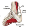
Label the diagram


Why is the achilles tendon vulnerable to pathology?
- Unlike other tendons it has no … …
- It is surrounded by a … (connective tissue sheath to ensure gliding )
- It has a poor blood supply
- i. Posterior tibial artery ( proximal and distal section)
- ii. Peroneal artery ( supplies midsection)
- Blood vascularity weakest at the bone –tendon interface
- Blood supply weakest at 2 to 6 cm form the calcaneal attachment
- Unlike other tendons it has no tendon sheath
- It is surrounded by a paratenon ( connective tissue sheath to ensure gliding )
- It has a poor blood supply
- i. Posterior tibial artery ( proximal and distal section)
- ii. Peroneal artery ( supplies midsection)
- Blood vascularity weakest at the bone –tendon interface
- Blood supply weakest at 2 to 6 cm form the calcaneal attachment
Why is the achilles tendon vulnerable to pathology?
- Unlike other tendons it has no tendon sheath
- It is surrounded by a paratenon ( connective tissue sheath to ensure …)
- It has a poor … …
- i. Posterior … artery (proximal and distal section)
- ii. … artery ( supplies midsection)
- Blood vascularity weakest at the bone –tendon interface
- Blood supply weakest at 2 to 6 cm form the calcaneal attachment
- Unlike other tendons it has no tendon sheath
- It is surrounded by a paratenon ( connective tissue sheath to ensure gliding )
- It has a poor blood supply
- i. Posterior tibial artery ( proximal and distal section)
- ii. Peroneal artery ( supplies midsection)
- Blood vascularity weakest at the bone –tendon interface
- Blood supply weakest at 2 to 6 cm form the calcaneal attachment
Why is the achilles tendon vulnerable to pathology?
- Unlike other tendons it has no tendon sheath
- It is surrounded by a paratenon ( connective tissue sheath to ensure gliding )
- It has a poor blood supply
- i. Posterior tibial artery ( proximal and distal section)
- ii. Peroneal artery ( supplies midsection)
- Blood vascularity weakest at the … –… interface
- Blood supply weakest at 2 to 6 cm from the … attachment
- Unlike other tendons it has no tendon sheath
- It is surrounded by a paratenon ( connective tissue sheath to ensure gliding )
- It has a poor blood supply
- i. Posterior tibial artery ( proximal and distal section)
- ii. Peroneal artery ( supplies midsection)
- Blood vascularity weakest at the bone –tendon interface
- Blood supply weakest at 2 to 6 cm form the calcaneal attachment
- Achilles tendon - Blood vascularity weakest at the … –tendon interface
- Blood supply weakest at .. to … cm form the calcaneal attachment
- Achilles tendon - Blood vascularity weakest at the bone –tendon interface
- Blood supply weakest at 2 to 6 cm form the calcaneal attachment
Achilles rupture
- Occurs after a sudden forced … to the foot
- Violent … in a planatar flexed foot
- Usually ruptures 4 to 6 cm above the calcaneal insertion in the hypovascular region
- Occurs after a sudden forced plantarflexion to the foot
- Violent dorsiflexion in a plantar flexed foot
- Usually ruptures 4 to 6 cm above the calcaneal insertion in the hypovascular region

Achilles rupture
- Occurs after a sudden forced … to the foot
- Violent dorsiflexion in a … foot
- Usually ruptures 4 to 6 cm above the … insertion in the hypovascular region
- Occurs after a sudden forced plantarflexion to the foot
- Violent dorsiflexion in a plantar flexed foot
- Usually ruptures 4 to 6 cm above the calcaneal insertion in the hypovascular region

Achilles Rupture - Treatment
- Most common - In Functional …
- Surgery
- … to … repair
- VY advancement
- Failure to heal- Tendon …
- Tendon used is the one closest in proximity – FLEXOR HALLUCIS LONGUS
- In Functional bracing
- Surgery
- End to end repair
- VY advancement
- Failure to heal- Tendon transfer
- Tendon used is the one closest in proximity – FLEXOR HALLUCIS LONGUS

Achilles Rupture - Treatment
- Most common - In Functional bracing
- Surgery
- End to end repair
- … advancement
- Failure to heal- … transfer
- Tendon used is the one closest in proximity – FLEXOR … …
- In Functional bracing
- Surgery
- End to end repair
- VY advancement
- Failure to heal- Tendon transfer
- Tendon used is the one closest in proximity – FLEXOR HALLUCIS LONGUS

What is tendon transfer?
This procedure repositions the flexor hallucis longus tendon, (commonly called the “FHL” tendon) to reinforce a diseased Achilles tendon.
When is tendon transfer used?
This procedure repositions the flexor hallucis longus tendon, (commonly called the “FHL” tendon) to reinforce a diseased Achilles tendon.
Surgical approach to the Achilles
- Patient is … (Requires intubation) or in lazy … position
- Landmarks: The malleoli and the Achilles tendon- which is easily …
- Incision: Longitudinal – slightly medially based ( but can go laterally as well)
- Structure to avoid : … nerve laterally
- Avoid going medial to Flexor hallucis longus (FHL) ( easily identifiable as has muscle fibres at this level ) - NEUROVASCULAR BUNDLE MEDIALLY
- Patient is prone (Requires intubation) or in lazy lateral position
- Landmarks: The malleoli and the Achilles tendon- which is easily palpable
- Incision: Longitudinal – slightly medially based ( but can go laterally as well)
- Structure to avoid : Sural nerve laterally
- Avoid going medial to Flexor hallucis longus (FHL) ( easily identifiable as has muscle fibres at this level ) - NEUROVASCULAR BUNDLE MEDIALLY
Surgical approach to the Achilles
- Patient is prone (Requires intubation) or in lazy lateral position
- Landmarks: The … and the Achilles tendon- which is easily palpable
- Incision: Longitudinal – slightly … based ( but can go … as well)
- Structure to avoid : Sural nerve laterally
- Avoid going medial to Flexor … … (FHL) ( easily identifiable as has muscle fibres at this level ) - NEUROVASCULAR BUNDLE MEDIALLY
- Patient is prone (Requires intubation) or in lazy lateral position
- Landmarks: The malleoli and the Achilles tendon- which is easily palpable
- Incision: Longitudinal – slightly medially based ( but can go laterally as well)
- Structure to avoid : Sural nerve laterally
- Avoid going medial to Flexor hallucis longus (FHL) ( easily identifiable as has muscle fibres at this level ) - NEUROVASCULAR BUNDLE MEDIALLY
Surgical approach to the Achilles
- Patient is prone (Requires …) or in lazy lateral position
- Landmarks: The malleoli and the Achilles tendon- which is easily palpable
- Incision: … – slightly medially based ( but can go laterally as well)
- Structure to avoid : Sural nerve laterally
- Avoid going medial to Flexor hallucis longus (FHL) ( easily identifiable as has muscle fibres at this level ) - … BUNDLE MEDIALLY
- Patient is prone (Requires intubation) or in lazy lateral position
- Landmarks: The malleoli and the Achilles tendon- which is easily palpable
- Incision: Longitudinal – slightly medially based ( but can go laterally as well)
- Structure to avoid : Sural nerve laterally
- Avoid going medial to Flexor hallucis longus (FHL) ( easily identifiable as has muscle fibres at this level ) - NEUROVASCULAR BUNDLE MEDIALLY
Surgical approach to the Achilles
- Patient is prone (Requires intubation) or in lazy lateral position
- Landmarks: The … and the Achilles tendon- which is easily palpable
- Incision: Longitudinal – slightly medially based ( but can go laterally as well)
- Structure to avoid : Sural nerve laterally
- Avoid going …. to Flexor hallucis longus (FHL) ( easily identifiable as has muscle fibres at this level ) - NEUROVASCULAR BUNDLE MEDIALLY
- Patient is prone (Requires intubation) or in lazy lateral position
- Landmarks: The malleoli and the Achilles tendon- which is easily palpable
- Incision: Longitudinal – slightly medially based ( but can go laterally as well)
- Structure to avoid : Sural nerve laterally
- Avoid going medial to Flexor hallucis longus (FHL) ( easily identifiable as has muscle fibres at this level ) - NEUROVASCULAR BUNDLE MEDIALLY
Tibialis Posterior Tendon
- … aspect of …. membrane, fibula and tibia and has … insertions in the foot
- Action-
- Plantarflexes the … joint
- Principal invertor of the foot
- Adducts and supinates the foot
- Arterial supply form the Posterior tibial, peroneal and sural nerve
- Has a watershed area around the medial malleoli
- Posterior aspect of interosseous membrane, fibula and tibia and has 9 insertions in the foot
- Action-
- Plantarflexes the ankle joint
- Principal invertor of the foot
- Adducts and supinates the foot
- Arterial supply form the Posterior tibial, peroneal and sural nerve
- Has a watershed area around the medial malleoli
Tibialis Posterior Tendon
- Posterior aspect of interosseous membrane, fibula and tibia and has 9 insertions in the foot
- Action-
- …. the ankle joint
- Principal … of the foot
- Adducts and … the foot
- … supply form the Posterior tibial, peroneal and sural nerve
- Has a watershed area around the medial …
- Posterior aspect of interosseous membrane, fibula and tibia and has 9 insertions in the foot
- Action-
- Plantarflexes the ankle joint
- Principal invertor of the foot
- Adducts and supinates the foot
- Arterial supply form the Posterior tibial, peroneal and sural nerve
- Has a watershed area around the medial malleoli

What tendon is this?

Tibialis posterior tendon
Function of Tibialis Posterior Tendon
- … lower leg
- Facilitates foot …
- Supports the foot’s … arch
- Plays a critical role in hindfoot inversion during the gait cycle
- Stabilise lower leg
- Facilitates foot inversion
- Supports the foot’s medial arch
- Plays a critical role in hindfoot inversion during the gait cycle
Function of Tibialis Posterior Tendon
- Stabilise … leg
- Facilitates foot inversion
- Supports the foot’s medial arch
- Plays a critical role in … inversion during the … cycle
- Stabilise lower leg
- Facilitates foot inversion
- Supports the foot’s medial arch
- Plays a critical role in hindfoot inversion during the gait cycle
Tibialis Posterior Insufficiency
- Presenting symptoms/signs
- Post-… pain
- … pain + aching
- Progressive … foot deformity
- Forefoot problems: progressive hallux valgus, metatarsalgia, lesser toe deformities
- Rarely, tarsal tunnel syndrome
- Presenting symptoms/signs
- Post-malleolar pain
- Arch pain + aching
- Progressive flat foot deformity
- Forefoot problems: progressive hallux valgus, metatarsalgia, lesser toe deformities
- Rarely, tarsal tunnel syndrome
Tibialis Posterior Insufficiency
- Presenting symptoms/signs
- Post-malleolar pain
- Arch pain + aching
- Progressive flat foot deformity
- … problems: progressive hallux …, metatarsalgia, … toe deformities
- Rarely, … tunnel syndrome
- Presenting symptoms/signs
- Post-malleolar pain
- Arch pain + aching
- Progressive flat foot deformity
- Forefoot problems: progressive hallux valgus, metatarsalgia, lesser toe deformities
- Rarely, tarsal tunnel syndrome
Tibialis Posterior Insufficiency


Tibialis Posterior Insufficiency- Treatment
- Non-surgical
- …
- … wear modification
- Orthotics- … arch supports
- Physiotherapy
- Surgery
- Reconstruction (tendon transfer)
- Fusion (if secondary arthritis)
- Non-surgical
- Analgesics
- Shoe wear modification
- Orthotics - medial arch supports
- Physiotherapy
- Surgery
- Reconstruction (tendon transfer)
- Fusion (if secondary arthritis)
Tibialis Posterior Insufficiency- Treatment
- Non-surgical
- Analgesics
- Shoe wear modification
- …- medial arch supports
- …therapy
- Surgery
- Reconstruction (tendon transfer)
- Fusion (if secondary arthritis)
- Non-surgical
- Analgesics
- Shoe wear modification
- Orthotics- medial arch supports
- Physiotherapy
- Surgery
- Reconstruction (tendon transfer)
- Fusion (if secondary arthritis)
Tibialis Posterior Insufficiency- Treatment
- Non-surgical
- Analgesics
- Shoe wear modification
- Orthotics- medial arch supports
- Physiotherapy
- Surgery
- … (… transfer)
- … (if secondary arthritis)
- Non-surgical
- Analgesics
- Shoe wear modification
- Orthotics- medial arch supports
- Physiotherapy
- Surgery
- Reconstruction (tendon transfer)
- Fusion (if secondary arthritis)
Tibialis Posterior Insufficiency- Treatment
- Non-…
- Analgesics
- Shoe wear modification
- Orthotics - … arch supports
- Physiotherapy
- …
- Reconstruction (tendon transfer)
- Fusion (if secondary arthritis)
- Non-surgical
- Analgesics
- Shoe wear modification
- Orthotics - medial arch supports
- Physiotherapy
-
Surgery
- Reconstruction (tendon transfer)
- Fusion (if secondary arthritis)
What are the two surgical options for tibialis posterior insufficiency?
reconstruction (tendon transfer) or fusion (if secondary arthritis)
Tibialis Posterior Dysfunction - Which tendon to use for the transfer?
Tibialis Posterior Dysfunction - Which tendon to use for the transfer? (flexor digitorum)

Surgical Approach to Tibialis Posterior tendon
- Position: …
- Landmark: Tip of medial … and the base of the …
- Incision : 10 cm longitudinal incision from tip of MM
- Internervous plane : None
- Dissection : Avoid damage to the long saphenous vein and nerve
- Divide the retinaculum and identify the tibialis posterior tendon by pulling on it
- (FHL will plantar flex the great toe and FDL will plantarflex the lesser toes)
- Dangers: Saphenous nerve and the tibialis posterior tendon are particularly vulnerable
- Position: Supine
- Landmark: Tip of medial malleoli and the base of the navicular
- Incision : 10 cm longitudinal incision from tip of MM
- Internervous plane : None
- Dissection : Avoid damage to the long saphenous vein and nerve
- Divide the retinaculum and identify the tibialis posterior tendon by pulling on it
- (FHL will plantar flex the great toe and FDL will plantarflex the lesser toes)
- Dangers: Saphenous nerve and the tibialis posterior tendon are particularly vulnerable
Surgical Approach to Tibialis Posterior tendon
- Position: Supine
- Landmark: Tip of medial malleoli and the base of the navicular
- Incision : … cm longitudinal incision from tip of MM
- Internervous plane : None
- Dissection : Avoid damage to the long … vein and nerve
- Divide the retinaculum and identify the tibialis posterior tendon by pulling on it
- (FHL will plantar flex the great toe and FDL will plantarflex the lesser toes)
- Dangers: Saphenous nerve and the tibialis posterior tendon are particularly vulnerable
- Position: Supine
- Landmark: Tip of medial malleoli and the base of the navicular
- Incision : 10 cm longitudinal incision from tip of MM
- Internervous plane : None
- Dissection : Avoid damage to the long saphenous vein and nerve
- Divide the retinaculum and identify the tibialis posterior tendon by pulling on it
- (FHL will plantar flex the great toe and FDL will plantarflex the lesser toes)
- Dangers: Saphenous nerve and the tibialis posterior tendon are particularly vulnerable
Surgical Approach to Tibialis Posterior tendon
- Position: Supine
- Landmark: Tip of … malleoli and the base of the navicular
- Incision : 10 cm longitudinal incision from tip of MM
- Internervous plane : None
- Dissection : Avoid damage to the long saphenous vein and nerve
- Divide the retinaculum and identify the tibialis posterior tendon by pulling on it
- (FHL will plantar flex the … toe and FDL will plantarflex the … toes)
- Dangers: Saphenous nerve and the … … tendon are particularly vulnerable
- Position: Supine
- Landmark: Tip of medial malleoli and the base of the navicular
- Incision : 10 cm longitudinal incision from tip of MM
- Internervous plane : None
- Dissection : Avoid damage to the long saphenous vein and nerve
- Divide the retinaculum and identify the tibialis posterior tendon by pulling on it
- (FHL will plantar flex the great toe and FDL will plantarflex the lesser toes)
- Dangers: Saphenous nerve and the tibialis posterior tendon are particularly vulnerable
Danger of surgical approach to tibialis posterior tendon - … nerve and the tibialis posterior tendon are particularly vulnerable
Danger of surgical approach to tibialis posterior tendon - Saphenous nerve and the tibialis posterior tendon are particularly vulnerable
Tibialis Posterior Dysfunction
- Identifying it

Ankle Arthritis
- Usually post-…
- Presentation:
- Pain
- …
- Deformity
- Usually post-traumatic
- Presentation:
- Pain
- Swelling
- Deformity
Ankle Arthritis
- Usually post-traumatic
- Presentation:
- …
- Swelling
- …
- Usually post-traumatic
- Presentation:
- Pain
- Swelling
- Deformity
Pathology - Ankle Arthritis
- Nasty … – cartilage damage
- Malalignment – leads to abnormal loading
- … altered in the ankle joint
- Leads to abnormal point loading
- Eventual joint space narrowing and pain
- Nasty fracture – cartilage damage
- Malalignment – leads to abnormal loading
- Biomechanics altered in the ankle joint
- Leads to abnormal point loading
- Eventual joint space narrowing and pain
Pathology - Ankle Arthritis
- Nasty fracture – cartilage damage
- … – leads to abnormal loading
- Biomechanics altered in the ankle joint
- Leads to abnormal … loading
- Eventual joint space narrowing and pain
- Nasty fracture – cartilage damage
- Malalignment – leads to abnormal loading
- Biomechanics altered in the ankle joint
- Leads to abnormal point loading
- Eventual joint space narrowing and pain
Pathology - Ankle Arthritis
- Nasty fracture – cartilage damage
- Malalignment – leads to abnormal …
- Biomechanics altered in the ankle joint
- Leads to abnormal point loading
- Eventual joint space … and pain
- Nasty fracture – cartilage damage
- Malalignment – leads to abnormal loading
- Biomechanics altered in the ankle joint
- Leads to abnormal point loading
- Eventual joint space narrowing and pain
Pathology - Ankle Arthritis
- Nasty fracture – cartilage …
- … – leads to abnormal loading
- Biomechanics altered in the ankle joint
- Leads to abnormal point loading
- Eventual joint space narrowing and pain
- Nasty fracture – cartilage damage
- Malalignment – leads to abnormal loading
- Biomechanics altered in the ankle joint
- Leads to abnormal point loading
- Eventual joint space narrowing and pain
Surgical Management of Ankle Arthritis
- Failed medical / non-operative control…………
- “Early” disease - joint …
- Arthroscopy (or open procedure)
- Debridement / synovectomy
- Arthroscopy (or open procedure)
- “Late” disease – joint … or replacement
- Arthrodesis (fusion)
- Arthroplasty (replacement)
- Excision Arthroplasty (excision joint)
- Failed medical / non-operative control…………
- “Early” disease - joint preservation
- Arthroscopy (or open procedure)
- Debridement / synovectomy
- Arthroscopy (or open procedure)
- “Late” disease – joint abolition or replacement
- Arthrodesis (fusion)
- Arthroplasty (replacement)
- Excision Arthroplasty (excision joint)
Surgical Management of Ankle Arthritis
- Failed medical / non-operative control…………
- “…” disease - joint preservation
- Arthroscopy (or open procedure)
- Debridement / synovectomy
- Arthroscopy (or open procedure)
- “…” disease – joint abolition or …
- Arthrodesis (…)
- Arthroplasty (replacement)
- Excision Arthroplasty (excision joint)
- Failed medical / non-operative control…………
- “Early” disease - joint preservation
- Arthroscopy (or open procedure)
- Debridement / synovectomy
- Arthroscopy (or open procedure)
- “Late” disease – joint abolition or replacement
- Arthrodesis (fusion)
- Arthroplasty (replacement)
- Excision Arthroplasty (excision joint)
Surgical Management of Ankle Arthritis
- Failed medical / non-operative control…………
- “Early” disease - joint preservation
- Arthroscopy (or open procedure)
- … / synovectomy
- Arthroscopy (or open procedure)
- “Late” disease – joint abolition or replacement
- Arthrodesis (fusion)
- … (replacement)
- Excision … (excision joint)
- Failed medical / non-operative control…………
- “Early” disease - joint preservation
- Arthroscopy (or open procedure)
- Debridement / synovectomy
- Arthroscopy (or open procedure)
- “Late” disease – joint abolition or replacement
- Arthrodesis (fusion)
- Arthroplasty (replacement)
- Excision Arthroplasty (excision joint)
Ankle Arthroplasty
- … relief
- Preservation of joint …
- Preservation of function
- Polyarthropathy; Subtalar / Triple complex
- Compare with ankle arthrodesis
- Pain relief
- Preservation of joint mobility
- Preservation of function
- Polyarthropathy; Subtalar / Triple complex
- Compare with ankle arthrodesis
Ankle Arthroplasty
- Pain relief
- Preservation of joint mobility
- Preservation of …
- …; Subtalar / Triple complex
- Compare with ankle …
- Pain relief
- Preservation of joint mobility
- Preservation of function
-
Polyarthropathy; Subtalar / Triple complex
- Compare with ankle arthrodesis
Ankle Fusion/ Arthrodesis
- Indications
- … Relief
- Severe …
- TAR not appropriate
- Indications
- Pain Relief
- Severe Deformity
- TAR not appropriate

Ankle Fusion/ Arthrodesis
- Indications
- Pain Relief
- Severe Deformity
- … not appropriate
- Indications
- Pain Relief
- Severe Deformity
- TAR not appropriate

Anterior Approach to the Ankle for TAR
- Position : …
- Landmarks: Both the … which are subcutaneous
- Incision : 15 cm longitudinal incision midway between the malleoli
- Internervous Plane: None
- Intermuscular plane : Between EHL and EDL ( both supplied by deep peroneal but receive their supply proximal to the incision)
- Dissection : Find the … bundle and mobilise laterally
- Dangers: i. Superficial peroneal nerve ii. Deep peroneal nerve iii. Anterior tibial artery
- Position : Supine
- Landmarks: Both the malleoli which are subcutaneous
- Incision : 15 cm longitudinal incision midway between the malleoli
- Internervous Plane: None
- Intermuscular plane : Between EHL and EDL ( both supplied by deep peroneal but receive their supply proximal to the incision)
- Dissection : Find the neurovascular bundle and mobilise laterally
- Dangers: i. Superficial peroneal nerve ii. Deep peroneal nerve iii. Anterior tibial artery
Anterior Approach to the Ankle for TAR
- Position : Supine
- Landmarks: Both the malleoli which are sub…
- Incision : … cm longitudinal incision midway between the malleoli
- Internervous Plane: None
- Intermuscular plane : Between EHL and EDL ( both supplied by deep peroneal but receive their supply proximal to the incision)
- Dissection : Find the neurovascular bundle and mobilise …
- Dangers: i. Superficial peroneal nerve ii. Deep peroneal nerve iii. Anterior tibial artery
- Position : Supine
- Landmarks: Both the malleoli which are subcutaneous
- Incision : 15 cm longitudinal incision midway between the malleoli
- Internervous Plane: None
- Intermuscular plane : Between EHL and EDL ( both supplied by deep peroneal but receive their supply proximal to the incision)
- Dissection : Find the neurovascular bundle and mobilise laterally
- Dangers: i. Superficial peroneal nerve ii. Deep peroneal nerve iii. Anterior tibial artery
Anterior Approach to the Ankle for TAR
- Position : Supine
- Landmarks: Both the malleoli which are subcutaneous
- Incision : 15 cm longitudinal incision midway between the malleoli
- … Plane: None
- … plane : Between EHL and EDL (both supplied by deep peroneal but receive their supply proximal to the incision)
- Dissection : Find the neurovascular bundle and mobilise laterally
- Dangers: i. Superficial … nerve ii. Deep … nerve iii. Anterior tibial artery
- Position : Supine
- Landmarks: Both the malleoli which are subcutaneous
- Incision : 15 cm longitudinal incision midway between the malleoli
- Internervous Plane: None
- Intermuscular plane : Between EHL and EDL ( both supplied by deep peroneal but receive their supply proximal to the incision)
- Dissection : Find the neurovascular bundle and mobilise laterally
- Dangers: i. Superficial peroneal nerve ii. Deep peroneal nerve iii. Anterior tibial artery
Anterior Approach to the Ankle for TAR - dangers (Which nerves (2) and artery)
Dangers: i. Superficial peroneal nerve ii. Deep peroneal nerve iii. Anterior tibial artery
Tibiotalocalcaneal Arthrodesis (TTC)
- Severe …
- … ankle fractures
- Complex failed ankle fixation
- Failed TAR
- * If TNJ / CCJ involved – consider pantalar arthrodesis
- Severe deformity
- Osteoporotic ankle fractures
- Complex failed ankle fixation
- Failed TAR
- * If TNJ / CCJ involved – consider pantalar arthrodesis

Tibiotalocalcaneal Arthrodesis (TTC)
- Severe deformity
- Osteoporotic ankle fractures
- Complex failed ankle …
- Failed TAR
- * If TNJ / CCJ involved – consider pantalar …
- Severe deformity
- Osteoporotic ankle fractures
- Complex failed ankle fixation
- Failed TAR
- * If TNJ / CCJ involved – consider pantalar arthrodesis

Tibiotalocalcaneal Arthrodesis (TTC)
- Severe deformity
- Osteoporotic ankle fractures
- Complex … ankle fixation
- Failed …
- * If TNJ / CCJ involved – consider pantalar arthrodesis
- Severe deformity
- Osteoporotic ankle fractures
- Complex failed ankle fixation
- Failed TAR
- * If TNJ / CCJ involved – consider pantalar arthrodesis

Ankle “Sprains”
- … ligament
- Passes form anterior margin of the fibular malleolus, to the talus bone

- Lateral ligament
- Passes form anterior margin of the fibular malleolus, to the talus bone

Ankle “Sprains”
- Lateral ligament
- Passes from anterior margin of the … malleolus, to the … bone

- Lateral ligament
- Passes form anterior margin of the fibular malleolus, to the talus bone

ATFL (Anterior talofibular ) /CFL (Calcaneofibular)
- In ankle ‘…’ ligaments affected (+posterior talofibular)
- Weakest and commonly …
- Commonly gets bruised and stretched during inversion injuries
- Prevents talar tilt
- If weak then the ankle feels …
- Positive anterior drawer test
- Positive talar tilt test
- In ankle ‘sprain’ ligaments affected (+posterior talofibular)
- Weakest and commonly injured
- Commonly gets bruised and stretched during inversion injuries
- Prevents talar tilt
- If weak then the ankle feels unstable
- Positive anterior drawer test
- Positive talar tilt test
ATFL (Anterior talofibular ) /CFL (Calcaneofibular)
- In ankle ‘sprain’ ligaments affected (+posterior talofibular)
- Weakest and commonly injured
- Commonly gets … and stretched during inversion injuries
- Prevents … tilt
- If weak then the ankle feels unstable
- Positive … drawer test
- Positive talar tilt test
- In ankle ‘sprain’ ligaments affected (+posterior talofibular)
- Weakest and commonly injured
- Commonly gets bruised and stretched during inversion injuries
- Prevents talar tilt
- If weak then the ankle feels unstable
- Positive anterior drawer test
- Positive talar tilt test
ATFL (Anterior talofibular ) /CFL (Calcaneofibular)
- In ankle ‘sprain’ ligaments affected (+posterior talofibular)
- Weakest and commonly injured
- Commonly gets bruised and … during … injuries
- Prevents talar tilt
- If weak then the ankle feels unstable
- Positive anterior drawer test
- Positive … … test
- In ankle ‘sprain’ ligaments affected (+posterior talofibular)
- Weakest and commonly injured
- Commonly gets bruised and stretched during inversion injuries
- Prevents talar tilt
- If weak then the ankle feels unstable
- Positive anterior drawer test
- Positive talar tilt test
Ankle “Sprains”
- Majority sprains recover within 3 months
- Beware the sprain that …
- Back to basics – clinical …
- Majority sprains recover within 3 months
- Beware the sprain that persists…..
- Back to basics – clinical reassessment
The majority of ankle sprains recover within … months
3 months
Ankle “Sprains” - Acute lateral ligament strain (ATFL) / CFL
- RICE (… … COMPRESSION ELEVATION)
- Physiotherapy directed …
- Loading injured ligaments
- Proprioception
- Strength and return to …
- RICE (REST ICE COMPRESSION ELEVATION)
- Physiotherapy directed rehabilitation
- Loading injured ligaments
- Proprioception
- Strength and return to function
Ankle “Sprains” - Acute lateral ligament strain (ATFL) / CFL
- RICE (REST ICE … …)
- Physiotherapy directed rehabilitation
- … injured ligaments
- …ception
- Strength and return to function
- RICE (REST ICE COMPRESSION ELEVATION)
- Physiotherapy directed rehabilitation
- Loading injured ligaments
- Proprioception
- Strength and return to function
After ankle sprain - may get Diagnosis of Chronic …
After ankle sprain, may be left with chronic instability - may get a diagnosis: Chronic Instability

Diagnosis: Chronic Instability (ankle)
- Examine - … … draw (sulcus sign)
- Diagnose - further investigations - used to do stress …
but MRI more common now (very useful to demonstrate related pathology)
- Examine - positive anterior draw (sulcus sign)
- Diagnose - further investigations - used to do stress radiographs
but MRI more common now (very useful to demonstrate related pathology)

Diagnosis: Chronic Instability (ankle)
- Examine - positive anterior draw (… sign)
- Diagnose - further investigations - used to do stress radiographs
but … more common now (very useful to demonstrate related pathology)
- Examine - positive anterior draw (sulcus sign)
- Diagnose - further investigations - used to do stress radiographs
but MRI more common now (very useful to demonstrate related pathology)

Indications For Surgery - ankle sprain
- Do we in acute rupture?
- In chronic mechanical instability symptoms not responding to non operative rehabilitation?
- Do we in acute rupture? - no
- In chronic mechanical instability symptoms not responding to non operative rehabilitation? - yes

Surgical Options - Ankle sprain
- … … tissue over eachother with … to tighten - sometimes tissue too flimsy = use anchors within bone to gain strength
- Can use … also - adjunct
- If fail - tendon transfer (peroneus brevis tendon)
- Double breast tissue over eachother with sutures to tighten - sometimes tissue too flimsy = use anchors within bone to gain strength
- Can use tapes also - adjunct
- If fail - tendon transfer (peroneus brevis tendon)

Surgical Options - Ankle sprain
- Double breast tissue over eachother with sutures to tighten - sometimes tissue too flimsy = use … within bone to gain strength
- Can use tapes also - adjunct
- If fail - tendon transfer (… … tendon)
- Double breast tissue over eachother with sutures to tighten - sometimes tissue too flimsy = use anchors within bone to gain strength
- Can use tapes also - adjunct
- If fail - tendon transfer (peroneus brevis tendon)

Hallux Valgus
- Hallux valgus - deformity - big toe into valgus
- Often called … - which is the prominent median eminence
- Commonly … finding
- Occur in females more than males
- Family history +/- …
- No symptom = no surgery
- Hallux valgus - deformity - big toe into valgus
- Often called bunion - prominent median eminence is the bunion
- Commonly incidental finding
- Occur in females more than males
- Family history +/- footwear
- No symptom = no surgery

Hallux Valgus
- Hallux valgus - deformity - big toe into valgus
- Often called bunion - prominent median eminence is the bunion
- Commonly incidental finding
- Occur in … more than …
- … history +/- footwear
- No … = no surgery
- Hallux valgus - deformity - big toe into valgus
- Often called bunion - prominent median eminence is the bunion
- Commonly incidental finding
- Occur in females more than males
- Family history +/- footwear
- No symptom = no surgery

Hallux Valgus - Symptoms and Signs (5)
- Pain
- Deformity
- Modification of shoe wear
- Nerve irritation
- Lesser toe deformity
Hallux Valgus - Symptoms and Signs (5)
- Pain
- Deformity
- … of shoe wear
- … irritation
- … toe deformity
- Pain
- Deformity
- Modification of shoe wear
- Nerve irritation
- Lesser toe deformity
Hallux Valgus - Treatment
- Non- Surgical
- Surgical
- …nectomy
- …tomy
- Proximal
- Metatarsal shaft
- Distal
- Ist TMT joint fusion
- Technique used depends on the cause and the amount of correction required
- Non- Surgical
- Surgical
- Bunionectomy
-
Osteotomy
- Proximal
- Metatarsal shaft
- Distal
- Ist TMT joint fusion
- Technique used depends on the cause and the amount of correction required
Hallux Valgus - Treatment
- Non- Surgical
- Surgical
- Bunionectomy
- Osteotomy
- Proximal
- … shaft
- Distal
- Ist … joint fusion
- Technique used depends on the cause and the amount of correction required
- Non- Surgical
- Surgical
- Bunionectomy
- Osteotomy
- Proximal
- Metatarsal shaft
- Distal
- Ist TMT joint fusion
- Technique used depends on the cause and the amount of correction required
Biomechanics - Hallux Valgus Deformity
- … deviation of the 1st ray (The first ray is the segment of the foot composed of the first metatarsal and first cuneiform bones)
- Medial deviation of the 1st ray (The first ray is the segment of the foot composed of the first metatarsal and first cuneiform bones)

Dorsomedial Approach to the Great Toe (hallux valgus deformity)
- Position: …
- Landmarks: easily palpable 1st … joint
- Incision: Proximal to the IP joint and curve over the medial eminence staying medial to EHL
- Internervous Plane : none
- Dissection: divide fascia and then periosteum
- Dangers: i. Dorsal cutanous nerve ii. Extensor hallucis tendon iii, Flexor hallucis longus

- Position: Supine
- Landmarks: easily palpable 1st MTP joint
- Incision: Proximal to the IP joint and curve over the medial eminence staying medial to EHL
- Internervous Plane : none
- Dissection: divide fascia and then periosteum
- Dangers: i. Dorsal cutanous nerve ii. Extensor hallucis tendon iii, Flexor hallucis longus
Dorsomedial Approach to the Great Toe (hallux valgus deformity)
- Position: Supine
- Landmarks: easily palpable 1st MTP joint
- Incision: … to the IP joint and curve over the … eminence staying medial to EHL
- Internervous Plane : none
- Dissection: divide fascia and then periosteum
- Dangers: i. Dorsal cutanous nerve ii. Extensor hallucis tendon iii, Flexor hallucis longus

- Position: Supine
- Landmarks: easily palpable 1st MTP joint
- Incision: Proximal to the IP joint and curve over the medial eminence staying medial to EHL
- Internervous Plane : none
- Dissection: divide fascia and then periosteum
- Dangers: i. Dorsal cutanous nerve ii. Extensor hallucis tendon iii, Flexor hallucis longus
Dorsomedial Approach to the Great Toe (hallux valgus deformity)
- Position: Supine
- Landmarks: easily … 1st MTP joint
- Incision: Proximal to the IP joint and curve over the medial eminence staying medial to EHL
- Internervous Plane : none
- Dissection: divide … and then periosteum
- Dangers: i. Dorsal cutanous nerve ii. Extensor hallucis tendon iii, Flexor hallucis longus

- Position: Supine
- Landmarks: easily palpable 1st MTP joint
- Incision: Proximal to the IP joint and curve over the medial eminence staying medial to EHL
- Internervous Plane : none
- Dissection: divide fascia and then periosteum
- Dangers: i. Dorsal cutanous nerve ii. Extensor hallucis tendon iii, Flexor hallucis longus
Dorsomedial Approach to the Great Toe (hallux valgus deformity)
- Position: Supine
- Landmarks: easily palpable … MTP joint
- Incision: Proximal to the IP joint and curve over the medial eminence staying medial to EHL
- Internervous Plane : none
- Dissection: divide fascia and then periosteum
- Dangers: i. Dorsal … nerve ii. Extensor … tendon iii, Flexor … longus

- Position: Supine
- Landmarks: easily palpable 1st MTP joint
- Incision: Proximal to the IP joint and curve over the medial eminence staying medial to EHL
- Internervous Plane : none
- Dissection: divide fascia and then periosteum
- Dangers: i. Dorsal cutanous nerve ii. Extensor hallucis tendon iii, Flexor hallucis longus
Dorsomedial Approach to the Great Toe (hallux valgus deformity) - Dangers
- i. … … nerve ii. … hallucis tendon iii, … hallucis longus

- i. Dorsal cutanous nerve ii. Extensor hallucis tendon iii, Flexor hallucis longus
Dorsomedial Approach to the Great Toe (hallux valgus deformity)
- Position: Supine
- Landmarks: easily palpable 1st MTP joint
- Incision: Proximal to the IP joint and curve over the medial eminence staying … to EHL
- Internervous Plane : …
- Dissection: divide fascia and then periosteum
- Dangers: i. Dorsal cutanous nerve ii. Extensor hallucis tendon iii, Flexor hallucis longus

- Position: Supine
- Landmarks: easily palpable 1st MTP joint
- Incision: Proximal to the IP joint and curve over the medial eminence staying medial to EHL
- Internervous Plane : none
- Dissection: divide fascia and then periosteum
- Dangers: i. Dorsal cutanous nerve ii. Extensor hallucis tendon iii, Flexor hallucis longus
Take home message - surgery of foot and ankle
- Do not rely on x rays to define the injury
- History taking and examination is the key to an accurate diagnosis
- Think of the anatomy to understand the pathology
- Exhaust non-surgical options first before considering surgical intervention
- Before you operate : think of using the best surgical approach for that case