Innate Immune Defences & Inflammation 2: The Induced Response Flashcards
Cells of the innate & adaptive immune system
- The discovery of innate lymphoid cells (ILCs) is blurring the traditional boundaries between innate and adaptive immune systems. Invariant natural killer T cells, some B cells at epithelial barriers (B1 cells) and gamma delta T cells have innate qualities whereas NK cells may adapt after their first encounter with a pathogen due to innate immune memory. T and B cells also have innate immune receptors such as TLRs.
- The discovery of innate lymphoid cells (ILCs) is blurring the traditional boundaries between innate and adaptive immune systems. Invariant natural killer T cells, some B cells at epithelial barriers (B1 cells) and gamma delta T cells have innate qualities whereas NK cells may adapt after their first encounter with a pathogen due to innate immune memory. T and B cells also have innate immune receptors such as TLRs.

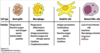
Innate immune cells
- What cells are involved? What do they do?
- Neutrophils - Phagocytosis, Antimicrobial peptides, Reactive oxygen and nitrogen species
- Macrophages - Phagocytosis, Antimicrobial peptides, Reactive oxygen and nitrogen species, Inflammatory mediators, antigen presentation, cytokines, complement proteins
- Dendritic cells - Antigen presentation, Costimulatory signals, Reactive oxygen species, Interferon, Cytokines
- Natural Killer cells - Lysis of viral-infected cells, Interferon, Macrophage activation, Granzyme, Perforin

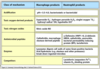
What are the functions of a neutrophil?
- Phagocytosis, Antimicrobial peptides, Reactive oxygen and nitrogen species
What are the functions of a macrophage?
- Phagocytosis, Antimicrobial peptides, Reactive oxygen and nitrogen species, Inflammatory mediators, antigen presentation, cytokines, complement proteins

What are the functions of dendritic cells?
- Antigen presentation, Costimulatory signals, Reactive oxygen species, Interferon, Cytokines (Plasmacytoid dendritic cells (DCs) are of lymphoid origin, Myeloid DCs are of myeloid origin - plasmacytoid DCs produce large amounts of type 1 IFN whereas for myeloid DCs the main role is antigen presentation)

What are the functions of natural killer cells?
- Lysis of viral-infected cells, Interferon, Macrophage activation, Granzyme, Perforin (Perforin is pore forming permitting entry of granzyme into cells where it induces apoptosis)

… is pore forming permitting entry of granzyme into cells where it induces apoptosis.
Perforin is pore forming permitting entry of granzyme into cells where it induces apoptosis.
Plasmacytoid dendritic cells (DCs) are of … origin
Plasmacytoid dendritic cells (DCs) are of lymphoid origin
Myeloid DCs are of … origin
Myeloid DCs are of myeloid origin
… DCs produce large amounts of type 1 IFN whereas for … DCs the main role is antigen presentation
plasmacytoid DCs produce large amounts of type 1 IFN whereas for myeloid DCs the main role is antigen presentation
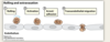
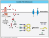
Phagocyte recruitment
- … produced by macrophages dilate local blood vessels and increase endothelial adhesion molecule expression.
- … attract monocytes and neutrophils to the infection
- Cell adhesion molecules (ICAM-1 and VCAM-1) are upregulated on the endothelium which bind to integrins (family of adhesion molecules) on the leukocytes
- Cytokines produced by macrophages dilate local blood vessels and increase endothelial adhesion molecule expression.
- Chemokines attract monocytes and neutrophils to the infection
- Cell adhesion molecules (ICAM-1 and VCAM-1) are upregulated on the endothelium which bind to integrins (family of adhesion molecules) on the leukocytes

Phagocyte recruitment
- Cytokines produced by macrophages … local blood vessels and … endothelial adhesion molecule expression.
- Chemokines attract monocytes and … to the infection
- Cell adhesion molecules (ICAM-1 and VCAM-1) are upregulated on the endothelium which bind to integrins (family of adhesion molecules) on the leukocytes
- Cytokines produced by macrophages dilate local blood vessels and increase endothelial adhesion molecule expression.
- Chemokines attract monocytes and neutrophils to the infection
- Cell adhesion molecules (ICAM-1 and VCAM-1) are upregulated on the endothelium which bind to integrins (family of adhesion molecules) on the leukocytes

Phagocyte recruitment
- Cytokines produced by macrophages dilate local blood vessels and increase endothelial adhesion molecule expression.
- Chemokines attract monocytes and neutrophils to the infection
- Cell … molecules (ICAM-1 and VCAM-1) are upregulated on the endothelium which bind to integrins (family of … molecules) on the leukocytes
- Cytokines produced by macrophages dilate local blood vessels and increase endothelial adhesion molecule expression.
- Chemokines attract monocytes and neutrophils to the infection
- Cell adhesion molecules (ICAM-1 and VCAM-1) are upregulated on the endothelium which bind to integrins (family of adhesion molecules) on the leukocytes
Phagocyte recruitment
- Cytokines produced by macrophages dilate local blood vessels and increase endothelial adhesion molecule expression.
- Chemokines attract monocytes and neutrophils to the infection
- Cell adhesion molecules (…-1 and …-1) are upregulated on the endothelium which bind to integrins (family of adhesion molecules) on the leukocytes
- Cytokines produced by macrophages dilate local blood vessels and increase endothelial adhesion molecule expression.
- Chemokines attract monocytes and neutrophils to the infection
- Cell adhesion molecules (ICAM-1 and VCAM-1) are upregulated on the endothelium which bind to integrins (family of adhesion molecules) on the leukocytes

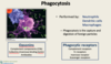
Phagocytosis is performed by …, … cells and …
Phagocytosis is performed by neutrophils, dendritic cells and macrophages
Neutrophils, Dendritic Cells and Macrophages all perform…
phagocytosis
Phagocytosis is the … and … of … particles
Phagocytosis is the capture and digestion of foreign particles
Phagocytosis is the … and … of … particles
Phagocytosis is the capture and digestion of foreign particles
What is phagocytosis?
Phagocytosis is the capture and digestion of foreign particles
… are proteins of the innate and adaptive immune system that facilitate phagocytosis and cell lysis by “marking” antigen.
Opsonins are proteins of the innate and adaptive immune system that facilitate phagocytosis and cell lysis by “marking” antigen.
Complement components (C3b) and Collectins (Mannose-binding lectin) and antibodies are all good examples of …
opsonins (opsonins are proteins of the innate and adaptive immune system that facilitate phagocytosis and cell lysis by “marking” antigen.)

Opsonins engage with … receptors (complement receptors, fc receptors, Mannose receptor, Scavenger receptors)
Opsonins engage with phagocytic receptors (complement receptors, fc receptors, Mannose receptor, Scavenger receptors)
… receptors recognize bacteria, viruses and apoptotic cells
Scavenger receptors recognize bacteria, viruses and apoptotic cells
The complement receptor CR1 binds to … (complement component)
The complement receptor CR1 binds to C3b (complement component)
C-type-lectin receptors (Dectin-1 & mannose receptor) help … bacteria. Mannose Receptor binds mannose and fructose residues of glycans (polysaccharides).
C-type-lectin receptors (Dectin-1 & mannose receptor) help phagocytose bacteria. Mannose Receptor binds mannose and fructose residues of glycans (polysaccharides).
Receptor mediated phagocytosis
- … receptors bind to opsonins
- Microorganisms are internalised by receptor-mediated endocytosis
- Fusion of … with lysosome forms a phagolysosome in which microorganisms are degraded
- Phagocytic receptors bind to opsonins
- Microorganisms are internalised by receptor-mediated endocytosis
- Fusion of endosome with lysosome forms a phagolysosome in which microorganisms are degraded

Receptor mediated phagocytosis
- Phagocytic receptors bind to opsonins
- Microorganisms are internalised by receptor-mediated endocytosis
- Fusion of endosome with … forms a … in which microorganisms are degraded
- Phagocytic receptors bind to opsonins
- Microorganisms are internalised by receptor-mediated endocytosis
- Fusion of endosome with lysosome forms a phagolysosome in which microorganisms are degraded

Receptor mediated phagocytosis
- Phagocytic receptors bind to …
- Microorganisms are internalised by receptor-mediated endocytosis
- Fusion of endosome with lysosome forms a phagolysosome in which microorganisms are degraded
- Phagocytic receptors bind to opsonins
- Microorganisms are internalised by receptor-mediated endocytosis
- Fusion of endosome with lysosome forms a phagolysosome in which microorganisms are degraded

Receptor mediated phagocytosis
- Phagocytic receptors bind to opsonins
- Microorganisms are internalised by receptor-mediated …
- Fusion of endosome with lysosome forms a phagolysosome in which microorganisms are degraded
- Phagocytic receptors bind to opsonins
- Microorganisms are internalised by receptor-mediated endocytosis
- Fusion of endosome with lysosome forms a phagolysosome in which microorganisms are degraded

Lysosomes can fuse with … to form a …
Lysosomes can fuse with phagosomes to form a phagolysosome
Antimicrobial mechanisms of phagocytes
- Within phagolysosome:
- … environment - … pH
- Oxygen and nitrogen derived products - break down pathogens
- Antimicrobial … are present
- Enzymes such as lysozyme - digests cell walls of some gram+ bacteria
- Lactoferrin - binds Fe2+ which is needed for bacterial growth (competitor)
- Within phagolysosome:
- Acidic environment - Low pH
- Oxygen and nitrogen derived products - break down pathogens
- Antimicrobial peptides are present
- Enzymes such as lysozyme - digests cell walls of some gram+ bacteria
- Lactoferrin - binds Fe2+ which is needed for bacterial growth (competitor)

Antimicrobial mechanisms of phagocytes
- Within phagolysosome:
- Acidic environment - Low pH
- Oxygen and nitrogen derived products - break down pathogens
- Antimicrobial peptides are present
- Enzymes such as … - digests cell walls of some gram+ bacteria
- Lactoferrin - binds Fe2+ which is needed for bacterial growth (competitor)
- Within phagolysosome:
- Acidic environment - Low pH
- Oxygen and nitrogen derived products - break down pathogens
- Antimicrobial peptides are present
- Enzymes such as lysozyme - digests cell walls of some gram+ bacteria
- Lactoferrin - binds Fe2+ which is needed for bacterial growth (competitor)
Antimicrobial mechanisms of phagocytes
- Within phagolysosome:
- Acidic environment - Low pH
- Oxygen and nitrogen derived products - break down pathogens
- Antimicrobial peptides are present
- Enzymes such as lysozyme - digests cell walls of some gram… bacteria
- … - binds Fe2+ which is needed for bacterial growth (competitor)
- Within phagolysosome:
- Acidic environment - Low pH
- Oxygen and nitrogen derived products - break down pathogens
- Antimicrobial peptides are present
- Enzymes such as lysozyme - digests cell walls of some gram+ bacteria
- Lactoferrin - binds Fe2+ which is needed for bacterial growth (competitor)

Antimicrobial mechanisms of phagocytes
- Within phagolysosome:
- Acidic environment - Low pH
- … and … derived products - break down pathogens
- Antimicrobial peptides are present
- Enzymes such as lysozyme - digests cell walls of some gram+ bacteria
- Lactoferrin - binds Fe2+ which is needed for … …
- Within phagolysosome:
- Acidic environment - Low pH
- Oxygen and nitrogen derived products - break down pathogens
- Antimicrobial peptides are present
- Enzymes such as lysozyme - digests cell walls of some gram+ bacteria
- Lactoferrin - binds Fe2+ which is needed for bacterial growth (competitor)

Neutrophil Extracellular Traps (NETs)
- When activated some neutrophils undergo a special form of cell death termed ‘…’
- During … nuclear chromatin is released from cells trapping microorganisms thus aiding phagocytosis
- When activated some neutrophils undergo a special form of cell death termed ‘NETosis’
- During NETosis nuclear chromatin is released from cells trapping microorganisms thus aiding phagocytosis

Neutrophil Extracellular Traps (NETs)
- When activated some neutrophils undergo a special form of cell … termed ‘NETosis’
- During NETosis nuclear chromatin is released from cells trapping microorganisms thus aiding phagocytosis
- When activated some neutrophils undergo a special form of cell death termed ‘NETosis’
- During NETosis nuclear chromatin is released from cells trapping microorganisms thus aiding phagocytosis

Neutrophil Extracellular Traps (NETs)
- When activated some neutrophils undergo a special form of cell death termed ‘NETosis’
- During NETosis nuclear … is released from cells trapping microorganisms thus aiding phagocytosis
- When activated some neutrophils undergo a special form of cell death termed ‘NETosis’
- During NETosis nuclear chromatin is released from cells trapping microorganisms thus aiding phagocytosis
Neutrophil Extracellular Traps (NETs)
- When activated some neutrophils undergo a special form of cell death termed ‘NETosis’
- During NETosis nuclear chromatin is released from cells trapping microorganisms thus aiding …
- When activated some neutrophils undergo a special form of cell death termed ‘NETosis’
- During NETosis nuclear chromatin is released from cells trapping microorganisms thus aiding phagocytosis

During NETosis nuclear … is released from cells trapping microorganisms thus aiding phagocytosis
During NETosis nuclear chromatin is released from cells trapping microorganisms thus aiding phagocytosis
During NETosis nuclear chromatin is released from cells trapping microorganisms thus aiding …
During NETosis nuclear chromatin is released from cells trapping microorganisms thus aiding phagocytosis
When activated some neutrophils undergo a special form of cell death termed ‘…’
When activated some neutrophils undergo a special form of cell death termed ‘NETosis’
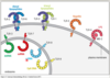
Pattern recognition receptors (PRRs)
- How many families of PRRs are there?
- What are they called?
- 5 families
- C type lectin receptors (CLRs), Toll-like receptors (TLRs), NOD-like receptors (NLRs), Rig-I like receptors (RLRs), Cytosolic DNA sensors (CDS)
Pattern recognition receptors (PRRs)
- How many families of PRRs are there?
- What are they called?
- 5 families
- C type lectin receptors (CLRs), Toll-like receptors (TLRs), NOD-like receptors (NLRs), Rig-I like receptors (RLRs), Cytosolic DNA sensors (CDS)
Below are all different families of what?
- C type lectin receptors (CLRs)
- Toll-like receptors (TLRs)
- NOD-like receptors (NLRs)
- Rig-I like receptors (RLRs)
- Cytosolic DNA sensors (CDS)
Pattern recognition receptors (PRRs)
… … … (…s) are receptors able to recognise conserved structures
Pattern recognition receptors (PRRs) are receptors able to recognise conserved structures

Pattern recognition receptors (PRRs) recognise patterns termed: …-associated … … (…s)
Pattern recognition receptors (PRRs) recognise patterns termed: pathogen-associated molecular patterns (PAMPs)

What do Pattern recognition receptors (PRRs) recognise ?
patterns termed: pathogen-associated molecular patterns (PAMPs)
Pattern-associated molecular patterns (PAMPs) & DAMPs
- PAMPs - Microbes evolve rapidly, so innate immunity must focus on highly … and … components of microbes (cell wall structures; nucleic acids)
- DAMPs – Damage associated molecular patterns; molecules released from necrotic cells
- PAMPs - Microbes evolve rapidly, so innate immunity must focus on highly conserved and essential components of microbes (cell wall structures; nucleic acids)
- DAMPs – Damage associated molecular patterns; molecules released from necrotic cells

Pattern-associated molecular patterns (PAMPs) & DAMPs
- PAMPs - Microbes evolve rapidly, so innate immunity must focus on highly conserved and essential components of microbes (cell wall structures; nucleic acids)
- DAMPs – … associated molecular patterns; molecules released from … cells
- PAMPs - Microbes evolve rapidly, so innate immunity must focus on highly conserved and essential components of microbes (cell wall structures; nucleic acids)
- DAMPs – Damage associated molecular patterns; molecules released from necrotic cells

DAMPs – … associated … patterns; molecules released from necrotic cells
DAMPs – Damage associated molecular patterns; molecules released from necrotic cells
C type lectin receptors (CLRs)
- CLRs are expressed by most cell type that phagocytose glycoproteins and microbes for antigen presentation to T lymphocytes
- CLRs bind to … in a calcium-dependent manner
- Type I CLRs assist with antigen uptake by phagocytes
- Type II CLRs are involved in fungal recognition
- Soluble CLRs include MBL that binds … on pathogen surfaces
- CLRs are expressed by most cell type that phagocytose glycoproteins and microbes for antigen presentation to T lymphocytes
- CLRs bind to carbohydrates in a calcium-dependent manner
- Type I CLRs assist with antigen uptake by phagocytes
- Type II CLRs are involved in fungal recognition
- Soluble CLRs include MBL that binds carbohydrates on pathogen surfaces

C type lectin receptors (CLRs)
- CLRs are expressed by most cell type that phagocytose glycoproteins and microbes for antigen presentation to T lymphocytes
- CLRs bind to carbohydrates in a …-dependent manner
- Type I CLRs assist with antigen uptake by phagocytes
- Type II CLRs are involved in … recognition
- Soluble CLRs include MBL that binds carbohydrates on pathogen surfaces
- CLRs are expressed by most cell type that phagocytose glycoproteins and microbes for antigen presentation to T lymphocytes
- CLRs bind to carbohydrates in a calcium-dependent manner
- Type I CLRs assist with antigen uptake by phagocytes
- Type II CLRs are involved in fungal recognition
- Soluble CLRs include MBL that binds carbohydrates on pathogen surfaces

C type lectin receptors (CLRs)
- CLRs are expressed by most cell type that phagocytose glycoproteins and microbes for antigen presentation to T lymphocytes
- CLRs bind to carbohydrates in a calcium-dependent manner
- Type I CLRs assist with … uptake by phagocytes
- Type II CLRs are involved in fungal recognition
- Soluble CLRs include MBL that binds carbohydrates on pathogen surfaces
- CLRs are expressed by most cell type that phagocytose glycoproteins and microbes for antigen presentation to T lymphocytes
- CLRs bind to carbohydrates in a calcium-dependent manner
- Type I CLRs assist with antigen uptake by phagocytes
- Type II CLRs are involved in fungal recognition
- Soluble CLRs include MBL that binds carbohydrates on pathogen surfaces

CLRs are expressed by most cell type that … glycoproteins and microbes for … presentation to T lymphocytes
CLRs are expressed by most cell type that phagocytose glycoproteins and microbes for antigen presentation to T lymphocytes
CLRs bind to … in a calcium-dependent manner
CLRs bind to carbohydrates in a calcium-dependent manner
Type I CLRs assist with antigen uptake by …
Type I CLRs assist with antigen uptake by phagocytes
Type II CLRs are involved in … recognition
Type II CLRs are involved in fungal recognition
Soluble CLRs include … that binds carbohydrates on pathogen surfaces
Soluble CLRs include MBL that binds carbohydrates on pathogen surfaces
Drosophila Toll Receptors
- Mutagenesis work on Drosophila revealed two members of the Toll family, dToll and 18-wheeler (dont need to know)
- Important for …
- Important for immunity to the fungal and bacterial infections
- Mammalian equivalent are the Toll-like receptors (TLRs)
- Mutagenesis work on Drosophila revealed two members of the Toll family, dToll and 18-wheeler (dont need to know)
- Important for development
- Important for immunity to the fungal and bacterial infections
- Mammalian equivalent are the Toll-like receptors (TLRs)

Drosophila Toll Receptors
- Mutagenesis work on Drosophila revealed two members of the Toll family, dToll and 18-wheeler (dont need to know)
- Important for development
- Important for immunity to the fungal and bacterial infections
- Mammalian equivalent are the …-… receptors
- Mutagenesis work on Drosophila revealed two members of the Toll family, dToll and 18-wheeler (dont need to know)
- Important for development
- Important for immunity to the fungal and bacterial infections
- Mammalian equivalent are the Toll-like receptors (TLRs)

Drosophila Toll Receptors
- Mutagenesis work on Drosophila revealed two members of the Toll family, dToll and 18-wheeler (dont need to know)
- Important for development
- Important for immunity to the … and … infections
- Mammalian equivalent are the Toll-like receptors (TLRs)
- Mutagenesis work on Drosophila revealed two members of the Toll family, dToll and 18-wheeler (dont need to know)
- Important for development
- Important for immunity to the fungal and bacterial infections
- Mammalian equivalent are the Toll-like receptors (TLRs)

Toll-like receptor structure
- Extracellular:
- … domain – site of pathogen binding
- Cytosolic side:
- TIR-domain - conserved stretch of ~200 amino acids
- Extracellular:
- LRR domain – site of pathogen binding
- Cytosolic side:
- TIR-domain - conserved stretch of ~200 amino acids

Toll-like receptor structure
- Extracellular:
- LRR domain – site of … binding
- Cytosolic side:
- TIR-domain - conserved stretch of ~200 amino acids
- Extracellular:
- LRR domain – site of pathogen binding
- Cytosolic side:
- TIR-domain - conserved stretch of ~200 amino acids

Toll-like receptor structure
- Extracellular:
- LRR domain – site of pathogen binding
- Cytosolic side:
- …-domain - conserved stretch of ~200 amino acids
- Extracellular:
- LRR domain – site of pathogen binding
- Cytosolic side:
- TIR-domain - conserved stretch of ~200 amino acids

Toll-like receptor structure
- Extracellular:
- LRR domain – site of pathogen binding
- … side:
- TIR-domain - conserved stretch of ~200 amino acids
- Extracellular:
- LRR domain – site of pathogen binding
-
Cytosolic side:
- TIR-domain - conserved stretch of ~200 amino acids

TLRs form functional …/…dimers
TLRs form functional hetero/homodimers

Cellular location of TLRS
- TLR-2 and TLR-6 recognise … lipopeptides (heterodimer)
- TLR-2 and TLR-1 recognise … lipopeptides (heterodimer)
- TLR-5 recognise flagellin
- TLR-4 recognise LPS on coat of gram- bacteria - works with MD-2
- The rest within endosome - recognise nucleic acid structures (TLRs 3, 7, 8, and 9 are situated in the membranes of endosomes and lysosomes)
- TLR10 is predominantly endosomal recognising dsRNA
- TLR-2 and TLR-6 recognise diacyl lipopeptides (heterodimer)
- TLR-2 and TLR-1 recognise triacyl lipopeptides (heterodimer)
- TLR-5 recognise flagellin
- TLR-4 recognise LPS on coat of gram- bacteria - works with MD-2
- The rest within endosome - recognise nucleic acid structures (TLRs 3, 7, 8, and 9 are situated in the membranes of endosomes and lysosomes)
- TLR10 is predominantly endosomal recognising dsRNA

Cellular location of TLRS
- TLR-2 and TLR-6 recognise diacyl lipopeptides (heterodimer)
- TLR-2 and TLR-1 recognise triacyl lipopeptides (heterodimer)
- TLR-5 recognise …
- TLR-4 recognise LPS on coat of gram- bacteria - works with MD-2
- The rest within endosome - recognise nucleic acid structures (TLRs 3, 7, 8, and 9 are situated in the membranes of endosomes and lysosomes)
- TLR10 is predominantly endosomal recognising dsRNA
- TLR-2 and TLR-6 recognise diacyl lipopeptides (heterodimer)
- TLR-2 and TLR-1 recognise triacyl lipopeptides (heterodimer)
- TLR-5 recognise flagellin
- TLR-4 recognise LPS on coat of gram- bacteria - works with MD-2
- The rest within endosome - recognise nucleic acid structures (TLRs 3, 7, 8, and 9 are situated in the membranes of endosomes and lysosomes)
- TLR10 is predominantly endosomal recognising dsRNA

Cellular location of TLRS
- TLR-2 and TLR-6 recognise diacyl lipopeptides (heterodimer)
- TLR-2 and TLR-1 recognise triacyl lipopeptides (heterodimer)
- TLR-5 recongise flagellin
- TLR-4 recognise … on coat of gram- bacteria - works with MD-2
- The rest within endosome - recognise nucleic acid structures (TLRs 3, 7, 8, and 9 are situated in the membranes of endosomes and lysosomes)
- TLR10 is predominantly endosomal recognising dsRNA
- TLR-2 and TLR-6 recognise diacyl lipopeptides (heterodimer)
- TLR-2 and TLR-1 recognise triacyl lipopeptides (heterodimer)
- TLR-5 recongise flagellin
- TLR-4 recognise LPS on coat of gram- bacteria - works with MD-2
- The rest within endosome - recognise nucleic acid structures (TLRs 3, 7, 8, and 9 are situated in the membranes of endosomes and lysosomes)
- TLR10 is predominantly endosomal recognising dsRNA

Cellular location of TLRS
- TLR-2 and TLR-6 recognise diacyl lipopeptides (heterodimer)
- TLR-2 and TLR-1 recognise triacyl lipopeptides (heterodimer)
- TLR-5 recongise flagellin
- TLR-4 recognise LPS on coat of gram- bacteria - works with MD-2
- The rest within … - recognise nucleic acid structures (TLRs 3, 7, 8, and 9 are situated in the membranes of … and lysosomes)
- TLR10 is predominantly endosomal recognising dsRNA
- TLR-2 and TLR-6 recognise diacyl lipopeptides (heterodimer)
- TLR-2 and TLR-1 recognise triacyl lipopeptides (heterodimer)
- TLR-5 recongise flagellin
- TLR-4 recognise LPS on coat of gram- bacteria - works with MD-2
- The rest within endosome - recognise nucleic acid structures (TLRs 3, 7, 8, and 9 are situated in the membranes of endosomes and lysosomes)
- TLR10 is predominantly endosomal recognising dsRNA

The TLRs include TLR1, TLR2, TLR3, TLR4, TLR5, TLR6, TLR7, TLR8, TLR9, TLR10, TLR11, TLR12, and TLR13, though the last … are not found in humans.
The TLRs include TLR1, TLR2, TLR3, TLR4, TLR5, TLR6, TLR7, TLR8, TLR9, TLR10, TLR11, TLR12, and TLR13, though the last three are not found in humans.
TLR-… and TLR-… recognise diacyl lipopeptides (heterodimer)
TLR-2 and TLR-6 recognise diacyl lipopeptides (heterodimer)
TLR-… and TLR-… recognise triacyl lipopeptides (heterodimer)
TLR-2 and TLR-1 recognise triacyl lipopeptides (heterodimer)
TLR-… recognise flagellin
TLR-5 recognise flagellin
TLR-… recognise LPS on coat of gram- bacteria - works with MD-2
TLR-4 recognise LPS on coat of gram- bacteria - works with MD-2
TLRs 3, 7, 8, and 9 are situated in the membranes of … and …
TLRs 3, 7, 8, and 9 are situated in the membranes of endosomes and lysosomes
TLR… is predominantly endosomal recognising dsRNA
TLR10 is predominantly endosomal recognising dsRNA

TLR-3 recognises …-stranded RNA
TLR-3 recognises double-stranded RNA

TLR-… recognises double-stranded RNA
TLR-3 recognises double-stranded RNA
TLR-… recognises single-stranded RNA
TLR-7 recognises single-stranded RNA

TLR-7 recognises …-stranded RNA
TLR-… recognises single-stranded RNA

TLR-8 recognises …-stranded RNA
TLR-8 recognises single-stranded RNA

TLR-… recongises CpG DNA
TLR-9 recongises CpG DNA

Cell Surface TLRs (1,2,4,5,6) mainly recognise … products, but on our own host molecules recognise mainly … or … molecules
Cell Surface TLRs (1,2,4,5,6) mainly recognise bacterial products, but on our own host molecules recognise mainly lipid or protein molecules

Cell Surface TLRs (which 5?) mainly recognise bacterial products, but on our own host molecules recognise mainly lipid or protein molecules
Cell Surface TLRs (1,2,4,5,6) mainly recognise bacterial products, but on our own host molecules recognise mainly lipid or protein molecules

… TSRs (3,7,8,9,10) mainly recognise viral products, but on our host molecules recognise our own DNA or RNA (dsRNA, ssRNA, DNA)
Endosomal TSRs (3,7,8,9,10) mainly recognise viral products, but on our host molecules recognise our own DNA or RNA (dsRNA, ssRNA, DNA)

Endosomal TSRs (which 5?) mainly recognise viral products, but on our host molecules recognise our own DNA or RNA (dsRNA, ssRNA, DNA)
Endosomal TSRs (3,7,8,9,10) mainly recognise viral products, but on our host molecules recognise our own DNA or RNA (dsRNA, ssRNA, DNA)

Endosomal TSRs (3,7,8,9,10) mainly recognise viral products, but on our host molecules recognise our own … and …
Endosomal TSRs (3,7,8,9,10) mainly recognise viral products, but on our host molecules recognise our own DNA or RNA (dsRNA, ssRNA, DNA)

Endosomal TSRs (3,7,8,9,10) mainly recognise … products, but on our host molecules recognise our own DNA or RNA (dsRNA, ssRNA, DNA)
Endosomal TSRs (3,7,8,9,10) mainly recognise viral products, but on our host molecules recognise our own DNA or RNA (dsRNA, ssRNA, DNA)

TLRs recognise … and … ligands
TLRs recognise exogenous and endogenous ligands

TLR signalling cascade
- TLR signalling induces genes that function in host defense:
- Pro-inflammatory & anti-inflammatory …
- MHC & co-stimulatory molecules
- antimicrobial peptides & complement components
- TLR signalling induces genes that function in host defense:
- Pro-inflammatory & anti-inflammatory cytokines
- MHC & co-stimulatory molecules
- antimicrobial peptides & complement components

TLR signalling cascade
- TLR signalling induces genes that function in host defense:
- Pro-inflammatory & anti-inflammatory cytokines
- … & co-stimulatory molecules
- antimicrobial peptides & complement components
- TLR signalling induces genes that function in host defense:
- Pro-inflammatory & anti-inflammatory cytokines
- MHC & co-stimulatory molecules
- antimicrobial peptides & complement components

TLR signalling cascade
- TLR signalling induces genes that function in host defense:
- Pro-inflammatory & anti-inflammatory cytokines
- MHC & co-stimulatory molecules
- antimicrobial peptides & complement components
- TLR signalling induces genes that function in host defense:
- Pro-inflammatory & anti-inflammatory cytokines
- MHC & co-stimulatory molecules
- antimicrobial peptides & complement components

TLR signalling cascade
- TLR signalling induces genes that function in host defense:
- Pro-inflammatory & anti-inflammatory cytokines
- MHC & co-stimulatory molecules
- … peptides & … components
- TLR signalling induces genes that function in host defense:
- Pro-inflammatory & anti-inflammatory cytokines
- MHC & co-stimulatory molecules
- antimicrobial peptides & complement components

TLR adaptor proteins
- Not all Toll-like receptors use all of the adaptor molecules
- TLR-… uses all of them
- TLR 3 only uses TRIF
- All apart from TLR3 use Myd88
- Not all Toll-like receptors use all of the adaptor molecules
- TLR-4 uses all of them
- TLR 3 only uses TRIF
- All apart from TLR3 use Myd88

At the top of the TLR signalling cascade 4 different adaptive proteins are used. These are …
At the top of the TLR signalling cascade 4 different adaptive proteins are used. These are MyD88, MAL, TRIF and TRAM

At the top of the TLR signalling cascade 4 different adaptive proteins are used. These are …
At the top of the TLR signalling cascade 4 different adaptive proteins are used. These are MyD88, MAL, TRIF and TRAM

TLR adaptor proteins
- Not all Toll-like receptors use all of the adaptor molecules
- TLR-4 uses all of them
- TLR 3 only uses …
- All apart from TLR3 use Myd88
- Not all Toll-like receptors use all of the adaptor molecules
- TLR-4 uses all of them
- TLR 3 only uses TRIF
- All apart from TLR3 use Myd88

TLR adaptor proteins
- Not all Toll-like receptors use all of the adaptor molecules
- TLR-4 uses all of them
- TLR 3 only uses TRIF
- All apart from TLR3 use …
- Not all Toll-like receptors use all of the adaptor molecules
- TLR-4 uses all of them
- TLR 3 only uses TRIF
- All apart from TLR3 use Myd88

TLR adaptor proteins
- Not all Toll-like receptors use all of the … molecules
- TLR-4 uses all of them
- TLR 3 only uses TRIF
- All apart from TLR3 use Myd88
- Not all Toll-like receptors use all of the adaptor molecules
- TLR-4 uses all of them
- TLR 3 only uses TRIF
- All apart from TLR3 use Myd88

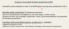
MyD88 gain of function mutation
- Waldenström macroglobulinemia (rare type of non-Hodgkin lymphoma)
- MyD88 mutation is present in 90% of patients causing cell growth and survival
- B cells make large amounts of … that can cause excess bleeding, vision problems and headaches
- Lymphoma cells proliferating in the bone marrow cause anaemia, neutropenia and thrombocytopenia
- Waldenström macroglobulinemia (rare type of non-Hodgkin lymphoma)
- MyD88 mutation is present in 90% of patients causing cell growth and survival
- B cells make large amounts of IgM that can cause excess bleeding, vision problems and headaches
- Lymphoma cells proliferating in the bone marrow cause anaemia, neutropenia and thrombocytopenia
MyD88 gain of function mutation
- Waldenström … (rare type of non-Hodgkin lymphoma)
- MyD88 mutation is present in 90% of patients causing cell growth and survival
- B cells make large amounts of IgM that can cause excess bleeding, vision problems and headaches
- Lymphoma cells proliferating in the bone marrow cause anaemia, neutropenia and thrombocytopenia
- Waldenström macroglobulinemia (rare type of non-Hodgkin lymphoma)
- MyD88 mutation is present in 90% of patients causing cell growth and survival
- B cells make large amounts of IgM that can cause excess bleeding, vision problems and headaches
- Lymphoma cells proliferating in the bone marrow cause anaemia, neutropenia and thrombocytopenia
MyD88 gain of function mutation
- Waldenström macroglobulinemia (rare type of non-Hodgkin lymphoma)
- MyD88 mutation is present in …% of patients causing cell growth and survival
- B cells make large amounts of IgM that can cause excess bleeding, vision problems and headaches
- Lymphoma cells … in the bone marrow cause anaemia, neutropenia and thrombocytopenia
- Waldenström macroglobulinemia (rare type of non-Hodgkin lymphoma)
- MyD88 mutation is present in 90% of patients causing cell growth and survival
- B cells make large amounts of IgM that can cause excess bleeding, vision problems and headaches
- Lymphoma cells proliferating in the bone marrow cause anaemia, neutropenia and thrombocytopenia
MyD88 gain of function mutation
- Waldenström macroglobulinemia (rare type of non-… lymphoma)
- MyD88 mutation is present in 90% of patients causing cell growth and survival
- B cells make large amounts of IgM that can cause excess bleeding, vision problems and headaches
- … cells proliferating in the bone marrow cause anaemia, neutropenia and thrombocytopenia
- Waldenström macroglobulinemia (rare type of non-Hodgkin lymphoma)
- MyD88 mutation is present in 90% of patients causing cell growth and survival
- B cells make large amounts of IgM that can cause excess bleeding, vision problems and headaches
- Lymphoma cells proliferating in the bone marrow cause anaemia, neutropenia and thrombocytopenia
MyD88 mutation is present in …% of patients with Waldenström macroglobulinemia (rare type of non-Hodgkin lymphoma) causing cell growth and survival
MyD88 mutation is present in 90% of patients with Waldenström macroglobulinemia (rare type of non-Hodgkin lymphoma) causing cell growth and survival
In Waldenström macroglobulinemia, B cells make large amounts of IgM that can cause 3 symptoms - what are they?
excess bleeding, vision problems and headaches (MyD88 mutation is present in 90% of patients causing cell growth and survival)
In Waldenström macroglobulinemia, Lymphoma cells proliferating in the bone marrow cause …, … and thrombocytopenia
In Waldenström macroglobulinemia, Lymphoma cells proliferating in the bone marrow cause anaemia, neutropenia and thrombocytopenia
A life without MyD88?
- Nine MyD88 deficient children suffered from life-threatening, often recurrent … bacterial infections, but were otherwise healthy, with normal resistance to other microbes.
- Their clinical status improved with …, possibly due to a compensatory effect of adaptive immunity or other innate immune mechanisms.
- Nine MyD88 deficient children suffered from life-threatening, often recurrent pyogenic bacterial infections, but were otherwise healthy, with normal resistance to other microbes.
- Their clinical status improved with age, possibly due to a compensatory effect of adaptive immunity or other innate immune mechanisms.

Are TLRs important?
- Of the 10 TLRs only deficiency in … has been linked to immunodeficiency.
- TLR-… Deficiency in Patients with Herpes Simplex Encephalitis (HSE) - Inflammation of the brain due to infection with herpes simplex virus (HSV-1)
- HSV-1 is a dsDNA virus, but during viral replication it produces dsRNA
- Defects in other signalling molecules involved in the TLR3 signalling pathway have also been associated with HSE
- Of the 10 TLRs only deficiency in TLR3 has been linked to immunodeficiency.
- TLR-3 Deficiency in Patients with Herpes Simplex Encephalitis (HSE) - Inflammation of the brain due to infection with herpes simplex virus (HSV-1)
- HSV-1 is a dsDNA virus, but during viral replication it produces dsRNA
- Defects in other signalling molecules involved in the TLR3 signalling pathway have also been associated with HSE
Are TLRs important?
- Of the 10 TLRs only deficiency in TLR3 has been linked to immunodeficiency.
- TLR-3 Deficiency in Patients with Herpes Simplex Encephalitis (HSE) - Inflammation of the brain due to infection with herpes simplex virus (HSV-1)
- HSV-1 is a …DNA virus, but during viral replication it produces …RNA
- Defects in other signalling molecules involved in the TLR3 signalling pathway have also been associated with HSE
- Of the 10 TLRs only deficiency in TLR3 has been linked to immunodeficiency.
- TLR-3 Deficiency in Patients with Herpes Simplex Encephalitis (HSE) - Inflammation of the brain due to infection with herpes simplex virus (HSV-1)
- HSV-1 is a dsDNA virus, but during viral replication it produces dsRNA
- Defects in other signalling molecules involved in the TLR3 signalling pathway have also been associated with HSE
Of the 10 TLRs only deficiency in … has been linked to immunodeficiency.
Of the 10 TLRs only deficiency in TLR3 has been linked to immunodeficiency.
TLRs in disease

TLRs in disease

TLRs in Infection
- … – TLR8
- … – TLR2 and 4
- T.. – TLR2 and 4
- HIV – TLR8
- Sepsis – TLR2 and 4
- Tuberculosis – TLR2 and 4
TLRs in Inflammation (Diseases)
- Systemic … … – TLR7, 8 and 9
- … Disease – TLR2 and 4
- A… – TLR2 and 4
- Systemic Lupus Erythematosus – TLR7, 8 and 9
- Alzheimer’s Disease – TLR2 and 4
- Atherosclerosis – TLR2 and 4
TLR agonists - Diseases
- Infection- genital warts (TLR… ligand - Aldara)
- Cancer - Melanoma (TLR… ligand - Aldara)
- Allergy – Ragweed pollen (TLR9)
- Vaccine adjuvant
- Infection- genital warts (TLR7 ligand - Aldara)
- Cancer - Melanoma (TLR7 ligand - Aldara)
- Allergy – Ragweed pollen (TLR9)
- Vaccine adjuvant
TLR agonists - Diseases
- Infection- genital warts (TLR7 ligand - Aldara)
- Cancer - Melanoma (TLR7 ligand - Aldara)
- Allergy – Ragweed pollen (TLR…)
- Vaccine adjuvant
- Infection- genital warts (TLR7 ligand - Aldara)
- Cancer - Melanoma (TLR7 ligand - Aldara)
- Allergy – Ragweed pollen (TLR9)
- Vaccine adjuvant
TLR agonists - Diseases
- Infection- genital warts (TLR7 ligand - Aldara)
- Cancer - Melanoma (TLR7 ligand - Aldara)
- Allergy – Ragweed pollen (TLR9)
- … adjuvant
- Infection- genital warts (TLR7 ligand - Aldara)
- Cancer - Melanoma (TLR7 ligand - Aldara)
- Allergy – Ragweed pollen (TLR9)
- Vaccine adjuvant
TLR antagonists - Disease
- … diseases (TLR7, 8 & 9)
- S… (TLR4)
- Autoimmunity (TLR7, 8 & 9)
- Sepsis (TLR4)
Nod-like receptors (NLRs)
- NLR = Nucleotide-binding … Rich
- Cytoplasmic pattern recognition molecules
- Two major groups- NLRCs and NLRPs – ‘C’ stands for ‘caspase recruitment domain (CARD)’ and the ‘P’ stands for pyrin domain.
- NLR = Nucleotide-binding Leucine Rich
- Cytoplasmic pattern recognition molecules
- Two major groups- NLRCs and NLRPs – ‘C’ stands for ‘caspase recruitment domain (CARD)’ and the ‘P’ stands for pyrin domain.
Nod-like receptors (NLRs)
- NLR = Nucleotide-binding Leucine Rich
- … pattern recognition molecules
- Two major groups- NLRCs and NLRPs – ‘C’ stands for ‘caspase recruitment domain (CARD)’ and the ‘P’ stands for pyrin domain.
- NLR = Nucleotide-binding Leucine Rich
- Cytoplasmic pattern recognition molecules
- Two major groups- NLRCs and NLRPs – ‘C’ stands for ‘caspase recruitment domain (CARD)’ and the ‘P’ stands for pyrin domain.
Nod-like receptors (NLRs)
- NLR = Nucleotide-binding Leucine Rich
- Cytoplasmic pattern recognition molecules
- Two major groups- NLRCs and NLRPs – ‘C’ stands for ‘caspase recruitment domain (CARD)’ and the ‘P’ stands for … domain.
- NLR = Nucleotide-binding Leucine Rich
- Cytoplasmic pattern recognition molecules
- Two major groups- NLRCs and NLRPs – ‘C’ stands for ‘caspase recruitment domain (CARD)’ and the ‘P’ stands for pyrin domain.
What PRRs are only found in the cytoplasm?
Nod-like receptors (NLRs) and RIG-I-like receptors (RLRs)
What are the two major groups of NLRs?
NLRCs and NLRPs

NLRCs
- Two examples: NLRC1 (…) and NLRC2 (…)
- They have a leucine rich domain which can bind to peptidoglycan which is present on the cell membrane of most bacteria
- Two examples: NLRC1 (NOD1) and NLRC2 (NOD2)
- They have a leucine rich domain which can bind to peptidoglycan which is present on the cell membrane of most bacteria

NLRCs
- Two examples: NLRC1 (NOD1) and NLRC2 (NOD2)
- They have a … rich domain which can bind to … which is present on the cell membrane of most bacteria
- Two examples: NLRC1 (NOD1) and NLRC2 (NOD2)
- They have a leucine rich domain which can bind to peptidoglycan which is present on the cell membrane of most bacteria

NOD1 and NOD2
- NOD1 and NOD2 detect similar yet distinct peptides of …
- NOD1 binds γ-glutamyl diaminopimelic acid (iE-DAP) (Mainly Gm-ve Bacteria)
- NOD2 binds muramyl dipeptide (both Gram+ve and Gram-ve bacteria)
- NOD1 and NOD2 detect similar yet distinct peptides of peptidoglycan
- NOD1 binds γ-glutamyl diaminopimelic acid (iE-DAP) (Mainly Gm-ve Bacteria)
- NOD2 binds muramyl dipeptide (both Gram+ve and Gram-ve bacteria)

NOD1 and NOD2
- NOD1 and NOD2 detect similar yet distinct peptides of peptidoglycan
- NOD1 binds γ-glutamyl diaminopimelic acid (…-DAP) (Mainly Gm… Bacteria)
- NOD2 binds muramyl dipeptide (both Gram+ve and Gram-ve bacteria)
- NOD1 and NOD2 detect similar yet distinct peptides of peptidoglycan
- NOD1 binds γ-glutamyl diaminopimelic acid (iE-DAP) (Mainly Gm-ve Bacteria)
- NOD2 binds muramyl dipeptide (both Gram+ve and Gram-ve bacteria)

NOD1 and NOD2
- NOD1 and NOD2 detect similar yet distinct peptides of peptidoglycan
- NOD1 binds γ-glutamyl diaminopimelic acid (iE-DAP) (Mainly Gm-ve Bacteria)
- NOD2 binds … dipeptide (both Gram+ve and Gram-ve bacteria)
- NOD1 and NOD2 detect similar yet distinct peptides of peptidoglycan
- NOD1 binds γ-glutamyl diaminopimelic acid (iE-DAP) (Mainly Gm-ve Bacteria)
- NOD2 binds muramyl dipeptide (both Gram+ve and Gram-ve bacteria)

NOD2 gain of function mutation linked to early onset … where granulomas develop in the organs of the body.
NOD2 gain of function mutation linked to early onset sarcoidosis where granulomas develop in the organs of the body.
NOD… gain of function mutation linked to early onset sarcoidosis where granulomas develop in the organs of the body.
NOD2 gain of function mutation linked to early onset sarcoidosis where granulomas develop in the organs of the body.
NOD2 gain of function mutation linked to early onset sarcoidosis where … develop in the organs of the body.
NOD2 gain of function mutation linked to early onset sarcoidosis where granulomas develop in the organs of the body.
NOD… loss of function mutation is associated with susceptibility to Crohn’s disease, a chronic intestinal inflammatory disorder
NOD2 loss of function mutation is associated with susceptibility to Crohn’s disease, a chronic intestinal inflammatory disorder
NOD2 loss of function mutation is associated with susceptibility to … disease, a chronic intestinal inflammatory disorder
NOD2 loss of function mutation is associated with susceptibility to Crohn’s disease, a chronic intestinal inflammatory disorder
NOD2 … of function mutation is associated with susceptibility to Crohn’s disease, a chronic intestinal inflammatory disorder
NOD2 loss of function mutation is associated with susceptibility to Crohn’s disease, a chronic intestinal inflammatory disorder
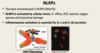
NLRPs
- The best characterised is NLRP… (NALP…)
- NLRP3 is activated by cellular stress; K+ efflux, ATP, reactive oxygen species and lysosomal damage
- Inflammasome activation is essential for IL-1 and IL-18 secretion
- The best characterised is NLRP3 (NALP3)
- NLRP3 is activated by cellular stress; K+ efflux, ATP, reactive oxygen species and lysosomal damage
- Inflammasome activation is essential for IL-1 and IL-18 secretion

NLRPs
- The best characterised is NLRP… (NALP…)
- NLRP3 is activated by … stress; K+ efflux, ATP, reactive oxygen species and lysosomal damage
- Inflammasome activation is essential for IL-1 and IL-18 secretion
- The best characterised is NLRP3 (NALP3)
- NLRP3 is activated by cellular stress; K+ efflux, ATP, reactive oxygen species and lysosomal damage
- Inflammasome activation is essential for IL-1 and IL-18 secretion
NLRPs
- The best characterised is NLRP… (NALP…)
- NLRP3 is activated by … stress; K+ efflux, ATP, reactive oxygen species and lysosomal damage
- … activation is essential for IL-1 and IL-18 secretion
- The best characterised is NLRP3 (NALP3)
- NLRP3 is activated by cellular stress; K+ efflux, ATP, reactive oxygen species and lysosomal damage
- Inflammasome activation is essential for IL-1 and IL-18 secretion

Activation of the inflammasomes results in the processing and subsequent secretion of the pro-inflammatory cytokines IL-… and IL-…
Activation of the inflammasomes results in the processing and subsequent secretion of the pro-inflammatory cytokines IL-1 and IL-18.

NLRP3 is activated by cellular stress; …+ efflux, A…, … oxygen species and lysosomal damage
NLRP3 is activated by cellular stress; K+ efflux, ATP, reactive oxygen species and lysosomal damage

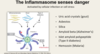
The Inflammasome senses danger
- Activated by cellular infection or cell …
- Stress caused by crystals getting stuck or bursting the phagosome during endocytosis (recognised - Uric acid crystals (gout))
- Activated by cellular infection or cell stress
- Stress caused by crystals getting stuck or bursting the phagosome during endocytosis (recognised - Uric acid crystals (gout))

The Inflammasome senses danger
- Activated by cellular infection or cell stress
- Stress caused by … getting stuck or bursting the phagosome during endocytosis (recognised - Uric acid crystals (…))
- Activated by cellular infection or cell stress
- Stress caused by crystals getting stuck or bursting the phagosome during endocytosis (recognised - Uric acid crystals (gout))

NALP3 Inflammasome
- Sensor of damage and cellular …
- … can form and are taken up by cells - this then drives activation and formation of the NALP3 inflammasome
- Conditions/Diseases/Infections such as: Gout, Asbestos, Silica, Amyloid beta (Alzheimer’s), Islet amyloid polypetide (T2 diabetes), Hemozoin (Malaria)
- Sensor of damage and cellular stress
-
Crystals can form and are taken up by cells - this then drives activation and formation of the NALP3 inflammasome
- Conditions/Diseases/Infections such as: Gout, Asbestos, Silica, Amyloid beta (Alzheimer’s), Islet amyloid polypetide (T2 diabetes), Hemozoin (Malaria)

NALP3 Inflammasome
- Sensor of damage and cellular stress
- Crystals can form and are taken up by cells - this then drives activation and formation of the NALP3 inflammasome
- Conditions/Diseases/Infections such as: G…, As…, Silica, Amyloid beta (Alzheimer’s), Islet amyloid polypetide (T2 diabetes), Hemozoin (Malaria)
- Sensor of damage and cellular stress
- Crystals can form and are taken up by cells - this then drives activation and formation of the NALP3 inflammasome
- Conditions/Diseases/Infections such as: Gout, Asbestos, Silica, Amyloid beta (Alzheimer’s), Islet amyloid polypetide (T2 diabetes), Hemozoin (Malaria)
NALP3 Inflammasome
- Sensor of damage and cellular stress
- Crystals can form and are taken up by cells - this then drives activation and formation of the NALP3 inflammasome
- Conditions/Diseases/Infections such as: Gout, Asbestos, Silica, Amyloid beta (…), Islet amyloid polypetide (… diabetes), Hemozoin (Malaria)
- Sensor of damage and cellular stress
- Crystals can form and are taken up by cells - this then drives activation and formation of the NALP3 inflammasome
- Conditions/Diseases/Infections such as: Gout, Asbestos, Silica, Amyloid beta (Alzheimer’s), Islet amyloid polypetide (T2 diabetes), Hemozoin (Malaria)

NALP3 Inflammasome
- Sensor of damage and cellular stress
- Crystals can form and are taken up by cells - this then drives activation and formation of the NALP3 inflammasome
- Conditions/Diseases/Infections such as: Gout, Asbestos, Silica, Amyloid beta (Alzheimer’s), Islet amyloid polypetide (T2 diabetes), Hemozoin (M…)
- Sensor of damage and cellular stress
- Crystals can form and are taken up by cells - this then drives activation and formation of the NALP3 inflammasome
- Conditions/Diseases/Infections such as: Gout, Asbestos, Silica, Amyloid beta (Alzheimer’s), Islet amyloid polypetide (T2 diabetes), Hemozoin (Malaria)

Frustrated phagocytosis
- … hip - fragments break off after grinding over years
- … by a macrophage - gets stuck in the process and puts cell under stress, leading to inflammasome activation and production of IL-1 and IL-18
- These people get inflamed hip, revision and tissue removed
- Artificial hip - fragments break off after grinding over years
- Phagocytosed by a macrophage - gets stuck in the process and puts cell under stress, leading to inflammasome activation and production of IL-1 and IL-18
- These people get inflamed hip, revision and tissue removed
Frustrated phagocytosis
- Artificial hip - fragments break off after grinding over years
- Phagocytosed by a macrophage - gets stuck in the process and puts cell under stress, leading to inflammasome activation and production of IL-.. and IL-..
- These people get inflamed hip, revision and tissue removed
- Artificial hip - fragments break off after grinding over years
- Phagocytosed by a macrophage - gets stuck in the process and puts cell under stress, leading to inflammasome activation and production of IL-1 and IL-18
- These people get inflamed hip, revision and tissue removed

Inflammasome cleavage of pro-IL-1 and pro-IL-18
- Activation of either IL-1 receptor family or toll-receptors - driving transcription and translation of IL-1B - made as pro-IL-1B - cleaved by …-1 released from inflammasome complex - makes mature active form of IL-1B
- Same process for IL-18 (requires cleavage by NALP3 inflammasome)
- Activation of either IL-1 receptor family or toll-receptors - driving transcription and translation of IL-1B - made as pro-IL-1B - cleaved by caspase-1 released from inflammasome complex - makes mature active form of IL-1B
- Same process for IL-18 (requires cleavage by NALP3 inflammasome)

Inflammasome cleavage of pro-IL-1 and pro-IL-18
- Activation of either IL-1 receptor family or toll-receptors - driving transcription and translation of IL-1B - made as pro-IL-1B - cleaved by caspase-1 released from inflammasome complex - makes mature … form of IL-1B
- Same process for IL-18 (requires cleavage by NALP3 inflammasome)
- Activation of either IL-1 receptor family or toll-receptors - driving transcription and translation of IL-1B - made as pro-IL-1B - cleaved by caspase-1 released from inflammasome complex - makes mature active form of IL-1B
- Same process for IL-18 (requires cleavage by NALP3 inflammasome)

Inflammasome cleavage of pro-IL-1 and pro-IL-18
- Activation of either IL-1 receptor family or toll-receptors - driving transcription and translation of IL-1B - made as pro-IL-1B - cleaved by caspase-1 released from inflammasome complex - makes mature active form of IL-1B
- Same process for IL-… (requires cleavage by NALP3 inflammasome)
- Activation of either IL-1 receptor family or toll-receptors - driving transcription and translation of IL-1B - made as pro-IL-1B - cleaved by caspase-1 released from inflammasome complex - makes mature active form of IL-1B
- Same process for IL-18 (requires cleavage by NALP3 inflammasome)
Inflammasome cleavage of pro-IL-1 and pro-IL-18
- Activation of either IL-1 receptor family or toll-receptors - driving transcription and translation of IL-1B - made as pro-IL-1B - cleaved by …-1 released from inflammasome complex - makes mature active form of IL-1B
- Same process for IL-18 (requires cleavage by NALP3 inflammasome)
- Activation of either IL-1 receptor family or toll-receptors - driving transcription and translation of IL-1B - made as pro-IL-1B - cleaved by caspase-1 released from inflammasome complex - makes mature active form of IL-1B
- Same process for IL-18 (requires cleavage by NALP3 inflammasome)

Gain of function mutations in NLRP3
- Cryopyrin-Associated Periodic Syndromes (…) - Caused by rare mutations in exon 3 of NLRP3 gene causing over production of IL-1
- 2 - Muckle wells syndrome (Prevalence unknown) and Familial cold autoinflammatory syndrome (1: 1000000)
- Cryopyrin-Associated Periodic Syndromes (CAPS) - Caused by rare mutations in exon 3 of NLRP3 gene causing over production of IL-1
- 2 - Muckle wells syndrome (Prevalence unknown) and Familial cold autoinflammatory syndrome (1: 1000000)

Gain of function mutations in NLRP3
- Cryopyrin-Associated Periodic Syndromes (CAPS) - Caused by rare mutations in exon 3 of NLRP3 gene causing over production of IL-…
- 2 - Muckle wells syndrome (Prevalence unknown) and Familial cold autoinflammatory syndrome (1: 1000000)
- Cryopyrin-Associated Periodic Syndromes (CAPS) - Caused by rare mutations in exon 3 of NLRP3 gene causing over production of IL-1
- 2 - Muckle wells syndrome (Prevalence unknown) and Familial cold autoinflammatory syndrome (1: 1000000)

Gain of function mutations in NLRP3
- Cryopyrin-Associated Periodic Syndromes (CAPS) - Caused by rare mutations in exon 3 of NLRP3 gene causing over production of IL-1
- 2 - … … syndrome (Prevalence unknown) and Familial cold autoinflammatory syndrome (1: 1000000)
- Cryopyrin-Associated Periodic Syndromes (CAPS) - Caused by rare mutations in exon 3 of NLRP3 gene causing over production of IL-1
- 2 - Muckle wells syndrome (Prevalence unknown) and Familial cold autoinflammatory syndrome (1: 1000000)

Gain of function mutations in NLRP3
- Cryopyrin-Associated Periodic Syndromes (CAPS) - Caused by rare mutations in exon 3 of NLRP3 gene causing over production of IL-1
- 2 - Muckle wells syndrome (Prevalence unknown) and … … autoinflammatory syndrome (1: 1000000)
- Cryopyrin-Associated Periodic Syndromes (CAPS) - Caused by rare mutations in exon 3 of NLRP3 gene causing over production of IL-1
- 2 - Muckle wells syndrome (Prevalence unknown) and Familial cold autoinflammatory syndrome (1: 1000000)
Muckle wells syndrome (Prevalence unknown)
- Can occur spontaneously or be triggered by …, …, fatigue, or other stresses.
- Symptoms of fever, rash, arthralgia, conjunctivitis, uveitis, sensorineural deafness, and potentially life-threatening amyloidosis
- (Abnormal deposits of a protein called amyloid (amyloidosis) cause progressive kidney damage in about one-third of people with Muckle-Wells syndrome; )
- Can be treated with Anakinra (IL-1RA)
- Can occur spontaneously or be triggered by cold, heat, fatigue, or other stresses.
- Symptoms of fever, rash, arthralgia, conjunctivitis, uveitis, sensorineural deafness, and potentially life-threatening amyloidosis
- (Abnormal deposits of a protein called amyloid (amyloidosis) cause progressive kidney damage in about one-third of people with Muckle-Wells syndrome; )
- Can be treated with Anakinra (IL-1RA)
Muckle wells syndrome (Prevalence unknown)
- Can occur spontaneously or be triggered by cold, heat, fatigue, or other stresses.
- Symptoms of fever, rash, arthralgia, conjunctivitis, uveitis, sensorineural deafness, and potentially life-threatening …
- Can be treated with Anakinra (IL-1RA)
- Can occur spontaneously or be triggered by cold, heat, fatigue, or other stresses.
- Symptoms of fever, rash, arthralgia, conjunctivitis, uveitis, sensorineural deafness, and potentially life-threatening amyloidosis
- (Abnormal deposits of a protein called amyloid (amyloidosis) cause progressive kidney damage in about one-third of people with Muckle-Wells syndrome; )
Familial cold autoinflammatory syndrome (1: 1000000)
- Triggered by exposure to …
- Symptoms of fever urticarial rash with headache, arthralgia, and sometimes conjunctivitis
- Can be treated with Anakinra (IL-1RA)
- Triggered by exposure to cold
- Symptoms of fever urticarial rash with headache, arthralgia, and sometimes conjunctivitis
- Can be treated with Anakinra (IL-1RA)
Familial cold autoinflammatory syndrome (1: 1000000)
- Triggered by exposure to cold
- Symptoms of … urticarial rash with headache, arthralgia, and sometimes conjunctivitis
- Can be treated with … (IL-1RA)
- Triggered by exposure to cold
- Symptoms of fever urticarial rash with headache, arthralgia, and sometimes conjunctivitis
- Can be treated with Anakinra (IL-1RA)
Muckle wells syndrome (Prevalence unknown)
- Can occur spontaneously or be triggered by cold, heat, fatigue, or other stresses.
- Symptoms of fever, rash, arthralgia, conjunctivitis, uveitis, sensorineural deafness, and potentially life-threatening …
- Can be treated with … (IL-1RA)
- Can occur spontaneously or be triggered by cold, heat, fatigue, or other stresses.
- Symptoms of fever, rash, arthralgia, conjunctivitis, uveitis, sensorineural deafness, and potentially life-threatening amyloidosis
- (Abnormal deposits of a protein called amyloid (amyloidosis) cause progressive kidney damage in about one-third of people with Muckle-Wells syndrome; )
- Can be treated with Anakinra (IL-1RA)
Both Muckle wells syndrome and Familial cold autoinflammatory syndrome can be treated with … (IL-1RA)
Both Muckle wells syndrome and Familial cold autoinflammatory syndrome can be treated with Anakinra (IL-1RA) (Both are Cryopyrin-Associated Periodic Syndromes (CAPS) - Caused by rare mutations in exon 3 of NLRP3 gene causing over production of IL-1)
Both Muckle wells syndrome and Familial cold autoinflammatory syndrome can be treated with Anakinra (IL-…)
Both Muckle wells syndrome and Familial cold autoinflammatory syndrome can be treated with Anakinra (IL-1RA) (Both are Cryopyrin-Associated Periodic Syndromes (CAPS) - Caused by rare mutations in exon 3 of NLRP3 gene causing over production of IL-1)
RIG-I-like receptors (RLRs)
- RIG-I and MDA5 are sensors of cytoplasmic …, a replication intermediate for viruses. They signal to induce pro-inflammatory … and IFN.
- RIG-I and MDA5 are sensors of cytoplasmic RNA, a replication intermediate for viruses. They signal to induce pro-inflammatory cytokines and IFN.
- RIG-I - Binds to single stranded RNA containing 5’-triphosphate (our 5’ RNA is capped so not recognised)
- MDA5 - Preferentially recognizes long double stranded RNA, Critical for picornavirus detection, Mutations are rare but have been associated with IFN related diseases, e.g. systemic lupus erythematosus and Aicardi–Goutières syndrome.

RIG-I-like receptors (RLRs)
- RIG-I and MDA5 are sensors of cytoplasmic RNA, a replication intermediate for viruses. They signal to induce pro-inflammatory cytokines and IFN.
- RIG-I - Binds to … stranded RNA containing 5’-triphosphate (our 5’ RNA is capped so not recognised)
- MDA5 - Preferentially recognizes long … stranded RNA, Critical for picornavirus detection, Mutations are rare but have been associated with IFN related diseases, e.g. systemic lupus erythematosus and Aicardi–Goutières syndrome.
- RIG-I and MDA5 are sensors of cytoplasmic RNA, a replication intermediate for viruses. They signal to induce pro-inflammatory cytokines and IFN.
- RIG-I - Binds to single stranded RNA containing 5’-triphosphate (our 5’ RNA is capped so not recognised)
- MDA5 - Preferentially recognizes long double stranded RNA, Critical for picornavirus detection, Mutations are rare but have been associated with IFN related diseases, e.g. systemic lupus erythematosus and Aicardi–Goutières syndrome.

RIG-I
- RIG-I and MDA5 are sensors of … …, a replication intermediate for viruses. They signal to induce pro-inflammatory cytokines and IFN.
- RIG-I Binds to … stranded RNA containing 5’-triphosphate (our 5’ RNA is capped so not recognised)
- RIG-I and MDA5 are sensors of cytoplasmic RNA, a replication intermediate for viruses. They signal to induce pro-inflammatory cytokines and IFN.
- RIG-I Binds to single stranded RNA containing 5’-triphosphate (our 5’ RNA is capped so not recognised)
MDA5
- RIG-I and MDA5 are sensors of cytoplasmic RNA, a replication intermediate for viruses. They signal to induce pro-inflammatory cytokines and IFN.
- Preferentially recognizes long … stranded RNA
- Critical for … detection
- Mutations are rare but have been associated with IFN related diseases, e.g. systemic lupus erythematosus and Aicardi–Goutières syndrome.
- RIG-I and MDA5 are sensors of cytoplasmic RNA, a replication intermediate for viruses. They signal to induce pro-inflammatory cytokines and IFN.
- Preferentially recognizes long double stranded RNA
- Critical for picornavirus detection
- Mutations are rare but have been associated with IFN related diseases, e.g. systemic lupus erythematosus and Aicardi–Goutières syndrome.
MDA5
- RIG-I and MDA5 are sensors of cytoplasmic RNA, a replication intermediate for viruses. They signal to induce pro-inflammatory cytokines and IFN.
- Preferentially recognizes long … stranded RNA
- Critical for … detection
- Mutations are rare but have been associated with IFN related diseases, e.g. systemic … erythematosus and Aicardi–… syndrome.
- RIG-I and MDA5 are sensors of cytoplasmic RNA, a replication intermediate for viruses. They signal to induce pro-inflammatory cytokines and IFN.
- Preferentially recognizes long double stranded RNA
- Critical for picornavirus detection
- Mutations are rare but have been associated with IFN related diseases, e.g. systemic lupus erythematosus and Aicardi–Goutières syndrome.
Cytosolic DNA sensors
- Stimulator of interferon genes (STING)-associated vasculopathy with onset in infancy (SAVI) is an autoinflammatory disease caused by …-of-function mutations in the gene that codes for STING. Patients produce too much type 1 IFN causing abnormal inflammation throughout the body, especially in the skin, blood vessels, and lungs.
- Stimulator of interferon genes (STING)-associated vasculopathy with onset in infancy (SAVI) is an autoinflammatory disease caused by gain-of-function mutations in the gene that codes for STING. Patients produce too much type 1 IFN causing abnormal inflammation throughout the body, especially in the skin, blood vessels, and lungs.

Cytosolic DNA sensors
- Stimulator of interferon genes (STING)-associated vasculopathy with onset in infancy (SAVI) is an autoinflammatory disease caused by gain-of-function mutations in the gene that codes for STING. Patients produce too much type 1 … causing abnormal inflammation throughout the body, especially in the skin, blood vessels, and lungs.
- Stimulator of interferon genes (STING)-associated vasculopathy with onset in infancy (SAVI) is an autoinflammatory disease caused by gain-of-function mutations in the gene that codes for STING. Patients produce too much type 1 IFN causing abnormal inflammation throughout the body, especially in the skin, blood vessels, and lungs.

Cytosolic DNA sensors
- Stimulator of interferon genes (STING)-associated vasculopathy with onset in infancy (SAVI) is an autoinflammatory disease caused by gain-of-function mutations in the gene that codes for STING. Patients produce too much type … IFN causing abnormal inflammation throughout the body, especially in the skin, blood vessels, and lungs.
- Stimulator of interferon genes (STING)-associated vasculopathy with onset in infancy (SAVI) is an autoinflammatory disease caused by gain-of-function mutations in the gene that codes for STING. Patients produce too much type 1 IFN causing abnormal inflammation throughout the body, especially in the skin, blood vessels, and lungs.

Cytosolic DNA sensors
- Stimulator of interferon genes (…)-associated vasculopathy with onset in infancy (SAVI) is an autoinflammatory disease caused by gain-of-function mutations in the gene that codes for …. Patients produce too much type 1 IFN causing abnormal inflammation throughout the body, especially in the skin, blood vessels, and lungs.
- Stimulator of interferon genes (STING)-associated vasculopathy with onset in infancy (SAVI) is an autoinflammatory disease caused by gain-of-function mutations in the gene that codes for STING. Patients produce too much type 1 IFN causing abnormal inflammation throughout the body, especially in the skin, blood vessels, and lungs.

Stimulator of interferon genes (STING)-associated vasculopathy with onset in infancy (SAVI) is an autoinflammatory disease caused by …-of-function mutations in the gene that codes for STING
Stimulator of interferon genes (STING)-associated vasculopathy with onset in infancy (SAVI) is an autoinflammatory disease caused by gain-of-function mutations in the gene that codes for STING
Acute Phase Response
- Acute phase proteins are mainly produced by the …
- Induced by cytokines such as TNF, IL-6 and IL-1 during infection and inflammation
- Acute phase proteins can activate complement and induce opsonisation/phagocytosis
- Raised erythrocyte sedimentation rate (ESR) and C-reactive protein (CRP) are characteristic of an acute phase response and are used clinically to detect inflammation
- Acute phase proteins are mainly produced by the liver
- Induced by cytokines such as TNF, IL-6 and IL-1 during infection and inflammation
- Acute phase proteins can activate complement and induce opsonisation/phagocytosis
- Raised erythrocyte sedimentation rate (ESR) and C-reactive protein (CRP) are characteristic of an acute phase response and are used clinically to detect inflammation

Acute Phase Response
- Acute phase proteins are mainly produced by the …
- Induced by … such as TNF, IL-6 and IL-1 during infection and inflammation
- Acute phase proteins can activate complement and induce opsonisation/phagocytosis
- Raised erythrocyte sedimentation rate (ESR) and C-reactive protein (CRP) are characteristic of an acute phase response and are used clinically to detect inflammation
- Acute phase proteins are mainly produced by the liver
- Induced by cytokines such as TNF, IL-6 and IL-1 during infection and inflammation
- Acute phase proteins can activate complement and induce opsonisation/phagocytosis
- Raised erythrocyte sedimentation rate (ESR) and C-reactive protein (CRP) are characteristic of an acute phase response and are used clinically to detect inflammation

Acute Phase Response
- Acute phase proteins are mainly produced by the …
- Induced by cytokines such as TNF, IL-6 and IL-1 during infection and inflammation
- Acute phase proteins can activate … and induce opsonisation/phagocytosis
- Raised erythrocyte sedimentation rate (ESR) and C-reactive protein (CRP) are characteristic of an acute phase response and are used clinically to detect inflammation
- Acute phase proteins are mainly produced by the liver
- Induced by cytokines such as TNF, IL-6 and IL-1 during infection and inflammation
- Acute phase proteins can activate complement and induce opsonisation/phagocytosis
- Raised erythrocyte sedimentation rate (ESR) and C-reactive protein (CRP) are characteristic of an acute phase response and are used clinically to detect inflammation

Acute Phase Response
- Acute phase proteins are mainly produced by the liver
- Induced by cytokines such as …, IL-6 and IL-1 during infection and inflammation
- Acute phase proteins can activate complement and induce opsonisation/phagocytosis
- Raised … sedimentation rate (…) and C-reactive protein (CRP) are characteristic of an acute phase response and are used clinically to detect inflammation
- Acute phase proteins are mainly produced by the liver
- Induced by cytokines such as TNF, IL-6 and IL-1 during infection and inflammation
- Acute phase proteins can activate complement and induce opsonisation/phagocytosis
- Raised erythrocyte sedimentation rate (ESR) and C-reactive protein (CRP) are characteristic of an acute phase response and are used clinically to detect inflammation

Acute Phase Response
- Acute phase proteins are mainly produced by the liver
- Induced by cytokines such as TNF, IL-.. and IL-.. during infection and inflammation
- Acute phase proteins can activate complement and induce …/…
- Raised erythrocyte sedimentation rate (ESR) and C-… protein (…) are characteristic of an acute phase response and are used clinically to detect inflammation
- Acute phase proteins are mainly produced by the liver
- Induced by cytokines such as TNF, IL-6 and IL-1 during infection and inflammation
- Acute phase proteins can activate complement and induce opsonisation/phagocytosis
- Raised erythrocyte sedimentation rate (ESR) and C-reactive protein (CRP) are characteristic of an acute phase response and are used clinically to detect inflammation

Acute phase proteins can activate … and induce opsonisation/phagocytosis
Acute phase proteins can activate complement and induce opsonisation/phagocytosis
Raised … … rate (…) and …-… protein (…) are characteristic of an acute phase response and are used clinically to detect inflammation
Raised erythrocyte sedimentation rate (ESR) and C-reactive protein (CRP) are characteristic of an acute phase response and are used clinically to detect inflammation
Raised erythrocyte sedimentation rate (ESR) and C-reactive protein (CRP) are characteristic of an … … response and are used clinically to detect inflammation
Raised erythrocyte sedimentation rate (ESR) and C-reactive protein (CRP) are characteristic of an acute phase response and are used clinically to detect inflammation
Raised erythrocyte sedimentation rate (ESR) and C-reactive protein (CRP) are characteristic of an acute phase response and are used clinically to detect …
Raised erythrocyte sedimentation rate (ESR) and C-reactive protein (CRP) are characteristic of an acute phase response and are used clinically to detect inflammation
… increases as serum becomes more viscous due to the presence of extra protein.
ESR increases as serum becomes more viscous due to the presence of extra protein.
