D23 - Internal ear, acoustic and vestibular pathways Flashcards
1
Q
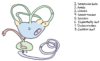
Internal ear
What is the inner ear composed of?
A
-
Membranous labyrinth
- A series om membranous fluid-filled ducts and chambers
- Surrounded by osseus labyrinth
-
Osseous labyrinth
- Within the pars petrosa of os temporale
- The membranous layrinth consists of:
- Vestibular labyrinth (with receptor organs for balance)
- Cochlear labyrinth (with organ of hearing)
- The osseous labyrinth consists of:
- Vestibulum
- 3 semicircular canals
- Cochlea
- Membranous labyrinth is filled with endolymph (K+)
- Osseous labyrinth is filled with perilymph (Na+)
-
Spatia perilympahtica:
- Between the membranous labyrinth and the osseous labyrinth
- Fluid-filled clefts
- Connected to subarachnoid space by the vestibular and cochlear aqueduts
- Filled with perilymph (Na+)
2
Q
Internal ear
Vestibule
A
- Central chamber of the osseuous labyrinth
- Communicates with:
- Rostrally: cochlea
- Caudally: semicircular canals
- The lateral wall of the vestibule has 2 windows:
- Vestibular window
- Cochlear window
3
Q
Inner ear
Vestibular labyrinth
A
- Sense and conduct impulses of balance bia n. vestibularis
- Composed of:
- Saccule
- Utricle
- Semicircular ducts
- Semicircular canals sense physical movement of the head
-
Otolitic organ sense linear acceleration (gravity)
- Otolitic organ are located within the saccule and utricle
- Innervation:
- Sensory (afferent)
- N. vestibulocochlearis → n. vestibularis
4
Q
Internal ear
Vestibular labyrinth: saccule & utricle
A
- 2 enlargements within the osseous vestibule
- Otolitic organ is located within the utricle and saccul
- Semicircular ducts: arise from the utricle (for balance)
- Spiral cochlear duct: arise from the saccule (for hearing)
-
Macule:
- Medial wall
- Macula sacculi
- Macula utriculi
5
Q
Internal ear
Vestibular labytinth: semicircular canals
A
- Arise from the utricle
- House the semicircular duct of the vestibular labyrinth
- 3 channels:
- Anterior channel
- Lateral channel
- Posterior channel
-
Amullae membranae:
- Enlargement of the semicircular ducts near their junction of with the utriclulus
- One for each duct
- Crista ampullaris:
- Projecting into the ampullae memebranae
-
Cupula:
- Gelatinous layer on crista ampullaris
-
Crura membrancea:
- End of semicircular ducts, where they join the utriculus
- Semicircular ducts are filled with endolymph
- When the head moves, endolymph flows to the ampulla
- Endolymph pushes the cupula with hair cells that transduce the mechanical movement to electrical signal

6
Q
Inner ear
Cochleae labyrinth
A
- Location: wall of membranous cochlear labyrinth
- Receptor organ: ggl. spiralis
-
Cochlea is a duct forming a spiral around a bone column, divided into three chambers (scala) by:
- Basilar membrane
- Lamina spiralis ossea
- Ressiner’s membrane / vestibular membrane
-
Scala vestibuli
- Perilymph (Na+)
- Begins at the vestibular window (oval window)
-
Scala tympani
- Perilymph (Na+)
-
Scala media / cochlear duct
- Endolymph (K+)
- Ends at the cochlear window (round window)
- Contains:
- Organ of corti (organ of hearing)
- Tympanic membrane (floor of the cochlear duct)
- Vestibular membrane (roof of cochlear duct)
- Lateral membrane (formed by lig. spirale)
-
Scala vestibuli and scala tympani communicate at the apex of the cochlea
- The apex is called helicotrema
- Turns of the spiral:
- Car: 3
- Eq: 2.5
- Su: 4
- Ru: 3.5

7
Q
Inner ear
Cochlear labyrinth: organ of Corti
A
- The hearing organ
- Within the cochlear duct
- Organum spirale
-
Organ of Corti includes two types of cells
- Sensory cells called hair cells
- Supporting cells
- The hair cells are resting on the basilar membrane and reaching into the space of the cochlear duct
- Role of supporting cells: during displacement of the basilar membrane, the hair cells touch the tectorial membrane → excitation

8
Q
Innervation
A
-
N. vestibulocochlearis (VIII) (afferent)
- N. vestibularis (from utricle and saccule
- N. cochlearis (from base of cochlea)
- Special sensory:
- Nucl. cochlearis
- Nucl. vestibularis
- Corpus trapezodium (location: pons)→ porus acousticus internus
9
Q
Vestibular pathway
A

10
Q
Acoustic pathway
A

