A29 - The vertebral column, epaxial and hypaxial muscles, muscles moving the head Flashcards
Give the general structures of a vertebrae
-
Corpus vertebrae
- Extremitas cranialis [=Caput vertebrae] (cranial covex)
- Extremitas caudalis [=Fossa vertebrae] (caudal concave)
- Crista ventralis
-
Arcus vertebrae
- Pediculus arcus vertebrae
- Lamina arcus vertebrae
- Foramen vertebrale
- Incisura vertebralis cranialis
- Incisura vertebralis caudalis
- Foramen intervertebrale
- Processus spinosus
- Processus transversus
- Processus articularis cranialis
- Processus articularis caudalis
- Processus mamillare (on the arch of Th and L vertebrae, between proc. articularis cranialis and proc. articularis caudalis)
-
Processus accessori
- Proc. articularis caudalis → proc. transversus
- Sus: caudal thoracic vertebrae
- Car: Thoracic and lumbar vertebrae

Give the number of vertebrae in different species

Curvatures in the vertebral column
- In domestic animals, the vertebrae form a chain arranged horizontally, along which three major curvatures are recognized:
- Dorsal convex curvature (head → neck)
- Dorsal concave curvature (neck → chest)
- Dorsal convex curvature (thorax → lumbar region)
Definition of epaxial muscles
- Muscles of the axial zone, which elevate, laterally flex and rotate the cervical, thoracic and lumbar vertebrae
- Innervation: dorsal branches of the spinal nerves

Definition of hypaxial muscles
- Mainly in the cervical region
- Muscles which depress the cervical vertebrae
- Number of muscles are small compared to epaxional musculature

Give the epaxial muscles
Long muscles:
-
M. splenius (superficial layer)
- Pars capitis
- Pars cervicalis
-
M. iliocostalis (middle layer)
- Pars thoracica
- Pars lumborum (ca)
- M. longissimus (middle layer)
- Pars lumborum
- Pars thoracis
- Pars cervicalis
- Pars atlantis
- Pars capitis
-
M. spinalis et semispinalis (deep layer)
- Pars thoracis
- Pars cervicalis
-
M. semispinalis capitis (deep layer)
- M. biventer cervicis
- M. complexus
-
M. multifidi (deep layer)
- Pars lumborum
- Pars thoracis
- Pars cervicalis
Short muscles:
- M. intertransversarii
- Mm. interspinales
-
Mm. rotatores
- Pars brevis
- Pars longus
M. splenius
- Divided into (ø ca):
- Pars capitis
- Pars cervicalis
-
Origin:
- Th1-3, processus spinosus
- Lig. nucahe, caudal end
-
Insertion:
- Crista nucha
- Mastoid part of os temporale
-
Action:
- Bilateral: Extend the neck, raises head
- Unilateral: Draw head laterally
- Eq: Movements of head when galopping, maintains balance when jumping
- Innervation: r. dorsalis ex nn. cervicalis et thoracis
Epaxial
M. iliocostalis (lateral)
- Ca, divided into:
- Pars lumborum (ca)
- Pars thoracica
- Origin: Crista iliaca and fascial sheet
-
Insertion:
- Eq, sus: Fuse with m. longissimus
- Others:
- Ribs on a**ngulus costae
- C7 vertebra, processus transversus
-
Action:
- Fix/stabilize the vertebral column
- Lateral flexion and expiration
- Innervation: r. dorsalis ex nn. cervialis et thoracis spinalis
Epaxial
M. longissimus (medial)
- Divided into:
- Pars lumborum
- Pars thoracis
- Pars cervicalis
- Pars atlantis
- Pars capitis
- Origin: Sacrum and ilium
-
Insertion:
- _Lumborum and thoraci_s:
- Processus transversus/mamillaris/spinosus of lumbar and thoracic vertebrae
- Angula costae
- Cervicis: Between processus transversus of thoracic vertebrae and cervical vertebrae
- Atlantis: Between thoracic/cervical vertebrae and ala atlantis
- Capitis: Processus mastoideus of temporal bone
- _Lumborum and thoraci_s:
-
Action:
- Bilateral: Stabilize and flex head, neck and back dorsally ! increase wightbearing capasity
- Unilateral: Flex the vertebral column laterally, rotate atlantoaxial joint
- Innervation: r. dorsalis ex nn. lumbales, throacis et cervicalis spinalis
Epaxial
M. spinalis or M. spinalis et semispinalis
- M. spinalis (eq, sus)
- M. spinalis et semispinalis (ca, ru)
- Divided into:
- Pars thoracis
- Pars cervicalis
- Origin: Lumbar vertebrae, processus spinosus
- Insertion: C2-C3, processus spinosus
-
Action:
- Bilateral: Extend/stabilize the back, elevate the neck
- Unilateral: Flex nec/back laterally
- Innervation: r. dorsalis ex nn. lumbales, thoracis et cervicalis spinalis
Epaxial
M. semispinalis capitis
- Divided into:
- M. biventer cervicis (dorsal)
- M. complexus (ventral)
-
Origin:
- M. biventer cervicis: Th2-4, processus transversus
- M. complexus: C3-Th1, processus articularis caudalis
- Insertion: Os occipitale (crista nuchae, protubiranta occipitalis externa)
-
Action:
- Bilateral: Extends neck
- Unilateral: Flexes head and neck laterally
- Innervaiton: rr. dorsalis ex nn. cranialis thoracis et cervialis spinalis
Epaxial
Mm. multifidi
- Divided into:
- Pars lumborum
- Pars thoracis
- Pars cervicalis
- Origin: Processus transversus from sacrum → axis
- Insertion: 3 segments cranial to vertebrae of origin, processus spinosus or arcus dorsalis
-
Action: Stabilize the vertebral column in junction with other muscles
- Lumbar region: Flex the back ventrally
- Thoracic region: Rotate the vertebral column
- Cervical region: Elevate/flex the neck laterally
- Innervation: Dorsal branches of segmental spinal nerves
Epaxial
Mm. intertransversarii
- Extend mainly between the processus transversus of adjacent vertebrae
- Action: Stabilize and flex cervical and lumbar vertebral column
- Innervation: Dorsal branches of segmental spinal nerves
Epaxial
Mm. interspinales
- Runs between the adjacent processus spinosus of lumbar, thoracic and cervical vertebrae
-
Action:
- Stabilize the vertebral column
- Ca: flex the spine ventrally
- Innervation: Dorsal branches of segmental spinal nerves
Epaxial
Mm. rotatores
- Divided into:
- Pars brevis
- Pars longus
- Cranial part of thoracic vertebrae, between processus spinosus/transversus
Epaxial
Give the hypaxial muscles
-
M. longus colli
- Pars thoracis
- Pars cervicis
-
M. scalenius
- Pars ventralis (ø ca)
- Pars medius
- Pars dorsalis (ø eq)
M. longus colli
- Divided into:
- Pars thoracis
- Pars cervicalis
- T**h5/6, corpus vertebrae → atlas
- Action: Flex the neck
- Innervation: R. ventralis ex nn. cervicalis spinalis
Hypaxial
M. scalenus
M. scalenus dorsalis: (ø eq)
- Origin: Processus transversus of C4-C6
-
Insertion:
- Dorsal muscular part: rib 1-4
- Ventral muscular part: rib 1-9
M. scalenus medialis
- Origin: Processus transversus of C6-C7
- Insertion: Rib 1
M. scalenus ventralis: (ø ca)
- Origin: Processus transversus of C4-6?
- Insertion: Rib 1
-
Action:
- Inspiration
- Unilateral contraction: flexes nack laterally
- Bilateral contraction: flexes neck ventrally
- Innervation: R. ventralis ex nn. spinalis cervicalis

Statics of the trunk
- Statics: Maintains the equilibrium of the body in a state of rest or in motion
- Dynamics: Analyses the movement of the body during locomotion
- Eq: More weight (55%) is carried by the forelimbs
“Bow and string” theory:
-
Bow: The thoracolumbar vertebrae, their articulations and the ligaments and muscles provides a flexible structure
- Dorsally it has a convex arch which gets its elasticity and nuchael and supraspinous ligaments
- The two ends of the bow are fixed by:
- Sternum
- Linea alba
- Abdominal muscles
- String: Abdominal muscles, which reaches from the thorax to the plevis
- The bow is indirectly attached to the string by interosition of the:
- Cranially: Thoracic skeleton
- Caudally: Pelvic bones
- The string attaches all along the bow
-
Abdominal wall region it is formed by:
- Linea alba
- Abdominal muscles
- Aponeurosis
-
Thorax it is formed by:
- Sternum
- Ribs
- Intercostl muscles
-
Abdominal wall region it is formed by:
- Contraction of the abdominal muscle → flexion of bow
- Contraction of the epaxial muscles → straightens the bow
- Weight of the viscera attached to the vertebral column → straightens the bow
- Weight of the viscera on the abdominal muscles → bends the bow
- “Bow and string theory” can extend into the cervical region, but the curve is in the opposite direction to that of the thoracolumbar region
- Cervical vertebrae and their articulations consititue the bow
- Nuchal ligament acts as the string
- Role of the cantilevers (utstikker): equalize centrifugal push
- Division of the cantilevers:
- Front: head and neck
- Hind: sacrum and tail
- Role: pusch against the bow, allowing the limb to reach gallop
- Sacrum serve as base of attachment of muscle mass of hindlimb, for mobile connection with last lumbar vertebra but immobile with pelvis
- Speed increases → thrust increases → lowering of head & lens neck muscles → reastablish equilibrium of body as a whole

Muscles moving the head
Give the muscles moving the head
- M. sternocephalicus
- M. brachiocephalicus
- M: longissimus capitis
-
M. semispinalis
- M. biventer
- M. complexus
- M. omotransversarius
Specific activator of the head (between cervical vertebrae and os occipitale):
- M. rectus capitis dorsalis major
- M. rectus capitis dorsalis minor
- M. rectus capitis lateralis
- M. rectus capitis ventralis
- M. obliquus capitis caudalis
- M. longus capitis
- M. splenius
M. longissimus capitis
- Origin: Continuation of the longus colli muscle, attach centrolaterally of cervical vertebrae
- Insertion: Base of skull
- Action: Depress head, incline cranial part of the neck
M. sternocephalicus
- Origin: manubrium sterni
-
Insertion:
-
Divided into:
- M. sternomastoideus: mastoid part of os temporale
- M. sternooccipitalis: c**rista nucha of os occipitale
-
Divided into:
- Action: Draw the head and neck to the side

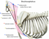
M. brachiocephalicus
From humerus to head and neck
- Divided into:
-
M. cleidobrachialis
- Origin: Clavicle
- Insertion: Distal part of christa humeri
-
M. cleidocephalicus
-
M. cleidomastoideus
- Origin: Processus mastoideus of os temporale
- Insertion: Iscription clavicularis
-
M. cleidocervicalis
- Origin: The dorsal midline over the cranial half of the neck
- Insertion: Inscription clavicularis
-
M. cleidomastoideus
-
M. cleidobrachialis
-
Action:
- If forelimb is fixed: unilateral contraction of the muscle moves head into the lateral direction, bilateral contraction fixes head and neck (or bends downward)
- If limb is free (it’s in an elevated position), it will move forelimb cranially
- Innervation: N. accessorius (XI)
M. semispinalis
- Divided into:
- M. biventer
- M. complexus
-
Origin:
- M. biventer: Proc. transversus of Th2-4
- M. complexus: Proc. articularis of C3-Th1
-
Insertion: Os occipitale on
- Crista nucae
- Protuberentia occipitalis externus
- Action: Extends, holds and ends the neck
M. rectus capitis dorsalis major
- Origin: Processus spinosus of axis
- Insertion: Squama occipitalis
- Action: Flex the atlantooccipital joint dorsally (raises head)
M. rectus capitis dorsalis minor
- Origin: Tuberculum dorsale of atlas
- Insertion: Squamosa occipitalis
- Action: Flex/extend the atlantooccipital joint horisontally
M. omotransversarius
- Origin: Spina scapula on ventral part
-
Insertion: Ala atlantis, caudal border
- Eq: also C2-4 on processus transversus
-
Action:
- Advance the limb (draw the shoulder cranially
- Flex the neck laterally
- Innervation: N. accessorius (XI)
M. obliquus capitis cranialis
- Origin: Ala atlantis
- Insertion: Squama occipitalis
- Action: Flex joints of the head dorsally, incline head to the sides
M. rectus capitis ventralis
- Origin: Arcus ventralis of the atlas
- Insertion: Base of the skull
- Action: Flex the atlantooccipital joint ventrally
M. obliquus capitis caudalis
- Origin: Processus spinosus of axis
- Insertion: Ala atlantis
-
Action:
- Unilaterally it rotates the head around dens of the axis
- Bilaterally it fixes the atlantoaxial joint
M. longus capitis
-
Origin:
- Continuation of the longus colli muscle
- Attach centrolaterally of cervical vertebrae
- Insertion: Base of the skull
- Action: Depress the head, incline cranial part of the neck
M. splenius
-
Origin:
- Thoracolumbar fascia
- Processus spinosus of Th1-3
-
Insertion:
- Christa nuchae
- Mastoid part of os temporale
- Action: Extends, holds and ends the neck
- Innervation: R. dorsalis ex nn. cervialis et thoracis
M. rectus capitis lateralis
-
Origin: Atlas from:
- Arcus ventralis
- A**la atlantis
- Insertion: Processus paracondylaris of os occipitale
- Action: Flex/extend the atlantooccipital joint horisontally
