Radiology quiz Flashcards
(16 cards)
What are the standard views for radiography of the small animal thorax?
The views you take will depend on the pathology you suspect:

Descriibe RL radiograph normals?
- Crura appear parallel (crura of diaphragm two fibroelastic bands that arise from the lumbar vertebrae and insert into the central tendon of the diaphragm.)
- CVC merges with the cranial (right) diaphragmatic crus
- Fundus of stomach (if seen) is caudal to the left (caudal) crus and often gas filled

Discuss normal radiographic appearance LR?
- Crura create a Y-shape
- Caudal vena cava crosses the cranial (left) crus to merge with the caudal (right) crus
- Fundus is caudal to the left crus but might be cranial to the right crus
–May be fluid filled

Discuss normal radiogrpahic appearance of DV?
- Diaphragm appears as a single dome
- Gas should be in the fundus
–Left and dorsal

Discuss normal radiogrpahic appearance of VD?
- Diaphragm often appears as 3 ‘humps’
- Distance between diaphragm & heart is greater than for DV
–Better visualisation of accessory lobe
•Gas should be in the pylorus
–Right and ventral

2.When checking for lung metastases, which views of the thorax should you take?
Both right and left lateral, and either a DV or VD views should be taken.
The dog in the attached picture had an abdominal mass and was being assessed for lung metastasis. Why is there such a difference between these views?

The metastasis are in the shadow on the heart on the RL
3.What is the cranio-caudal centring point for the primary beam when taking a lateral view of a small animal thorax?

4.When taking a lateral view of the thorax, for most dogs how should you prevent axial rotation?
For most dogs it is necessary to place a foam pad under the sternum to correct any axial rotation. (In some very shallow chested dogs, e.g. brachycepahlic breeds, a pad may be needed under the thoracic spine instead.)
4.Give another important aspect of patient positioning for a lateral thoracic radiograph in small animals?
It is also important to extend the forelimbs cranially and secure to prevent excessive tissue overlying the cranial thorax
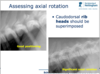
.What is the most reliable way to assess a lateral thoracic radiograph for axial rotation?
The most reliable indicator is superimposition of the rib heads of the caudal ribs.
7.What is the most reliable way to assess a DV or VD thoracic radiograph for axial rotation?
A well positioned DV view:
- The dorsal spinous processes are ovoid or fusiform in shape and lie centrally within the vertebral bodies
- The sternum overlies the spine (this cannot usually be seen due to the superimposition)
Significant axial rotation (DV):
- The dorsal spinous processes are elongated (finger-like) and lie to one side of the vertebral bodies
- The sternum is seen separate from the spine
8.At what point in the respiratory cycle should you make the exposure for a standard radiograph of the thorax?
Routine thoracic radiographs should be taken at peak inspiration. This gives best visualisation of the lung fields and an accurate portrayal of the size of the cardiac silhouette.
Discuss this expiratory radiograph?

Expiratory RLR radiograph:
The cardiac silhouette appears to fill the height of the thorax, there is tracheal and bronchial compression and increased opacity in the caudal lung fields. This could be mistaken for pulmonary oedema.
Discuss this inspiratory radiograph?

Inspiratory RLR radiograph:
The cardiac silhouette does not appear as big, there is less airway compression and the caudal lung fields have normal opacity.
This dog was not in congestive heart failure and the coughing was caused by the airway compression.
How can you assess if a lateral thoracic radiograph of a dog is inspiratory?
How can you assess if a DV or VD thoracic radiograph of a dog is inspiratory?

