Oral tumours, oral surgery & stick injuries Flashcards
Give an overview of oral tumours?
- Tumours can arise from bone, teeth or soft tissue structures of the lower (mandible) or upper (maxilla) jaw, or the tongue or pharynx
- Cat most common SCC
- Most tumours of the oral cavity are malignant
- Malignant melanoma and squamous cell carcinoma most common in dogs
- Squamous cell carcinoma most common in cats
- Other malignant tumours include:
- Fibrosarcoma
- Osteosarcoma
- Multilobular osteochondrosarcoma
Discuss tumour diagnosis?
You can’t just look at a tumour and decide what it is need to send off for histopath
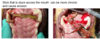
Look at some of these examples of tumours?

Look at some of these examples of tumours?

Look at some of these examples of tumours?

Look at some of these examples of tumours?

Benign tumours are also common and include (naming of benign tumours varies):
- Acanthomatous ameloblastoma (aka basal cell tumour by old vets)
- Peripheral odontogenic fibroma (aka epulis, fibrosing epulis)
Surgery is the mainstay treatment for the majority of malignant and benign tumours
Other treatments options (instead or in addition to surgery) include:
- Radiation therapy
- Chemotherapy
- Immunotherapy (there is a melanoma vaccine available but need to be qualified oncologist to order from USA and not all melanomas respond to the vaccine)
Oral tumours overview?
- Oral tumours are relative common in cats and dogs
- Benign and malignant tumours of the oral cavity account for 3-12% of all tumours in cats and 6% of all tumours in dogs
Oral tumour clinical signs?
- Presence of a mass in the oral cavity
- Increased salivation, blood in the saliva, odorous breath
- If involving alveolar bone teeth may be loose
- Swelling on the face or bulging of the eye (exophthalmos)
- Bloody nasal discharge
- Difficulty eating or pain on opening the mouth, weight loss and enlarged lymph nodes in the neck region
- Loose teeth, especially in animals with general good teeth, may be indicative of cancer-induced bone loss, especially in cats
How to diagnose oral tumours?
Physical examination
- Concomitant problems
- Size and site of oral mass
- Evaluation of regional lymph nodes
Blood tests
FNA
- Often non-diagnostic as requires the lesion to exfoliate (apart from SCC which may exfoliate)
Core biopsy
- Histopathology (bony lesions might prove difficult to obtain representative sample)
Imaging of the skull
- Conventional radiography
- Ideally, CT scan
- To assess bone involvement and degree of margins and aggressiveness
Staging
- Fibrosarcoma(local), osteosarcoma and SCC (will spread to peripheral sites e.glung)
- Conventional radiography
- Ideally, CT scan Oral tumours-diagnostics
Oral tumour treatment options?
Treatment options depend on the location of the tumour and on the type (biology) of the tumour
- benign tumours excised with 1 cm margins
- Malignant tumours excised with 2-3 cm margins
Mandibulectomy
- Unilateral rostral
- Bilateral rostral
- Segmental
- Caudal
- hemimandibuletomy
Maxillectomy
Immunotherapy for melanoma in dogs
Outline mandibulectomy surgery?

Discuss Epulis-peripheral odontogenic fibromas?
- Derived from cells of the periodontal ligament
- There is bone involvement. If you radiographed likely to see lysis around teeth root
- Benign tumour type
- Aim in removal get margin that includes local bone and teeth it is associated with
- Typically, dogs over the age of six (but can be seen at any age); rare in cats
- Common tumour type that is often misnamed as epulis when it should be called peripheral odontogenic fibromas
- Has a relatively good outcome post surgery
- Curative surgery requires taking away bone and teeth local to tumour
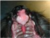
What can be seen here?


What can be seen here?


What can be seen here?


What can be seen here?


What can be seen here?


What can be seen here?


Discuss surgical aftercare for oral surgery?
- Most animals discharged 2-5 days after surgery, depending on level of surgery, comfort and ability to eat soft food
- Return for re-check 7-10 days postop
- Restrictions
- Analgesia
- Antibiotics
- Restrictive (Elizabethan) collar to prevent self-traumatisation
- Limited exercise
- Soft canned food or soaked kibble for 2-3 weeks postop
- No chews, raw hide or chewing toys for at least 3-4 weeks postop Surgical aftercare
Discuss postoperative complications?
- Incision breakdown requiring further surgery to repair
- Bleeding from the nose following maxillectomy
- Increased salivation –may persist for some weeks
- Mandibular drift following mandibulectomy
- Difficulties eating –usually not a problem in dogs but a common problem in cats (routine to put oesophagostomy tube placed at time of surgery)
- Recurrence of tumour
Discuss what to do with cat post oral surgery?
oesophagostomy tube placement

What has occurred here?


What has occurred here?

Salivary mucocele called rannula caused by obstruction to lingual drainage during surgery
Discuss outcome of oral tumour surgery?
Tumour type and staging dependent
- e.g., fibrosarcoma continues to have high local recurrence rate requiring adjunctive radiation therapy or further surgery
- Benign tumours may be cured as long as clean margins have been achieved
Use VSSO website for update information in the consulting room Veterinary surgical society of oncologists Outcome
Discuss oropharyngeal stick injuries?
- Relatively common condition in dogs
- Medium to large breed dogs are over-represented
- Relationship between large breed dogs and low head carriage has been postulated
- Two distinct presentations:
- Acute (< 7 days) or chronic (> 7 days)
- Chronic: injury wont have been observed by owner
- Acute: owner will have seen

Where do stick injuries classically penetrate?

Oropharyngeal stick injuries -diagnostics?
Clinical history –Observed or unobserved
Clinical signs
Acute (< 7 days old)
- Oral pain, dysphagia, blood stained saliva, etc.
Chronic (> 7 days old)
- Cervical swelling with or without discharging sinus
- Chronic cases constitute the majority of reported cases
Owner may have pulled it out and be aware that pieces may be left in if they have done this.
Oropharyngeal stick injuries -further diagnostics?
- Survey radiographs
- Skull/cervical & thoracic
- Ultrasonography
- Computed tomography (CT)
- Magnetic resonance imaging (MRI)
- Flexible endoscopy
- Rigid endoscopy
What can be seen here?

Hole under tongue with piece of wood sticking out problem is when you pull it out have you removed it all? All the fragments as well?
Owner has pulled stick out and can’t see if anything is left in there so could proceed to?
Use flexible endoscope in to check to localise further material. May need to proceed to open surgery with ventral midline cervical approach.
What has happened here?

Ventral cervical swelling from chronic stick injury
How can chronic stick injuries be investigated?

What can be seen on this CT?

Stick between vertical ramus of mandible
Discuss postoperative management of stick injuries?
Course of broad spectrum antibiotics with bactericidal action we don’t want this to progress to sepsis
- 7-14 days
- Clavulanate amoxicillin
- Cephalexin
- Fluoroquinolone Metronidazole
Analgesics
No collar and lead for 2-3 weeks –use a harness
Feeding
- Often feed as normal
- Consider canned food/moistened kibble
Complications of stick injuries?
- Recurrence/development of a discharging sinus. Cannot guarantee got all of it out.
- Pyrexia
- Neck pain
- Bacteraemia
- Nerve damage (recurrent laryngeal nerve damage)
- Dysphagia
What is the other type of presentation of stick injuries?
Discuss cleft palates?
Birth defect leading to abnormal opening between the mouth and nose
Lip (primary cleft palate, cleft lip, harelip)
- Unilateral
- Bilateral
Along roof of the mouth (secondary cleft palate)
- Affecting hard palate only
- Affecting soft palate only
- Affecting both hard and soft palate
Both

What is this?

Primary cleft palate
Repair 3-6 months of age when more skeletally mature and tissue more likely to hold sutures.
Anything in front of the incisive fissure.
Defect external nasal.
Mostly a cosmetic issue.
What is this?

Secondary cleft palate
- Anything that runs from the midline caudally.
- Hard palate involved left image and right image soft palate often both palates involved.
- Will show clinical signs.
What can be seen here?
Combined primary & secondary defects
What can be seen here?

Palatine hypoplasia
Soft palate defect which is bilateral. Ends up with a pseudouvela.
Clinical signs of congenital defects?
- Stunted growth due to poor weight gain
- Breathing difficulties upon exertion
- Coughing or gagging especially when eating or drinking
- Nasal discharge that may include food which may require flushing to clear
- Infection or pneumonia due to food aspiration
- Abnormal visual appearance with cleft lip defect
Management of congenital defects?
- Breeders commonly euthanase affected individuals
- Otherwise, management usually surgical
- Usually wait until affected individual is 3-6 months old
- Numerous ways on surgical management dependent on type of cleft, etc.
- Prone to dehiscence and requirement for repeat surgery
Describe secondary palate defect closures?

Aftercare and outcomes of secondary congenital defects?
- Give antibiotics for individuals with pneumonia or nasal infection
- Elizabethan collar for 2-3 weeks to stop self-trauma
- Soft food for 3-4 weeks
- No hard chews or toys, etc. that can be chewed
- Use of oesophagostomy feeding tube appears to make little or no difference to likelihood of dehiscence
- Do not breed from affected individuals, etc.
Complications of congenital defect surgery?
- Partial or complete dehiscence
- Nasal discharge or sneezing
- Continue coughing or gagging due to short soft palate
