[A] 1.17 Local hyperaemia Flashcards
Hyperaemia
Excess of blood in the vessels supplying an organ/body part
Regulation of circulation is by…
- Hydrodynamic factors
- Neurohormonal regulation → Vasomotion control
- Terminal circulatory bed
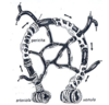
Terminal circulatory bed
Section between the arterioles and venules
- Nutrient, oxygen supply & waste removal
- Large capacity but only holds 5% of the blood
- Major place of circulatory disorders
The haemostatic function of the endothelium
- Maintains the blood in a fluid state
- Anti-thrombotic and pro-fibrinolytic properties
- Upon injury → Opposit effect
The endothelium is critical in…
- Fluid distribution
- Inflammation
- Immunity
- Angiogenesis (development of new blood vessels)
- Haemostasis
Local alterations in blood flow and perfusion
Hyperaemia
- Arteries
- Active hyperemia
- The slowdown of the arterial flow
- Veins:
- Local & systemic venous congestion
- Ischaemia
General alterations in the blood flow and perfusion
- Collapse
- Shock
Hyperaemia due to decreased blood flow: What is seen?
- Contracted arteriole
- ↓ Influx in the terminal bed
- Vasoconstrictor paralysis
- Prehaemostasis
- ↑ Vascular permeability

Prehaemostasis
- Plasma leakage
- Extravasation
- Inflammation produces strong stimuli:
- Mechanical
- Thermal
- Chemical
- Toxic
Pathologically increased blood flow: Signs
- Bright red
- Slightly swollen
- Warm area

Decreased blood flow: Pathological signs
- Bluish red
- Swollen
- Colder
The appearance of distended blood vessels is indicative of…
Vascular injection
Local congestion
- Passive process
- Blood accumulated and slows down in the venous circulation
- Prehaemostasis, hemostasis
- Vein is obstructed or compressed
- Torsion of the intestine
- Strangulation of the spleen
- Thrombosis

Pathology of local congestion: Organ
- Dark red, bluish-red, cyanotic
- Swollen
- Large amount of blood on the cut surface

Pathology of local congestion: Mucosal membranes (skin)
- Dark red, bluish-red, swollen
- Oedemic submucosa
- Wrinkled
- Serous fluid in the lumen of the organ
Infarceratio - Venous infarction
Caused by blocked outflow
- Infraceratio haemorrhagica
- Long-lasting/final obstruction of a vein
- No nutrient/oxygen supply
- Plasma & RBCs in the tissues
- Necrosis of large area
Slow occlusion
- Collaterals enlarge
- Remodel
- Drainage is maintained
- Liver cirrhosis
- Compressed portal veins
- Veins of oesophagus
- “Medusa head”
Systemic venous congestion
Not only the terminal circulatory bed is affected
- Caused by:
- Acute/chronic heart flailure
- Paralysis of the vasomotor centre
- Shock
- Appearance
- In all the organs
Pathology of venous congestion: Liver
- First centrilobular areas affected first
- Chronic case: nutmeg liver

Pathology of venous congestion: Spleen
Hyperemic septic splenitis
Pathology of venous congestion: Kidneys
Medulla affected only
Pathology of venous congestion: Lung
Oedema
Pathology of venous congestion: Intestines
Dark from the serous membranes
Congestive induration
- Chronic congestion
- Parenchyma is destroyed
- Replaced by collagenous fibrous connective tissues
Congestive induration: Pathological effect
- Firm
- Moist content is less on the cut surface

