DIT review - MSK 1 Flashcards

1
Q

Differentiate between the two types of bone formation: endochondral and membranous ossification

A
  • Endochondral ossification
    • Chondrocytes lay down cartilage matrix first
    • Osteoclasts and osteoblasts replace cartilage with woven bone, then remodel to lamellar bone
    • Occurs in axial skeleton, appendicular skeleton, and base of skull
  • Membranous ossification
    • Woven bone formed directly without cartilage, then later remodeled to lamellar bone
    • Occurs in skull and facial bones
2
Q

What is the mutated gene in achondroplasia, and what is its normal function

A
  • Achondroplasia is a defect in endochondral ossification
    • Defect of FGFR3 (gain-of-function)
      • Normally responsible for inhibition of cartilage proliferation
      • Constitutive activation of FGFR3 = inhibited chondrocyte proliferation
    • Autosomal dominant with full penetrance
      • Homozygosity is lethal
3
Q

What are the 2 main benign and 2 main malignant primary bone tumors?

A

Benign: Osteochondroma, Giant cell tumor

Malignant: Osteosarcoma, Ewing sarcoma

4
Q

What part of the bone (diaphysis, metaphysis, or epiphysis), do the following tumors occur:

  • Osteochondroma
  • Giant cell tumr
  • Osteosarcoma
  • Ewing sarcoma
A
  • Diaphysis:
    • Osteoid osteoma
    • Ewing sarcoma
  • Metaphysis:
    • Osteosarcoma
    • Osteochondroma
  • Epiphysis:
    • Giant cell tumor
5
Q

What bone tumor is a lateral projection with overyling cartilage cap?

A

Osteochondroma

6
Q

What is Codman triangle, and what tumor is it associated with

A

Elevation of the periosteum due to tumor

Seen in osteosarcoma

7
Q

What bone tumor is associated with “onion-skin” appearance

A
  • Ewing sarcoma
  • X-ray reveals “onion-skin” appearance – tumor grows within medullary center of bone, pushing outwards and causing periosteum (outer layer) to lay down new layers of bone
  • THINK: eWING = Chicken WINGS and onion rings
8
Q

What tumor is associated with “soap bubble” appearance

A

Giant cell tumor (“osteoclastoma”)

Soap bubbles are in the epiphysis and Giant cell tumor is the only one in the epiphysis

9
Q

What tumor is associated with sunburst appearance on x-ray

A

Osteosarcoma

  • THINK: osteoSarComa (S = sunburst and C = Codman)
10
Q

Which bones are usually affected in giant cell tumor

A
  • distal femur or proximal tibia (knee region)
11
Q

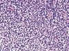
What are the proliferating cells involved in Ewing Sarcoma

A

Poorly differentiated cells derived from neuroectodem

Biopsy reveals small round blue cells resembling lymphocytes

12
Q

What translocation is associated with Ewing sarcoma

A

t(11;22)

THINK: 11 + 22 = 33 (Ewing’s jersey number)

13
Q

What is the defect in osteogenesis imperfecta

A
  • Defect in Collagen type I
14
Q

Presentation of osteogenesis imperfecta

A

Clinical features:

Multiple fractures

Blue sclera (exposure of choroidal veins)

Hearing loss (fracture of bones of middle ear

15
Q

What is the basic premise behind osteoporosis

A
  • Reduction in trabecular (spongy) and cortical bone mass loss
  • Usually due to increased bone resorption (osteoclast > osteoblast)
16
Q

Causes of osteoporosis

A
  • Old age
  • Decreased estrogen levels
17
Q

Describe the labs in osteoporosis (calcium, phosphate, PTH, and alkaline phosphatase)

A

All labs are normal

18
Q

What disease is due to disordered bone remodelling due to increased osteoclastic and osteoblatic activity

A
  • Paget disease of the bone
19
Q

What disease is due to disorder of bone resorption (decreased osteoclast function)

A

Osteopetrosis

20
Q

What disease is associated with increasing hat size

A

Paget disease of the bone

21
Q

Describe the clinical features of osteopetrosis

A
  • Fractures
  • Bone fills marrow space
    • Extramedullary hematopoiesis
    • Pancytopenia
    • “Bone-in-bone” x-ray
  • Vision and hearing loss
    • Impingement of cranial nerves
  • Hydrocephalus
    • Narrowing of foramen magnum
22
Q

What are the lab values in osteopetrosis (calcium, phosphate, PTH, ALP)

A
  • Usually normal
  • May have decreased Ca2+ in severe disease
23
Q

Treatment of osteopetrosis

A
  • Bone marrow transplant (osteoclasts derived from monocytes)
24
Q

Describe the presentation of rickets

A
  • Bow legs, rachitic rosary, frontal bossing (enlarged forehead), pigeon-breast deformity
25
What are the lab values in rickets/osteomalacia (calcium, phosphate, PTH, ALP)
* Decreased vitamin D * Decreased Ca2+ * Increased PTH * Decreased phosphate * Increased alkaline phosphatase (hyperactivity of osteoblasts)
26
What are the stages of Paget disease?
* Lytic – osteoclasts * Mixed – osteoclasts + osteoblasts * Sclerotic – osteoblasts * Quiescent – minimal osteoclast/osteoblast
27
Describe the clinical features of Paget's
* Mosaic pattern of woven and lamellar bone * Increased hat size due to skull thickening * Hearing loss due to impingement of cranial nerve
28
What are the labs in Paget's (calcium, phosphate, PTH, ALP)
* Isolated elevation of alkaline phosphatase (due to end osteoblastic phase) * All other labs are normal
29
What are complications of Paget
* High output cardiac failure – due to increased blood flow from formation of arteriovenous shunts in bone * Osteosarcoma – end-stage osteoblasts at risk for mutation
30
What is osteitis fibrosa cystica
* Bony manifestation of an endocrine disorder (e.g. elevated PTH causing bone resorption) * High PTH = excess osteoclastic activity = “brown tumors” in bone (cystic spaces lined by osteoclasts filled with fibrous stroma and blood)
31
Causes of Osteitis fibrosa cystica
* Hyperparathyroidism * High PTH causing high Ca2+, low PO43-, and high ALP * Pseudohypoparathyroidism (akak Albright hereditary osteodystrophy) * Renal tubules resistant to PTH * Low Ca2+ and high PO34- * High PTH in response to low Ca2+
32
What is polyostotic fibrous dysplasia?
* Bone is replaced by collagen and fibroblasts
33
What is the triad in polyostotic fibrous dysplasia
* Endocrine disorder (Precocious puberty) * Café-au-lait spots * Polyostotic fibrous dysplasia
34
What are the 4 muscles of the rotator cuff?
SItS: Supraspinatous Infraspinatous teres minor Subscapularis
35
What function does each muscle of the rotator cuff have in movement of the arm? Supraspinatous, Infraspinatous, teres minor, Subscapularis
* (1) Supraspinatus * Abducts arm initially (before deltoid kicks in) * Most common rotator cuff injury * Assessed with “empty can” test * (2) Infraspinatus * Laterally rotates arm * (3) Teres minor * Adducts and laterally rotates arm * (4) Subscapularis * Medially rotates and adducts arm
36
What structures are commonly damaged in anterior shoulder dislocation
Axiallary nerve Posterior circumflex artery
37
What are the bones of the wrist
* Wrist bones = So Long To Pinky, Here Comes The Thumb * Scaphoid, Lunate, Triquetrum, Pisiform, Hamate, Capitate, Trapezoid, Trapezium
38
Which nerve is affected in carpal tunnel syndrome
Median nerve
39
What are risk factors of osteoarthritis
Age, obesity, trauma
40
Which arthritis improves with use and which worsens with use
Osteoarthtis - pain worsens with use RA - pain worst in the morning and improves with use
41
Which arthritis is associated with eburnation of bone
Osteoarthritis
42
Differentiate between Heberden and Bouchard nodes
Heberden - DIP Bouchard - PIP
43
Which arthritis spares the DIP?
RA
44
Most common affected joints in osteoarthritis
* Mostly affects weight-bearing joints (hips, lumbar spine, knees), PIP and DIP
45
Which arthritis has symmetric and which has asymmetric joint involvement
OA = asymmetric RA = symmetric
46
What is pannus, and which arthritis is it associated with?
Pannus = inflamed granulation tissue in RA * RA is a systemic autoimmune disease that causes synovitis which leads to the creation of pannus (inflamed granulation tissue) * Recall that myofibroblasts within granulation tissue contract, which is what causes a lot of symptoms of RA
47
What HLA is associated with RA?
HLA-DR4
48
Which arthritis is associated with subluxation and wrist and finger deviation?
RA