Neuroscience Week 2: Organization and Cellular Components of the Nervous System Flashcards
(144 cards)
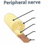
Identify


Superficial epineurium
The outer covering of the nerve

Perineurium
- A mechanically strong sheath that is dense and forms a protective barrier around the nerve fascicle: a blood-nerve barrier.
- It encases two separate nerve fascicles.

Endoneurium
A loose connective tissue.
Deep epineurium
- Accounts for the connective tissue sandwiched between the nerve fascicles.
- We find vasculature in this region.
Perineurial septa
Pass through the nerve fascicles and carry vasculature to the nerve fibers.
NERVE FIBERS
Nerve fiber axon.
Myelin sheath surrounds myelinated axons.
Schwann cells: each myelinates at most one axon internode.
Nerve Fascicles Histology
- Perineurium comprises a flattened form of epithelial cells. They are joined by special junctions, which helps it withstand tremendous pressure.
- Endoneurium comprises collagenous fibers.
- Superficial epineurium is a supporting coat: a cylindrical, dense connective tissue sheath.
- Deep epineurium lies between the fascicles.
Identify


Identify


Identify


Identify


Identify

Identify


Endoneurium
Invests single nerve fiber layers (inflammatory infiltrate in Guillain-Barre Syndrome)

Perineurium

(blood-nerve Permeability barrier)
Surrounds a fascicle of nerve fibers.
must be rejoined in microsurgery for limb reattachment

Identify


Resting membrane potential is?
At rest membrane potential is -60 to -80mV
Myelin sheath in PNS
Schwann Cells
Myelin Sheath in CNS
Oligodendrocytes
Identify


Identify


Identify


CNS Glial Cell types
- Astrocytes
- Microglial cells
- Ependymal cells
- Oligodendrocytes (“Macroglia”)