Exam 2 week 6 ppt 4 CN 7 & 8 Flashcards
Facial nerve number
CN VII
types of function of facial nerve
- –General sensory
- –Special sensory
- –Somatic (Branchial) motor
- –Visceral (Parasympathetic) efferents

List the cranial nerve nuclei in pons and medulla related to the facial nerve (VII): (4)
Facial Nucleus
Solitary nucleus
Superior salivatory nucleus
Nucleus of the spinal tract of V

Facial Nerve
general sensory functions
Tactile sensations from
- skin of external ear,
- wall of external auditory meatus
- outer surface of tympanic membrane
Facial Nerve
nucleus associated with general sensory functional class
Nucleus of spinal tract of V
General Sensory Functions
Tactile sensations from
- skin of external ear,
- wall of external auditory meatus &
- outer surface of tympanic membrane
Facial Nerve
Special sensory functions
Taste information from the anterior 2/3 of the tounge
(associated nucleus: gustatory division of the solitary nucleus)
Facial Nerve
Somatic (branchial) Motor innervations:
- Muscles of facial expression &
- stapedius muscle
(associated nucleus: facial nucleus)
Stapedius muscle
the smallest skeletal muscle in the human body. At just over one millimeter in length, its purpose is to stabilize the smallest bone in the body, the stapes in the inner ear.
innervated by the somatic (branchial) motor portion of the facial nerve
Facial Nerve
Parasympathetic innervations
- Lacrimal glands
- Salivary glands
- submandibular
- sublingual
(associated nucleus: superior salivatory nucleus)
Facial nerve: What nucleus is associated with the special sensory functions
gustatory division of the solitary nucleus
Facial nerve: What nucleus is associated with the somatic (branchial) motor functions
Facial nucleus
Facial nerve: What nucleus is associated with the parasympathetic functions
superior salivatory nucleus
draw the facial nerve chart

Palthway of the sensory (special & general) portion of facial nerve
–1st degree afferent cell bodies are in the geniculate ganglion (equivalent to Dorsal Root Ganglion)
–Synapse in appropriate nucleus
- General somantic in Nucleus of spinal tract of V
- special sensory in Gustatory nucleus (gustatory portion of the solitary nucleus)
The special and general sensory input have their primary afferent cell bodies in the geniculate ganglion (facial nerve equivalent to dorsal root ganglion) and then Synapse in appropriate CNS nucleus:
Nucleus of spinal tract of V for general somatic afference
Gustatory division of Solitary nucleus for the special sensory afferents of taste

describe the Palthway of the nerve branches of facial nerve
- –Full nerve leaves pontomedullary junction
- –It Passes thru internal acoustic meatus with CN VIII
- –then Enters facial canal where geniculate ganglion found
- –the Parasympathetic fibers then split off to form greater petrosal nerve and synapse in pterygo-palatine ganglion in the pterygo-palatine fossa
- –the Sensory & motor branches pass thru stylomastoid foramen
The specific path of the facial nerve involves the Full nerve leaving the brainstem at the pontomedullary junction and passing thru internal acoustic meatus with CN VIII to Enter the facial canal where geniculate ganglion is found.
Parasympathetic fibers then split off to form the greater petrosal nerve which synapse in pterygo-palatine ganglion in the pterygo-palatine fossa
General sensory & motor branches pass through stylomastoid foramen

Clinical Evaluation of the Facial Nerve: Special sensory
taste with one or more of the following:
- salty
- sweet
- sour
Clinical Evaluation of the Facial Nerve: branchial motor
Ask patient to make Make facial expressions
- –Observe for asymmetries
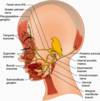
as you see here with this woman is asked to smile and has left facial nerve palsy

two things commonly tested in clinical evaluation of the facial nerve
- •Special sensory –
- taste (salty, sweet or sour)
- •Branchial motor
- –Make facial expressions
- –Observe for asymmetries
Bell’s Palsy:
prevalence
cause
acute or gradual onset?
characterized by (3)
- The most common disease affecting facial nerve
- •Often caused by herpes simplex virus
- •Acute onset
- •Characterized by
- paralysis of facial muscles,
- impaired corneal blink reflex, and
- hyperacusis
Bell’s Palsy is Most common disease affecting facial nerve and Often caused by herpes simplex virus. It has an Acute onset and is Characterized by paralysis of facial muscles, impaired corneal blink reflex, and hyperacusis (inability to tolerate normal sounds which seem abnormally loud)

what number is the vestibulocochlear nerve?
CN VIII
What type of functions does vestibulocochlear nerve have?
special sensory
special sensory functions of vestibulocochlear nerve
- Vestibular information
- balance/equilibrium
- Cochlear information
- hearing/auditory
The vestibulocochlear nerve (CN VIII) Special sensory conveys Vestibular (balance/equilibrium) information from the vestibular organs of the inner ear and Cochlear (hearing/auditory) information also from the inner ear
Vestibular Nerve:
where do nerve fibers end (I would say begin since it is sensory)?
where are cell bodies?
how does the nerve enter the cranium?
where does nerve enter brainstem
Vestibular portion of the vestibulocochlear nerve
- the nerve Endings are in the ampullae of semicircular canals & macular structures of the inner ear
- found in the petrous portion of the temporal bone
- the Cell bodies are in the vestibular ganglion
- nerve Enters the cranium thru internal acoustic meatus (like auditory nerve)
- nerve enters brainstem at pontomedulary junction (with auditory nerve()
Vestibular nerve portion of the 8th cranial nerve has Endings in ampullae of semicircular canals & macular structures of the inner ear. These structures are in the petrous portion of the temporal bone. The cell bodies of the primary afferent fibers are in the vestibular ganglion and the nerve Enters cranium thru internal acoustic meatus

Auditory Nerve:
where do nerve fibers end (I would say begin since it is sensory)?
where are cell bodies?
how does the nerve enter the cranium?
where does nerve enter brainstem?
- nerve Endings are in the cochlea
- found in the petrous portion of the temporal bone
- Cell bodies are in the spiral ganglion
- nerve Enters the cranium thru the internal acoustic meatus (so does vestibular nerve)
- nerve enters brainstem at pontomedulary junction (vestibular nerve does too)
Auditory nerve portion of the 8th Cranial nerve have their sensory endings in the organ of corti of the cochlea. The primary afferent axons have their Cell bodies in spiral ganglion and the nerve also enters the cranium thru internal acoustic meatus.

Nuclei associated with Vestibulochochlear Nerve (6)
The Nuclei are located dorsal caudal pons & rostral medulla (very lateral)
There are two pairs of cochlear nuclei (on each side of the brainstem along the very lateral aspect of the rostral medulla)
- dorsal cochlear
- ventral cochlear
–there are four pairs of vestibular nuclei (which span the pontomedullary junction)
- lateral vestibular (not shown in picture)
- medial vestibular
- inferior vestibular
- superior vestibular (not shown in picture)
The nuclei of the 8th cranial nerve are located dorsal caudal pons & rostral medulla. There are a Pair of cochlear nuclei (dorsal and ventral) on each side of the brainstem along the very lateral aspect of the rostral medulla. These nuclei relay the auditory information. There are 4 pairs of vestibular nuclei: which span the pontomedullary junction named the Lateral, Medial, Inferior, and Superior nuclei

Clinical evaluation of the Vestibulocochlear Nerve includes tests of: (3)
- –Ability to coordinate eye–head movements (vestibulo-ocular reflex)
- –Balance tasks
- –Hearing
Clinical evaluation of the vestibulocochlear nerve includes tests of: Ability to perform coordinated eye–head movements with movement of the head (vestibulo-ocular reflex), balance tasks and demonstrate appropriate hearing. We will discuss all of these test when we discuss the vestibular and auditory systems later in the course


