Workshop: Benign and Malignant Skin Lesions Flashcards
• What is the primary morphology of the lesion?
A. Macule B. Papule C. Patch D. Plaque E. Vesicle F. Nodule
cherry hemangioma- bright red raised papule
• What is the primary morphology of the lesion? A. Macule B. Papule C. Patch D. Plaque E. Vesicle F. Nodule

PAPULE-raised, purple, small
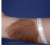
VENOUS LAKES
• What is the primary morphology of the lesion? A. Macule B. Papule C. Patch D. Plaque E. Vesicle F. Nodule

papule. dermatofibroma. raised + hyperpigmented

• What is the primary morphology of the lesion? A. Macule B. Papule C. Patch D. Plaque E. Vesicle F. Nodule
spider telangiectasia/nevi– a MACULE
• What is the primary morphology of the lesion? A. Macule B. Papule C. Patch D. Plaque E. Vesicle F. Nodule

varoucous, raised, possible a papule, maybe a plaque or nodule
seborrheic keratosis

seborrheic keratosis

seborrheic keratosis

seborrheic keratosis
primary morphology
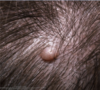
pedunculated papule. acrochordon


erythematous and yellow umbilicated papules on places with evident pores. sebacious hyperplasia
primary morphology

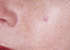
macule. solar lentigo

seborrhaic keratosis

actinic keratoses. papule, maybe a plaque

actinic keratosis
$

actinic keratosis

actinic keratosis–erythematous papules/macules with scale on sun- exposed sites
treatment for actinic keratosis
cryotherapy, maybe surgery, field therapy with fluorouracil

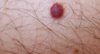
squamous cell carcinoma

squamous cell carcinoma

squamous cell carcinoma

squamous cell carcioma

Squamous cell carcinoma – erythematous crusted papules, plaques, nodules

basal cell carcioma– erythematous, umbilicated (sometimes), papule. doesn’t spread as fast as SCC

BCC- papule, telangiectasia