:L Flashcards
describe pathway of cutaneous afferents from the face? [1]
- Cutaneous afferents from the face travel in cranial nerves and enter the trigeminal nucleus
- Post-synaptic fibres from the trigeminal nucleus decussate and run alongside the medial lemniscal fibres from the body.
- the face afferents end in the VPM thalamus (ventro-postero-medial nucleus),
afferents from the face end come up through the medial lemniscus and terminate in which part of the thalamus? [1]
afferents from the body come up through the medial lemniscus and terminate in which part of the thalamus? [1]
afferents from the face end come up through the medial lemniscus and terminate in which part of the thalamus? [1]
VPM- ventro-postero-medial
afferents from the body come up through the medial lemniscus and terminate in which part of the thalamus? [1]
ventero-postero-lateral: VPL
(together they form complete somatosensory thalamus)
which of the following will show localised pain?
corticospinal tract
anterior spinothalamic tract
posterior spinothalamic tract
lateral reticulospinal tract
medial reticulospinal tract
which of the following will show localised pain?
corticospinal tract
anterior spinothalamic tract
posterior spinothalamic tract
lateral reticulospinal tract
medial reticulospinal tract
*** what are the VPM and VPL? [2] ***
VPL = Ventral Posterolateral Nucleus. primary thalamic relays for somatic sensation; that is, nociceptive and tactile/proprioceptive information from the body
VPM = ventral posteromedial nucleus. primary thalamic relays for somatic sensation; that is, nociceptive and tactile/proprioceptive information from the head
both nuclei in the thalamus !!
what is romberg’s test? what are you testing? how do you perform? what is a postive sign? [1]
Romberg’s test:
- tests proprioception
- standing patient and close eyes. instability & loss of balance is a positive sign
- called sensory ataxia (due to dorsal column damage)
why does inflammation produce long lasting pain?
tissue damage releases pro-inflam chemicals into extracellular space
these chemicals (e.g. bradykinin, K+) activate nociceptor & cause it to stay open for long depolariastion
a painful stimulus activates which receptors? [3]
touch receptor
wide dynamic range receptor
nociceptors
which of the lateral spinothalamic tracts causes perception of pain?
PAG
mediodorsal nuclei of thalamus
ventromedial (VM) & ventroposterior (VP) of thalamus
which of the lateral spinothalamic tracts causes perception of pain?
PAG
mediodorsal nuclei of thalamus
ventromedial (VM) & ventroposterior (VP) of thalamus
explain path of lateralspinothalamic tract
- which three nuclei does it terminate at? [3]
lateral spinothalamic tract:
decussates at site of entry and goes up
- axons reach midbrain, they branch to different nuclei
i) periaqueductal grey (PAG) - arousal
ii) mediodorsal nucleus
iii) ventromedial thalamic group
ii & iii = where concious perpection of pain is registered !
BUT NOT TO VPL / VPM (i.e. not somatosensory
which of the lateral spinothalamic tracts causes arousal & attention to pain?
PAG
mediodorsal nuclei of thalamus
ventromedial (VM) & ventroposterior (VP) of thalamus
which of the lateral spinothalamic tracts causes arousal & attention to pain?
PAG
mediodorsal nuclei of thalamus
ventromedial (VM) & ventroposterior (VP) of thalamus
:)
which of the lateral spinothalamic tracts causes unpleasant quality of painfulness?
PAG
mediodorsal nuclei of thalamus
ventromedial (VM) & ventroposterior (VP) of thalamus
which of the lateral spinothalamic tracts causes unpleasant quality of painfulness?
PAG
mediodorsal nuclei of thalamus
ventromedial (VM) & ventroposterior (VP) of thalamus
which of the lateral spinothalamic tracts causes unpleasant quality of painfulness?
PAG
mediodorsal nuclei of thalamus
ventromedial (VM) & ventroposterior (VP) of thalamus
which of the lateral spinothalamic tracts causes unpleasant quality of painfulness?
PAG
mediodorsal nuclei of thalamus
ventromedial (VM) & ventroposterior (VP) of thalamus
where do you find the insula? [1]
between which lobes? [2]
where do you find the insula? [1]
lateral fissure
between which lobes? [2]
frontal and temporal

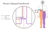
** what changes would be felt bc of this lesion (brown-sequard)? **

- loss of pain and temperature on right side of body below lesion (spinothalamic decussates at level of spinal cord entry)
- loss of motor movement on same side as lesion (corticospinal goes down ipsilateral side)
- loss of proprioception and vibration sense on the same side from damage (DCML has already decussated)
What are the two layers of the dura mater? [2]
- *periosteal layer** (which lines the inner surface of the bones) [1]
- *meningeal** layer which forms dural folds. [1]


posterior spinocerebellar tract pathway?
enters via dorsal root into dorsal horn: synapses with secondary neuron here and goes into posterior spinocerebella tract and goes up to cerebellum on SAME side (ipsilateral)
no decussation !!

describe pathway of anterior spinocerebellar tract xx enjoy
afferent nerve goes in via dorsal horn. synapse with secondary afferent here
a) MOST secondary fibres decussate and go up on the contralateral side
b) BUT, some fibres: stay on same side and go up ipsilateral side
aa) the controlateral ones: go to cerebellum, where they DECUSSATE AGAIN to get back to ipsilateral side
bb) ipsilateral side goes up and stays here
net effect is that both stay ipsilateral

which spinal tract carries the concious proprioception
Cortiocspinal
DCML
Spinothalamic
Spinocerebellar
which spinal tract carries the concious proprioception
Cortiocspinal
DCML
Spinothalamic
Spinocerebellar
which arteries supply the areas where the nerve roots enter and exit the spine dorsally and ventrally, respectively? [1]
radicular arteries
which part of spinal column does the artery of Adamkiewicz supply? [2]
lower thoracic or upper lumbar vertebrae
vertebral artery is a branch of which artery? [1]
subclavian
what is the difference between radicular artereis and segmental medullary arteries?
Radicular arteries
Supply the areas where the nerve roots enter and exit the spine dorsally and ventrally, respectively. These arise from anterior cervical arteries in the cervical region, from posterior intercostal arteries in the thoracic region, and from lumbar arteries in the lumbar region. They gain access to the cord via the intervertebral foramina, and important do not travel as far as the midline to anastomose with ASA or PSA.
Segmental medullary arteries
These are, to all intents and purposes, just like large radicular arteries. They arise similarly from the anterior cervical, intercostal and lumbar arteries, but the key difference is that they travel to the midline and anastomose with the ASA or PSA. The largest and most noteable segmental medullary artery is the great anterior segmental medullary artery of Adamkiewicz, which arises somewhere between T9 and T12, usually on the left, and anastomoses with the ASA to reinforce it as it supplies the lumbosacral spine.

Brown-Sequard Syndrome is damage to one side of spinal cord.
what paraylsis / loss of senses occurs at:
_ipsilaterally
1/2 levels below lesion ipsilaterally:
2 levels below lesion ipsilaterally
2 levels onwards below lesion on contralaterol sid_
ipsilaterally: get complete paralysis below the level of the lesion: due to blocking of corticospinal
1/2 levels below lesion ipsilaterally: complete anaethesia for 1/2 levels due to block of ispilateral spinothalamic.
2 levels below lesion ipsilaterally: loss of light touch, proprioception and vibration (due DCML)
2 levels onwards below lesion on contralaterol side: loss of pain, temp and crude touch 2 levels & below level of lesion - due to the nerves in spinothalamic tract crossing over over couple below

what are the three main branches of facial nerve (VII), what type of innervation do they provide?
greater pertrosal: parasympathetic innervation to lacrimal gland (eye fluid & tears)
- *chordae typmani:**
- special sensory innervation to anterior 2/3rd of tongue;
- parasympathetic innervation to sublingual and submandibular glands
motor root: TZBMC (two zulus bit my cat); temporal, zygomatic, buccal, marginal mandibular, cervical: motor muscles of facial expression
what are the three main branches of facial nerve (VII), what type of innervation do they provide?
greater pertrosal: parasympathetic innervation to lacrimal gland (eye fluid & tears)
chordae typmani:
- *- special sensory innervation to anterior 2/3rd of tongue;
- parasympathetic innervation to sublingual and submandibular glands**
motor root: TZBMC (two zulus bit my cat); temporal, zygomatic, buccal, marginal mandibular, cervical: motor muscles of facial expression
describe the difference in innervation from facial nerve between upper and lower muscles of face?
muscles of upper part of face - frontalis: bilateral innervation - gets innervation from both contralateral and ipsilateral motor cortex ! (upper motor neurons from L & R of brain synapse the lower motor neuron, which innervates the frontalis)
muscles of lower part of face: just contralteral innervation one upper motor neuron from L or R, travel down and decussate and synapses with lower motor neuron

what happens when have Bells palsy?
- which part of facial nerve is affected?
- what does this present as?
what happens when have stroke palsy?
- which part of facial nerve is affected?
- what does this present as?
what happens when have Bells palsy?
- which part of facial nerve is affected: lower motor neuron
- what does this present as: ipsilateral total paralysis
what happens when have stroke palsy?
- which part of facial nerve is affected: upper motor neuron
- what does this present as: contralateral lower muscles paralsis

describe what innervation the vagus nerve innervation does for the following x
- sensory
- parasympathetic
- motor [4]
- sensory: larynx (superior laryngeal and recurrent laryngeal nerve)
- parasympathetic: goes up to midgut
- motor: soft palate; pharynx; oesphagus; larynx (superior laryngeal and recurrent laryngeal nerve)
describe what innervation the vagus nerve innervation does for the following x
- sensory
- parasympathetic
- motor [4]
describe what innervation the vagus innervation does for the following x
- sensory: larynx (superior laryngeal and recurrent laryngeal nerve)
- parasympathetic: goes up to midgut
- motor: soft palate; pharynx; oesphagus; larynx (superior laryngeal and recurrent laryngeal nerve)


midbrain:
what is role of:
- superior colliculi [1]
- inferior colliculi [1]
- cerebral peduncles [1]
midbrain:
what is role of:
- superior colliculi [1]
- *eye movements and visual processing**
- inferior colliculi [1]
- *auditory processing**
- cerebral peduncles [1]
- *contains tracts descending from thalamus (CST & CBT)**
which level of the brainstaim did you find cerebral aquaduct?
midbrain
medulla
thalamus
pons
corpus callosum
which level of the brainstaim did you find cerebral aquaduct?
midbrain
medulla
thalamus
pons
corpus callosum
which CN spans all of the brainstem? [1]
facial nerve
where are the lesions?
Lesion 1:
Lesion 2:

where are the lesions?
Lesion 1: Right CN II
Lesion 2: **Right CN III
if things are same = CN II lesion (afferent is ruined)
if things are different = CN III lesions (efferent ruiend)**

which 3 reactions does the accomodation reflex test? [3]
constriction of pupils
thickening of lens (cilliary muscle constriction)
convergenece of eye balls ( CNIII: movement of eyeballs - medial rectus muscle !!)
Dolls eye reflex:
Afferent CN? [1]
Efferent CNs? [3]
how do u test? what is a normal reflex and abnormal reflex?
Dolls eye reflex:
Afferent CN? [1]: CN VIII
Efferent CNs? [3] CN III, CN IV, CN VI
- tested by turning head side to side
- normal reflex: head moves to right, eyes move left
- abnormal refelx: head moves to right, eyes follow

what structure connexts the cranial nerve nuclei controlling eye movement and the vestibular nuclei? [1]
medial longitudinal fasiculus:
CN III, IV and VI

blink reflex: afferent and efferent CNs?
gag reflex: afferent CN? efferent CN?
Blink reflex:
afferent CN: trigeminal - V1
(but also bright light (CN II & loud noise CN VIII))
efferent CN: CN VII - acts on orbicularis occuli muscle to close
gag reflex:
afferent CN: CN IX
efferent CN: CN X
which is main blood supply to pons? [1]
- branches? [3]
which is main blood supply to pons? [1]
basilar artery
- branches? [3]
paramedian branches
anterior iinferior cerebelllar artery
superior cerebellar artery
what is the blood supply to the medulla mainly from? [1]
- what are the 5 branches? [5]
medulla blood supply = verterbal arteries
- **anterior spinal artery
- posterior spinal artery
- posterior inferior cerebellar artery (PICA)
- anterir inferior cerebellar artery (AICA)
- basal artery**
which 2 arteries provide main blood supply to midbrain? [2]
basilar artery
- *- superior cerebellar artery
- posterior cerebellar artery**
choridal atery
Rule of 4:
which 4 midline structures do you get?
what are their deficits?

how do u check for raised intracranial pressure? [1]
opthalmascope - look for optic nerve where it enters retina (eyes are outgrwoth of brain). optic nerve gets squashed at high pressure - causes papilledema / optic discs bulge out

Rule of 4:
which 4 lateral structures do you get?
what are their deficits?
