Hearing Flashcards
what is the function of the
a) outer ear [2]
b) middle ear [3]
c) inner ear [2]
d) central auditory nS [1]
what is the function of the
a) outer ear: protec, localisation amplicafication
b) middle ear: impedance matching (allows collection of energy to move from larger tympanic window to much small er oval window), pressure equalisation, inner ear stimulation (energy contained within sound isnt reflected away from inner ear)
c) inner ear: sound filterting & signal transduction in nerve endings
d) central auditory nS: information processing
which of the following used electricochemical as its mode of operation?
a) outer ear
b) middle ear
c) inner ear
d) central auditory nS
which of the following used electricochemical as its mode of operation?
a) outer ear
b) middle ear
c) inner ear
* *d) central auditory nS**
which of the following used mechanical vibration as its mode of operation?
a) outer ear
b) middle ear
c) inner ear
d) central auditory nS
which of the following used mechanical vibration as its mode of operation?
a) outer ear
- *b) middle ear**
c) inner ear
d) central auditory nS
which of the following used air vibration as its mode of operation?
a) outer ear
b) middle ear
c) inner ear
d) central auditory nS
which of the following used air vibration as its mode of operation?
- *a) outer ear**
b) middle ear
c) inner ear
d) central auditory nS
which of the following uses mechanical, hydrodyanmic and electricochemical as its mode of operation?
a) outer ear
b) middle ear
c) inner ear
d) central auditory nS
which of the following uses mechanical, hydrodyanmic and electricochemical as its mode of operation?
a) outer ear
b) middle ear
* *c) inner ear**
d) central auditory nS
what is the function of the concha & pinna? [1]
gathers sound energy and focuses / concentrated it on the meatus

which part of the ear divides the outer ear with inner ear?
- eustachian tube?
- cochlea
- round window
- tympanic membrane
- malleus

which part of the ear divides the outer ear with inner ear?
- eustachian tube?
- cochlea
- round window
- tympanic membrane
- malleus
the malleus, incus and stapes are components of the middle ear, what is their function? [1]
transfer vibrational energy from tympanic membrane to oval window (tympanic membrane is greater in size than oval window, so needs amplfying

what are the middle ear osicles? [3]
function? [1]
- malleus
- incus
- stapes
transfer sound energy from tympanic membrane to oval window

which ossicle does tensor tympani muscle connect to? [1]
what does activation of tensor tympani muscle cause to occur? [2]
- connects to the malleus medially
- increased tension in tympanic membrane
- causes modification of sound transfer within middle ear!! dampens sound sensitivty (ear protection)
- *- suppresses sounds coming from within own head (**e.g. chewing)
what are name of auditory reflexes? [2]
what are the two functions of the auditory reflexes? [2]
what are name of auditory reflexes? [1]
- attentuation reflex
- startle reflex
what are the two functions of the auditory reflexes? [2]
- prevent damage to person & ear
- enable distinguish important sounds v background
attenuation reflex
a) activated by? [2]
b) relies on which muscles? [1]
c) function? [1]
attenuation reflex
a) activated by?: own voice / loud sounds
b) relies on which muscles: activation of middle ear muscles
c) function: dampens out low frequency sounds: improves ability to distinguish speech
what is the startle reflex characterised by/ [2]
what is the startle reflex characterised by [2]
- *- ducking down** **to protect neck
- blinking**
what is the cochlea? [2]
which canals does it contain? [3]
what is the cochlea?
- bone with spiral structure
- contains organ responsible for transducing sound energy
which canals does it contain? [3]
- scala tympani: allows send energy to ascend in the cochlea to the apex
- scala vestibuli: spirals otherway and ends at round window
- scala media
(spiral shape of the cochlea allows for differing frequencies to stimulate specific areas along the spiral)

what is the name of the organ in the cochlea that is responsible for sound transduction (converts mech signals into electrical signals)
***** how is perception of sound occured in this organ [2]? *****
**organ of corti
- contains inner hair cells: stereociliar of inner hair cells**
signal to afferent fibres in afferent nerve (provide sound sensation)
- contains outer hair cells: stereociliar of outer hair cellsmodify transduction of low level sounds (motor function: can modify vibrations sensed by inner hair cells & amplify low level sounds

what is structure of inner hair cells like found in the cochlea?
a) upper part [3]
b) lower part [3]
stereocilia of inner hair cells structure:
- *top part:
i) ** stereocilia that are interconnected by cell adhesion molecules
ii) contain K+ channels.
iii) surroundered by endolymph (more like ECF, more like ICF: high K, low Na) - *bottom part:**
i) surroundered by perilymph (more like ECF - low K, high Na)
ii) Ca2+ gate channels
iii) afferent nerve fibres close by

explain mech of how nerve impulse is sent from inner hair cell in cochlea?
- when stereocilia tether (cell adhesion bit) becomes strained, the ion gate opens and K+ moves into the sterocilia.
- occurs because at rest membrane potential of hair cell is negative (across the whole cell), so when the K+ channels open, K+ moves in. **depolarises the cells
-**
stereocilia become displaced / move
- opens K+ channels
- depolarises the cell
- causes Ca+2 channels to open lower down the cell
- causes exocytosis of vesicles
- fused with membrane
- causes NT to diffuse across and activate afferent nerve !
-

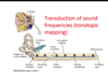
what does it mean that transduction of sound frequency occurs in tonotopic way in ear? [2]
transduction of sound freq occurs in tonotopic way:
- *high frequencies** generate maximum displacement near the base of the cochlea
- *lower frequencies** generate displacment near the end (caled the helicotrema)
WHY?
- due to mechinal and elastic properties of the membran
how do you measure loudness?
what is the scale ?
use decibels (dB)
decibel is a ratio: dB = 20log P/Pr, where P = pressure of incoming sound, Pr = pressure of reference of just audible sound
auditory threshold, 140 = loud rock group

* what is the minimum audibility curve? ! *
what are the units?
where do u see silence on it, where is painful?
why are we less sensitive to high & low frequency sounds?
why are we less sensitive to high & low frequency sounds?
bc energy of sounds doesnt make it into cochlea

what happens to sensitivity of high (&low?) frequncy sounds with age? [1]
what happens to sensitivity of high (&low?) frequncy sounds with age? [1]
decreases with age
explain pathway of hearing in spinal tracts xx
primary order neuron: spiral ganglia in cochlea, axons form auditory nerve & synapse in medulla
secondary order neuron: medulla –> pons. axons collaterals decussate = bilateral asecension. synapses form in the olivery nuclei (helps determine location of sound)
goes to ML & primary sensory cortex

how do u determine source of sound?
- which nuclei determine this
- what is an acoustic shadow?
location by:
- *timing and loudness of sound:**
- superior olivary nuclei use interaural time differences and interaural level differences (loudness) to localise sound
head casts an acoustic shadow: difference in sound pressure reaching the two ears - reduciton in sound level occurs for high freq. sounds for the far ear
how do synapses differ from L & R ear if the sound does not come from distance that is equidistant?
e.g:
sound reaches left ear first: left ear connects to neurons in olivary nuclei first
sound reaches right ear second: connects to neurons in olivary nuclei second
when comes from left ear first / louder on left: AP from left ear can penerate further in olivary nuclei c.f. from right.
if timing is similar: convergence occurs

in short, how can you determine sound location through timing?
SO: timing of the arrival of sound at each ear is being signalled at where excitation converges at medial superior olivary nuclei
how can you determine location through sound intensity?
inhibitory interneurons will inhbit activity of superior olivary nuclei that are coming from medulla: inhibit passage of information up auditory pathway
e.g. higher sound input from the left causes the inhibit sound input from the right - gives rise to ability to discrim where sound is coming from
( e.g. if sound absolutely infront of you: no inhib)
what does Rinne’s test test?
how do u do this?
what is normal and abnormal response (conduction deafness & sensorineural deafness)?
Rinne test: Place the base of a struck tuning fork on the mastoid bone behind the ear. Have the patient indicate when sound is no longer heard. Move fork (held at base) beside ear and ask if now audible. In a normal test, AC > BC; patient can hear fork at ear. With conductive loss, BC > AC; patient will not hear fork at ear.
- normal response: sound is heard louder and longer by air conduction. sound from tuning fork stops, but if move the fork closer - sound is still heard (bc easier to hear air conducted sound)
- conduction deafness: take tuning fork off mastoid proces, tuning fork wont be heard (bc bone conduction is better than air conduction of affected side)
sensorineural deafness: air conduction is better than bone conduction in affected ear. sound is loudest in unaffected ear
what is webers test?
how do u test?
vibrating tuning fork on middle of forehead & ask patient which ear it is heard.
normal patient = equally heard
conduction deafness = sound is louder in affected ear
sensorineural deafness = sound is louder in unaffected ear

what is glue ear / otitis media?
mucous build up in eustachain tube = limits motion of tympanic membrane = reduces transfer of energy.
why is perforation tympanic membrane bad?
what is otesclerosis? why bad?
why is perforation tympanic membrane bad?
affects ability of membrane to vibrate in response to sound
what is otesclerosis? why bad?
lack of motion within the ossicles - limits energy transfer
damage to which of the following would cause conduction aphasia rare form of aphasia in which both expression and comprehension remain intact, but the patient shows an isolated impairment in their ability to repeat simple phrases.
angular gyrus
arcuate fasiculus
broca’s area
wernickes area
damage to which of the following would cause conduction aphasia rare form of aphasia in which both expression and comprehension remain intact, but the patient shows an isolated impairment in their ability to repeat simple phrases.
angular gyrus
arcuate fasiculus
broca’s area
wernickes area
damage to which of the following would cause fluent, but meaningless speech in a patient?
angular gyrus
arcuate fasiculus
broca’s area
wernickes area
damage to which of the following would cause fluent, but meaningless speech in a patient?
angular gyrus
arcuate fasiculus
broca’s area
wernickes area
damage to which of the following would meaningful but abbrievated, ungrammatical speech
angular gyrus
arcuate fasiculus
broca’s area
wernickes area
damage to which of the following would meaningful but abbrievated, ungrammatical speech
angular gyrus
arcuate fasiculus
broca’s area
wernickes area
