:C Flashcards

1
Q

which nuclei in the hypothalamus do u need to know? [2]

A

paraventricular nucleus
supraoptic nucleus

2
Q

what is the difference in anterior and posterior pituitary development? [1]

A

what is the difference in anterior and posterior pituitary development? [1]

  • *posterior pit** = direct outgrowth of brain (specifically the hypothalamus)
  • *anterior pit** = indirect growth: develops from ectoderm in roof of mouth & migrates up
3
Q
A
4
Q

which part of the pituitary gland do the supraoptic and paraventricular nuclei project into? [1]

what occurs here? [1]

where do the other hypothalamic nuclei release their peptides? [1]

A

which part of the pituitary gland do the supraoptic and paraventricular nuclei project into? [1]
project into the posterior lobe

what occurs here? [1]
release peptides into the capillaries in the posterior pit

where do the other hypothalamic nuclei release their peptides? [1]
capillary plexus in the neck of the pit. stalk

5
Q
  • where do you find thermoreceptors? [2]
A
  • where do you find thermoreceptors? [2]
  • *i) cutaneous thermoreceptors
    ii) anterior nucleus of hypothalamus: blood temp**
6
Q

how does the hypothalamus regulate water balance?

  • where do you find osmoreceptors? [1]
  • which hypothalamic nuclei are stimulated to increase water in ur body ? how do they work?
A
  • where do you find osmoreceptors? [1]
  • *subfornical organ (wall of third ventricle): detects osmolarity**
  • subforrnical organ activates cells in the:
  • *i) medial preoptic nucleus**
  • this nucleus connects to the limbic system: regulates concious sense of thirst
  • *ii) paraventricular nucleus & supraoptic nucleus**
  • secrete ADH (makes more aquaporins in CD)
  • oxytocin
7
Q

what are three mechanisms of ADH reducing water loss? [3]

A
  • Increase aquaporins in CD
  • increases perm. of CD to urea (water follows)
  • stimulates sodium reab in thick loop of henle: Na/K/2Cl
8
Q

describe the mechanism of HPA axis (hypothalamus-pit-adrenal axis)

A

describe the mechanism of HPA axis (hypothalamus-pit-adrenal axis):

  • Cells in hypothalamus release CRH (Corticotropin releasing hormone)
  • CRH acts on anterior pit to releease ACTH (adrenocorticotropic hormone)
  • ACTH acts on adrenal cortex to release cortisol
  • *BUT: negative feedback system:**
  • cortisol inhbits release of above
9
Q

how is baby sucking on a breast an example of creating a neuro-hormonal reflex? [2]

how does oxytocin promote maternal bonding? [2]

A

baby sucking on a teat:

  • sucking action is transmitted to the hypothalamus via spinothalamic tract (neuro)
  • releases oyxtocin from posterior pit. (hormonal)
  • activates the milk down reflex

how does oxytocin promote maternal bonding? [2]

  • suckiling - causes oxytocin release in mothers brain
  • this is associated with reward - limbic system
10
Q
A
11
Q

why are anterior pituitary hormones released in ‘two hormone’ mechanism?

A
  • double negative feedback means hormones are released in cyclic fashion (diurnal system or montly cycles)

WORK IN CYCLES !!

12
Q

MESS:

  • how do supraoptic and paraventricular nuclei release peptides in circulation? [1]
  • how does hypothalamic cell bodies from anterior lobe release peptides into circulation? [1]
A

Posterior lobe: Hypothalamic cell bodies in the supraoptic and paraventricular nuclei have axons that project down the pituitary stalk to the posterior lobe of the pituitary. They release peptides into the capillaries in the posterior pituitary which circulate in the blood to other organs

•Anterior lobe: Hypothalamic cell bodies have shorter axons that release peptides on to the capillary plexus in the neck of the pituitary stalk

13
Q

what is growth hormone (GH) inhibited by? [1]

A

•Is inhibited by growth hormone inhibiting hormone = somatostatin.

14
Q

which anterior pituitary hormone is inhibited by its hypothalamic releasing hormone?

Adrenocorticotrophic hormone (ACTH)
Prolactin (PL)
Lutenising hormone (LH)
Follicle stimulating hormone (FSH)
Thyroid stimulating hormone (TSH)

A

which anterior pituitary hormone is inhibited by its hypothalamic releasing hormone?

Adrenocorticotrophic hormone (ACTH)
Prolactin (PL): inhibited by prolactin inhbiting factor (e.g. dopamine)
Lutenising hormone (LH)
Follicle stimulating hormone (FSH)
Thyroid stimulating hormone (TSH)

15
Q

which of the following stimulates production of sex hormones by gonads?

Adrenocorticotrophic hormone (ACTH)
Prolactin (PL)
Lutenising hormone (LH)
Follicle stimulating hormone (FSH)
Thyroid stimulating hormone (TSH)

A

which of the following stimulates production of sex hormones by gonads?

Adrenocorticotrophic hormone (ACTH)
Prolactin (PL)
Lutenising hormone (LH)
Follicle stimulating hormone (FSH)
Thyroid stimulating hormone (TSH)

16
Q

which of the following stimulates production of spem and eggs

Adrenocorticotrophic hormone (ACTH)
Prolactin (PL)
Lutenising hormone (LH)
Follicle stimulating hormone (FSH)
Thyroid stimulating hormone (TSH)

A

which of the following stimulates production of spem and eggs

Adrenocorticotrophic hormone (ACTH)
Prolactin (PL)
Lutenising hormone (LH)
Follicle stimulating hormone (FSH)
Thyroid stimulating hormone (TSH)

17
Q

which of the following regulates metabolism

Adrenocorticotrophic hormone (ACTH)
Prolactin (PL)
Lutenising hormone (LH)
Follicle stimulating hormone (FSH)
Thyroid stimulating hormone (TSH)

A

which of the following regulates metabolism

Adrenocorticotrophic hormone (ACTH)
Prolactin (PL)
Lutenising hormone (LH)
Follicle stimulating hormone (FSH)
Thyroid stimulating hormone (TSH)

18
Q

which of the following induces targers to produce insulin-like growth factors?

Adrenocorticotrophic hormone (ACTH)
Prolactin (PL)
Lutenising hormone (LH)
Follicle stimulating hormone (FSH)
Thyroid stimulating hormone (TSH)
Growth hormone (GH)

A

which of the following induces targers to produce insulin-like growth factors?

Adrenocorticotrophic hormone (ACTH)
Prolactin (PL)
Lutenising hormone (LH)
Follicle stimulating hormone (FSH)
Thyroid stimulating hormone (TSH)
Growth hormone (GH)

19
Q

which of the following regulates metabolism and the stress response?

Adrenocorticotrophic hormone (ACTH)
Prolactin (PL)
Lutenising hormone (LH)
Follicle stimulating hormone (FSH)
Thyroid stimulating hormone (TSH)
Growth hormone (GH)

A

which of the following regulates metabolism and the stress response?

Adrenocorticotrophic hormone (ACTH)
Prolactin (PL)
Lutenising hormone (LH)
Follicle stimulating hormone (FSH)
Thyroid stimulating hormone (TSH)
Growth hormone (GH)

20
Q

which of the following is released by gonadotrophin releasing hormone (GnRH)

Adrenocorticotrophic hormone (ACTH)
Prolactin (PL)
Lutenising hormone (LH)
Follicle stimulating hormone (FSH)
Thyroid stimulating hormone (TSH)
Growth hormone (GH)

A

which of the following is released by gonadotrophin releasing hormone (GnRH)

Adrenocorticotrophic hormone (ACTH)
Prolactin (PL)
Lutenising hormone (LH)
Follicle stimulating hormone (FSH)
Thyroid stimulating hormone (TSH)
Growth hormone (GH)

21
Q

which of the following is inhibited by prolactin inhbiting factor?

Adrenocorticotrophic hormone (ACTH)
Prolactin (PL)
Lutenising hormone (LH)
Follicle stimulating hormone (FSH)
Thyroid stimulating hormone (TSH)
Growth hormone (GH)

A

which of the following is inhibited by prolactin inhbiting factor?

Adrenocorticotrophic hormone (ACTH)
Prolactin (PL)
Lutenising hormone (LH)
Follicle stimulating hormone (FSH)
Thyroid stimulating hormone (TSH)
Growth hormone (GH)

22
Q

which of the following is released by Corticotrophin Releasing Hormone (CRH)

Adrenocorticotrophic hormone (ACTH)
Prolactin (PL)
Lutenising hormone (LH)
Follicle stimulating hormone (FSH)
Thyroid stimulating hormone (TSH)
Growth hormone (GH)

A

which of the following is released by Corticotrophin Releasing Hormone (CRH)

Adrenocorticotrophic hormone (ACTH)
Prolactin (PL)
Lutenising hormone (LH)
Follicle stimulating hormone (FSH)
Thyroid stimulating hormone (TSH)
Growth hormone (GH)

23
Q

which of the following is released gonadotrophin releasing hormone (GnRH) [2]

Adrenocorticotrophic hormone (ACTH)
Prolactin (PL)
Lutenising hormone (LH)
Follicle stimulating hormone (FSH)
Thyroid stimulating hormone (TSH)
Growth hormone (GH)

A

which of the following is released gonadotrophin releasing hormone (GnRH) [2]

Adrenocorticotrophic hormone (ACTH)
Prolactin (PL)
Lutenising hormone (LH)
Follicle stimulating hormone (FSH)
Thyroid stimulating hormone (TSH)

Growth hormone (GH)

24
Q
A
25
Q

spinal accessory nerve comes from which spinal levels? [1]

A

C1-5

26
Q

what makes up the posterior triangle of neck?

  • posterior border? [1]
  • anterior border? [1]
  • other border? lol [1]

what important structures run through it? [4]

A

what makes up the posterior triangle of neck? [3]

  • posterior border: sternocloidomastoid
  • anterior border: trapezius
  • other border: middle 1/3 clavicle

what important structures run through it? [4]

  • *- spinal accessory nerve (CN XI)** comes out of it and **innervates the trapezius
  • external jugular vein
  • brachail plexus (in inferior section)
  • part of subclavian**
27
Q

what is each section?

purple:
green:
red
yellow?

A

what is each section?

purple: digastric
green: submental
red: carotid
​yellow: muscular

28
Q

what is each section?

purple:
green:
red
yellow?

A

what is each section?

purple: digastric
green: submental
red: carotid
​yellow: muscular

29
Q

label the borders of the carotid triangle xx

A
30
Q

where do u find the carotid body? [1]
what type of receptors do u find in the carotid body? [1]

whre do u find carotid sinus?
​what type of receptors do u find in the carotid sinus? [1]

A

where do u find the carotid body? [1]
bifurcation of carotid artery (into internal CA & external CA)

what type of receptors do u find in the carotid body? [1]
chemorecptors

whre do u find carotid sinus?
bifurcation of carotid artery (into internal CA & external CA)
​what type of receptors do u find in the carotid sinus? [1]
baroreceptors

31
Q

MESS:

what are the borders of the carotid triangle? [3]

A

the posterior belly of the digastric muscle, the superior belly of the omohyoid muscle, and the anterior border of the SCM.

32
Q

where do u find the carotid body? [1]
what type of receptors do u find in the carotid body? [1]

whre do u find carotid sinus?
​what type of receptors do u find in the carotid sinus? [1]

A

where do u find the carotid body? [1]
bifurcation of carotid artery (into internal CA & external CA)

what type of receptors do u find in the carotid body? [1]
chemorecptors

whre do u find carotid sinus?
bifurcation of carotid artery (into internal CA & external CA)
​what type of receptors do u find in the carotid sinus? [1]
baroreceptors

33
Q
A
34
Q

which of the following is innervated by the superior laryngeal nerve?

thyroartyenoid
cricoartyenoid
cricothyroid
transverse arytenoid
olbique arytenoid

A

which of the following is innervated by the superior laryngeal nerve?

thyroartyenoid
cricoartyenoid
cricothyroid
transverse arytenoid
olbique arytenoid

others = recurrent laryngeal nerve

35
Q
A
36
Q

injury to superior laryngeal nerve causes? [1]

injury to recurrent laryngeal nerve causes? [1]

A

injury to superior laryngeal nerve causes? [1]
paralysed cricothyroid - cant ary length & tension = monotone voice

injury to recurrent laryngeal nerve causes? [1]
paralysed vocal folds: horseness (cant abduct the vocal folds)

37
Q

MESS:

for posterior triangle, what is the

  • posterior border
  • inferior border
  • anterior border
A

MESS:

for posterior triangle, what is the

  • posterior border: SCM
  • inferior border: middle 1/3 clavicle
  • anterior border: trapezius
38
Q

MESS
which important structures run through the posterior triangle? [3]

A
  • spinal accessory nerve
  • external jugular vein
  • part of subclavian artery
  • part of brachial plexus
39
Q
A
40
Q

where do you find the roots of the cervical plexus? [1]

which nerves are the roots from? [1]

A

where do you find the roots of the cervical plexus? [1]
around middle posterior border of SCM

which nerves are the roots from? [1]
C1-4

41
Q

what is inside [3]

leaves [5]

and surrounds [2]

the carotid sheath?

A

Inside:
Common carotid & internal carotid
Internal jugular vein
Vagus Nerve

Leaving:
External carotid artery
CN IX
CN XI
CN XII
Superior laryngeal

Surrounding:
Ansa Cervicalis (C1-3): innervates: most of the infrahyoid muscles, including the sternothyroid muscle, sternohyoid muscle and the omohyoid muscle.
Ascending sympathetic fibers

42
Q

describe the path for when enters light –> occipital lobe xx

A

ganglion cells in retina have axons that project down optic nerve

two optic nerves meet at the optic chiasm

the axons pass through the optic chiasm and form the optic tract

go to lateral geniculate nucleus and go to occipital lobe

43
Q

objects in left visual field go to which visual cortex? [1] in right and left eye?

describe the pathway info goes if looking at something in left visual field from right and left eye?

A

left visual field –> right visual cortex (& vice versa)

if seeing something in the left visual field:

left eye
pass through lens & stimulate the photoreceptors in nasal hemiretina in left eye. ganglion cell axons project out of optic nerve & cross over in optic chiasm; end up synapsing on lateral geniculate nucleus on right hand side

right eye
left visual field stimulates temporal hemiretina & stays on same side in optic chiasm go to lateral **geniculate nucleus on right hand side

both then go to right visual cortex**

44
Q

describe how eyesight is affected from each lesion

A

1: partial optic nerve lesion - ipsilateral scotoma = partial blind spot in eye easy to be unaware !
2: complete optic nerve lesion = complete blindess in that eye
3: optic chiasm lesion = bitemporal hemnianopia - cant see in lateral side of both eyes
4: optic tract lesion = homonymous hemianopia - cant see lateral on one side & medial on the other
5: damage to meyers loop = upper left visual field loss

45
Q

describe how eyesight is affected from each lesion

A

1: partial optic nerve lesion - ipsilateral scotoma = partial blind spot in eye easy to be unaware !
2: complete optic nerve lesion = complete blindess in that eye
3: optic chiasm lesion = bitemporal hemnianopia - cant see in lateral side of both eyes
4: optic tract lesion = homonymous hemianopia - cant see lateral on one side & medial on the other
5: damage to meyers loop = upper left visual field loss

46
Q

what would you see / not see in 6-8?

A
  1. optic radation lesion: homonymous hemianopia with foveal sparing - there is macula sparing bc of axons on other side of brain

7 visual cortex lesion in inner cortex: homonymous hemianopia (very similar to above)

8: visual cortex lesion in superfial striate cortex (central area 17): bilateral loss of macula vison

47
Q
A
48
Q

which part of the brain gives you the ability to track an object? [1]

what does ^ connect with?

which pathway mediates neck flexes triggered by seen objects? [1]

A

which part of the brain gives you the ability to track an object? [1]
superior colliculi

some optic nerve fibres go to the superior colliculi -> connects to the medial longitudinal fasciculi (MLF): links together and synchronises the oculomotor nuclei

which pathway mediates neck flexes triggered by seen objects? [1]
tectospinal tract

= together give synchronised eyes and neck movement

49
Q

vestibulo-ocular reflex - what does this allow? [1]

which is the afferent CN? [1]
which is the efferent CN? [2]

A

vestibulo-ocular reflex - what does this allow? [1]
head changes angle, eyes compensate for movement & keep gaze steady

which is the afferent CN? [1]
vestibulo-cochlear (VN III)

which is the efferent CN? [1]
**abducens (CN VI): drives the lateral rectus muscle
CN III: drives the medial rectus muscle

mediated by: medial longitudinl fasiculus**

50
Q

explain the caloric stimulation test xx

A
  • warm / cold water is irrigated into extenal auditory canal with a syringe
  • creates a convective current in the endolymph of the nearby horizontal semicircular canal
  • if warm: mimics a head turn to ipsilateral side (both eyes will turn slowly away from the irrigated ear towards to contralateal ear, then flick occurs towards irrigated ear_
  • if cold: mimics head turn to contralateral side: lick occurs away irrigated ear_

COWS: cold flick to other ear; warm: flick to same ear

direction of the flick is called nystagmus

51
Q

explain the caloric stimulation test xx

A
  • warm / cold water is irrigated into extenal auditory canal with a syringe
  • creates a convective current in the endolymph of the nearby horizontal semicircular canal
  • if warm: mimics a head turn to ipsilateral side (both eyes will turn slowly away from the irrigated ear towards to contralateal ear, then flick occurs towards irrigated ear_
  • if cold: mimics head turn to contralateral side: lick occurs away irrigated ear_

COWS: cold flick to other ear; warm: flick to same ear

direction of the flick is called nystagmus

52
Q

what are the two types of photoreceptors? [2]

which of ^ do you only find in the fovea? [1]

which of ^ are more active in dark? [1]

A

what are the two types of photoreceptors? [2]
rods & cones (red, blue, green)

which of ^ do you only find in the fovea? [1]
cones

which of ^ are more active in dark? [1]
rods

53
Q

describe the structre of rods & cones

A

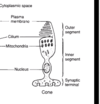
outer segment: **photoreceptive part

i) rods contain photopigment called rhodopsin
ii) cones contain photopigment called cone opsins**
* *occur in stacked plates**

inner segment: cell body (& mito)

54
Q

describe the structre of rods & cones

A

outer segment: **photoreceptive part

i) rods contain photopigment called rhodopsin
ii) cones contain photopigment called cone opsins**
* *occur in stacked plates**

inner segment: cell body (& mito)

55
Q

what happens to photoreceptors in the dark? [2]

what happens to photoreceptors in the light? [1]

A

what happens to photoreceptors in the dark? [1]
constant inward leak of sodium in outerpart of the receptor: keeps the cell depolarised. causes the release of glutamate from its synaptic ending

what happens to photoreceptors in the light? [1]
light hyperpolarises the tonic glutamate release

56
Q

after photoreception has occured, how does signal transduction occur?

A

light passes through the & meets intercalated discs of rods and cones: glutamate is continously released from synaptic endings of rods and cones onto bipolar cells in the dark

this release is supressed by light

bipolar cells then depolarise & release NTs onto ganglion cells

ganglion cells project their axons into the optic nerve and therefore signal this light sensation to the brain

57
Q

which 3 cells cause seeing stuff x

A
  • *photorceptors** (rods & cones - recive light signal & lose inhibitions) that connect to
  • *bipolar** cells that connect to
  • *ganglion** cels that send axons to optic nerve
58
Q
A
59
Q

what type of cells make up the conjuctiva? [1]

function? [1]

A

what type of cells make up the conjuctiva? [1]
stratified columnar epithelium; goblet cells

​function? [2]
mucous secreted: mixes with tears to make more viscous
covers sclera & inside of eyelids

60
Q

where is aq humour made in the eye? [1]

A

ciliary body

61
Q

what are the two subtypes of glaucoma?

how doe drug treatments for glaucoma work?

how do u treat? [5]

A

open angle glaucoma / type 1: slowly progressive condition. trabecular meshwork becomes gradually blocked. normal angle betweeen cornea and iris

primary angle close glaucoma: occurs when angle between cornea & iris is reduced. flow cannot go into canal of schlemm = rapid increase in pressure. sudden pain and visio n loss

62
Q

where are most photoreceptors found in the eye? [1]

* which bit of blood circulation can you see in eye using opthalmoscope? [1] *

A

where are most photoreceptors found in the eye? [1]
fovea

* which bit of blood circulation can you see in eye using opthalmoscope? [1] *
inner circulation