BB Histology Flashcards
what is the cytoplasm of the nerve cell body characterised by? [4]
- abundance of free ribosomes: appear as clumps called nissil bodies
- prominent **golgi apparatus
-**
neurotubules & neurofilaments
- large nucleus

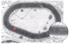
what is A? [1]
dendritic spine xx
what happens to cytoplasmic components in the axon hillock?
appears like there is no cytoplasmic components - its how u know that its the axon and not a dendritic spine !

what are the three types of axon junctions? [3]
what are the three types of axon junctions? [3]
- *axoaxonic:** axons - axons
- *axosomatic**: axon - cell body
- *axodentritic**: axon - dendrites
what type of staining do you use to demonstrate overall shape of neurons? [1]
silver staining !
which PNS cells ensure the rapid conduction of nerve impulses?
oligodendrocytes
ependymal
astrocytes
satellite cells
schwann cells
which PNS cells ensure the rapid conduction of nerve impulses?
oligodendrocytes
ependymal
astrocytes
satellite cells
schwann cells
what does myelin look like in myelin sheath?
lipid rich layer tha is wrapped around lots of times
what are the dots?
what is the light pink?
what is dark pink ?

what are the dots: axons
what is the light pink: myelin
what is dark pink: nuclei of schwan cells

label these parts of the schwann cell x


whar are the SL pictured here? [1]
what type of cell? [1]

whar are the white lines pictured here? [1]
schmidt-lanterman clefts
what type of cell? [1]
schwann cell

what occurs at the nodes of ranvier? [1]
what occurs at the nodes of ranvier? [1]
ions diffuse in & out of neuron: saltatory conduction
what is the role of satellite cells? [1]
where do you find? [1]
which type of staining? [1]
what do they look like? [1]
what is the role of satellite cells? [1]
help maintain the envrioment around neuronal body in the ganglion
where do you find? [1]
cells bodies of ganglia
which type of staining? [1]
H&E
what do they look like? [1]
cuboidal cells

what is the connective tissue of PNS? [3]
- *perineurium:** CT surrounds a group of nerve fibres. helps to form BBB
- *endoneurium**: collagen fibres secreted by schwann cells
- *epineurium**: outermost tissue. typical dense CT

which of the following helps to form BBB?
perineurium:
endosteum
periosteal
endoneurium:
epineurium:
which of the following helps to form BBB?
perineurium
endosteum
periosteal
endoneurium
epineurium
which of the following provide physical & metabolic support for the neurons
oligodendrocytes
ependymal
astrocytes
microglial
schwann
which of the following provide physical & metabolic support for the neurons
oligodendrocytes
ependymal
astrocytes
microglial
schwann
which of the following provide myelin in CNS
oligodendrocytes
ependymal
astrocytes
microglial
schwann
which of the following provide myelin in CNS
oligodendrocytes
ependymal
astrocytes
microglial
schwann
which of the following provide phagocytic properties in CNS
oligodendrocytes
ependymal
astrocytes
microglial
schwann
which of the following provide phagocytic properties in CNS
oligodendrocytes
ependymal
astrocytes
microglial
schwann
what is A?
oligodendrocytes
ependymal
astrocytes
microglial
schwann

what is A?
oligodendrocytes
ependymal
astrocytes
microglial
schwann
astrocytes:
function? [2]
name / types for astrocytes found in white and grey matter? [2]
astrocytes:
function:
- **support & modulate activity of neurons
- **maintain tight junctions of capillaries that form blood brain barrie
- *- buffer K+ conc in the ECF:** modualtes neuronal activity
name / types for astrocytes found in white and grey matter? [2]
- *protoplasmic:** gray matter
- *fibrous:** white matter

which of the following form blood brain barrier?
oligodendrocytes
ependymal
astrocytes
microglial
schwann
which of the following form blood brain barrier?
oligodendrocytes
ependymal
astrocytes
microglial
schwann
what is the difference between myeline sheath in CNS compared to produced in PNS? [4]
- different myelin-specific proteins
- fewer schmidt-lanterman clefts
- no external lamina
- nodes of ranvier larger
- thinner
which of the following line the ventricles of the brain and central canal of spinal cord
oligodendrocytes
ependymal
astrocytes
microglial
schwann
which of the following line the ventricles of the brain and central canal of spinal cord
oligodendrocytes
ependymal
astrocytes
microglial
schwann
what is B?
oligodendrocytes
ependymal
astrocytes
microglial
schwann

what is B?
oligodendrocytes
ependymal
astrocytes
microglial
schwann
how do you differentiate microglial cells?
elongated nuclei
small
when stained with heavy metals, exhbit short & twisted processes


