EX3 Endocrine Images Flashcards
What gland is this
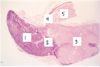
pituitary gland
- pars distalis
- pars intermedia
- pars nervosa
- pars tuberalis
- infundibular stalk

pituitary gland
- posterior lobe
- anterior lobe
what is this image depicting

formationg of the pituitary gland via the outpocketing of ectoderm from the roof of the mouth (rathke’s pouch) and outpocketing of the neuroectoderm lining the floor of the hypothalamus (neurohypophyseal bud)

- basophils
- acidophils
- chromophobes

- acidophils
- basophils
- chromophils
- sinusoid

- acidophils
- basophils
- chromophils
- sinusoid

- acidophils
- basophils
- chromophils
- sinusoids
- supporting reticular fibers; stroma

- pars distalis
- colloid filled space
- pars intermedia
- pars nervosa

posterior pituitary
- capillary
- neurosecretory bodies (harring bodies)
- pituitcytes

posterior pituitary
- herring bodies

- capillary
- blood vessel
- pituitcyte (nuclei)
- herring body
what is this an image of

adrenal gland
- capsule
- adrenal cortex
- adrenal medulla
- zona glomerulosa
- zona fasciculata
- zona reticularis

- capsule
- zona glomerulosa
- zona fasciculata
- zona reticularis
- adrenal medulla
which portion of the adrenal cortex is this image from

zona granulosa
what portion of the adrenal cortex is this image from

zona fasciculata
what portions of the adrenal cortex is this image from
zona reticularis
what is this an image of

adrenal medulla
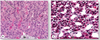
What are these images of; each one is at high magnification

islet of langerhans cells of the pancreas
What is than an image of; each at a high magnification

thyroid gland
A; follicular cell
B; parafollicular cell

- parafollicular cell
- basement membrane
- follicular cell
- thyroid follicle
- thyroglobulin (TGB)
what is this an image of

parathyroid glands
- chief cell
- blood vessel
- oxyphil cell
what is this an image of and what does the image on the right represent

parathyroid gland
- principal (chief) cells
- oxyphils
on thr right; fenestrated capillaries
