8.2: Neurovasculature of the Abdomen Flashcards
which vessel supplies the abdominal viscera and most of the anterior abdominal wall? Where does it enter through the spinal cord?
the abdominal aorta.

It enters the abdomen at T12 through the aortic hiatus of the diaphragm and descends along the vertebral column to the left of the midline.
It terminates at the L4 vertebral level, where it bifurcates into two common iliac arteries.
A single median sacral artery arises near the bifurcation.
which arteries (branches of the abdominal aorta) supply the structures of the posterior abdominal wall
Paired segmental parietal branches including inferior phrenic and lumbar arteries
Paired visceral branches supply organs of the retroperitoneum, these are:
- middle suprarenal
- testicular or ovarian
- renal arteries

The celiac trunk, a short trunk that arises at _____ and supplies the abdominal foregut. Its branches, the ___, left _____, and ____ ____arteries, anastomose extensively with each other
The celiac trunk, a short trunk that arises at T12/L1 and supplies the abdominal foregut. Its branches, the splenic, left gastric, and common hepatic arteries, anastomose extensively with each other
The superior mesenteric artery (SMA), which arises at ____, posterior to the neck of the pancreas. It supplies ___ structures, and its major branches include the ____ ____, ___ colic, ____ colic, and ____ arteries, as well as a series of ___ and ____branches
The superior mesenteric artery (SMA), which arises at L1, posterior to the neck of the pancreas. It supplies midgut structures, and its major branches include the inferior pancreaticoduodenal, middle colic, right colic, and ileocolic arteries, as well as a series of jejunal and ileal branches

The inferior mesenteric artery (IMA), which arises at ___ and has the smallest caliber of the three visceral trunks. It supplies the ____ through its left ___, ____, and ___ ____ branches
The inferior mesenteric artery (IMA), which arises at L3 and has the smallest caliber of the three visceral trunks. It supplies the hindgut through its left colic, sigmoidal, and superior rectal branches


2 major branches of the common iliac artiers that pass along the brim of the pelvis
- internal iliac artery, which descends into the pelvis
- external iliac artery, which gives off the inferior epigastric and deep circumfex iliac arteries before passing into the lower limb as the femoral artery.

outline the anastomoses of the celiac trunk, superior and inferior mesenteric arteries whihc provide collateral blood supply to the intestinal organs
celiac trunk and superior mesenteric artery: anastomose in the head of the pancreas through the pancreaticoduodenal arteries and in the body and tail of the pancreas through dorsal pancreatic and inferior pancreatic arteries.
superior and inferior mesenteric arteries anastomose near the junction of the transverse and descending colons through the middle and left colic arteries. The marginal artery runs along the mesenteric border of the largest intestine and connects the ileocolic, right colic, middle colic, and left colic arteries.
The inferior mesenteric artery anastomoses with arteries of the rectum through its superior rectal artery

where do abdominal aortic aneurysms most comonly occur
Abdominal aortic aneurysms (AAAs) most commonly occur between the renal arteries and the bifurcation of the aorta. When small they can remain asymptomatic, but large aneurysms can be palpated through the abdominal wall to the left of the midline. Ruptured AAAs present with severe abdominal pain that radiates to the abdomen or back. Mortality rates for ruptured aneurysms approach 90% due to overwhelming hemorrhage
outline the branches of the branches of the abdominal aorta:
- inferior phrenic artery (paired)
- celiac trunk
- middle suprarenal
- superior mesenteric artery
- renal artery
- lumbar artery
- testicular/ovarian artery
- inferior mesenteric artery
- common iliac artery
- median sacral artery
The celiac trunk, a short trunk that arises at T12/L1 and supplies the abdominal foregut. Its branches, the splenic, left gastric, and common hepatic arteries, anastomose extensively with each other (Figs. 8.10 and 8.11).
The superior mesenteric artery (SMA), which arises at L1, posterior to the neck of the pancreas. It supplies midgut structures, and its major branches include the inferior pancreaticoduodenal, middle colic, right colic, and ileocolic arteries, as well as a series of jejunal and ileal branches (Fig. 8.12).
The inferior mesenteric artery (IMA), which arises at L3 and has the smallest caliber of the three visceral trunks. It supplies the hindgut through its left colic, sigmoidal, and superior rectal branches

The ___ ___ ____ receives blood from retroperitoneal and pelvic organs, walls of the abdomen and pelvis, and the lower limbs
The inferior vena cava (IVC) receives blood from retroperitoneal and pelvic organs, walls of the abdomen and pelvis, and the lower limbs

the ingerior vena cava originates along the ____ vertebral level where the __ ___ veins merge
It originates at the L5 vertebral level where the common iliac veins merge.
It ascends along the right side of the vertebral column, passes posterior to the liver, and pierces the central tendon of the diaphragm at the T8 vertebral level, where it enters the right atrium of the heart.
direct tributaries of the inferior vena cava

Paired common iliac veins drain the _____ ____ and ___ ____ ___.
Paired ____ ____ and ____ veins drain the posterior abdominal wall and diaphragm and accompany the arteries of similar name.
Veins of the retroperitoneal organs include the right and left ____ veins, the right ___ vein, and the right ____ or ____ (gonadal) vein. The suprarenal and gonadal veins on the left side drain to the ____ ____ ____.
Typically three hepatic veins enter the IVC from the liver immediately below the diaphragm.
Paired common iliac veins drain the external iliac and internal iliac veins.
Paired inferior phrenic and lumbar veins drain the posterior abdominal wall and diaphragm and accompany the arteries of similar name.
Veins of the retroperitoneal organs include the right and left renal veins, the right suprarenal vein, and the right testicular or ovarian (gonadal) vein. The suprarenal and gonadal veins on the left side drain to the left renal vein.
Typically three hepatic veins enter the IVC from the liver immediately below the diaphragm.

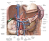
The ___ ___ vein (usually known as the portal vein), part of the ____ ____ ___, shunts nutrient-rich venous blood from the capillary beds of the gastrointestinal tract and its associated organs to sinusoids of the liver. This blood eventually enters the ___ ____ ___through the ____ veins.
The hepatic portal vein (usually known as the portal vein), part of the hepatic portal system, shunts nutrient-rich venous blood from the capillary beds of the gastrointestinal tract and its associated organs to sinusoids of the liver (Figs. 8.17 and 8.18). This blood eventually enters the inferior vena cava through the hepatic veins.

what’re the portosystemic pathways
normal connections between the systemic (caval) venous systems and portal venous systems.


the splenic vein drains most of fthe ____ structures, the superior mesenteric vein drains most of the ____ structures, and the inferior mesenteric vein drains most of the ____ structures and drains into the _____ vein
the splenic vein and the superior mesenteric vein meet to form the ___ ____ vein
the splenic vein drains most of fthe FOREGUT structures, the superior mesenteric vein drains most of the MIDGUT structures, and the inferior mesenteric vein drains most of the HINDGUT structures and drains into the SPLENIC vein
the splenic and superior mesenteric veins meet to form the hepatic portal vein.


8.17 label