Orthopaedic Hip Conditions Flashcards
what are some common conditions of the hip?
Osteoarthritis
Bursitis (causes lateral hip pain) - Gluteal tendinopathy (differential diagnosis)
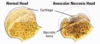
Avascular Necrosis (AVN)
Impingement
Labral Tear
what make sup the pelvis?
Each hemipelvis = fusion of 3 bones (Ischium, Ileum and Pubis)
joined by Sacrum posteriorly and pubic symphysis anteriorly
Acetabulum - Socket

Femur is long bone, what is its high clinical significance?
NoF
Bleeding
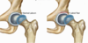
what are the different parts of the femur and their function?
Head - Articulates with Acetabulum, Head covered in smooth hyaline cartilage and if damaged wont regenerate and beginning of arthritis
Neck - Blood supply
Greater Trochanter - Attachment for Abductors and Rotators (external)
Lesser trochanter - Attachment for Psoas

what is the acetabulum?
Part of Pelvis
Cup-shape socket
Ligamentum teres in middle

what is the labrum and its function?
Fibrocartilagious lining of acetabulum
Deepen socket
Add stability

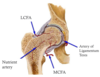
what is the blood supply?
Profunda femoris (branch of femoral)
Branches medial and lateral circumflex arteries
MFCA (Major contributor to femoral head) - 2 branches - Ascend to head and Transverse to form cruciate anastomosis
LFCA (lateral femoral circumflex artery) - 3 branches - Ascending branch to joint capsule, Transverse branch to cruciate anastomosis, Descending branch

hwat are other minor contributors to the blood supply?
Artery of Ligamentum Teres
Nutrient Arteries of Bone (vessels in the femur so when fractured blood supply to head is disrupted
where does the blood supply to the neck of the femur enter and what is its clinical significance?
Neck Of Femur - Primary blood supply enters via Capsule
Clinical Significance - Fracture Neck of Femur
Intracapsular Fracture = Blood supply disrupted
Extracapsular Fracture = Blood supply maintained
Anastomosis on the neck of femur and ascend to the head
Fractures out of the capsule don’t have risk of avascular necrosis

13 muscles around the hip joint which do what actions?
Flexors
Extensors
Abductors
Adductors
Rotators

what are bursae?
Fluid-filled sacs
Reduce friction between tissues
what is osteoarthritis?
Degenerative change of synovial joints
Progressive loss of articular cartilage
Secondary bony changes
Can be primary or secondary (a pathology that’s caused damage)
Hip replacement is main treatment for people with bad arthritis

how does osteoarthritis present?
Characterised by worsening pain and stiffness of the affected joint
Limiting everyday life
what is Trochanteric Bursitis?
Trochanteric Bursa is a Fluid-filled sac that is Sandwiched between hip abductors and ITB
Bursitis - Inflammation of the bursa, Swelling
Epidemiology - Females > Males

what are the causes of trochanteric bursitis?
Trauma
Over-use - Athletes, often runners, Repetitive movements
Abnormal movements:
- Distant problem, e.g. Scoliosis
- Local problem - Muscle wasting following surgery (so have to tense harder), Total Hip Replacement, Osteoarthritis

what is the presentation of trochanteric bursitis?
Pain (well localised)
Point tenderness
Lateral hip
what would be found on examination of trochanteric bursitis?
LOOK:
- May have scars from previous surgery
- May have muscle wasting - Gluteals
FEEL:
- Tenderness at Greater Tuberosity
MOVE:
- Worst pain in active abduction
how do you investigate trochanteric bursitis?
X-ray - May be normal, OA, THR, Spine abnormalities
MRI - Shows soft tissues and fluid
Ultrasound - Can be therapeutic as well as diagnostic, Guided injection

what is the treatment of trochanteric bursitis?
NSAIDs
Relative rest / Activity modification
Physiotherapy:
- Correct posture, abnormal movements
- Stretching
- Strengthen muscles around joint
Injection - Corticosteroids
Surgery - Bursectomy (leaves scar which may be painful and not helpful, divide ITB aswell), Rarely required
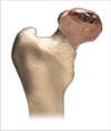
what is avascular necrosis?
Death of bone due to loss of blood supply
onset of hip pain

what is the epidemiology of avascular necrosis?
Males > Females
Average age 35-50 years old
80% = bilateral (May be offset in time)
3% = multifocal (3 or more joints)
what are the risk factos for avascular necrosis? (trauma)
Irradiation
Fracture
Dislocation
Iatrogenic

what are the risk factos for avascular necrosis? (systemic)
Idiopathic
Hypercoaguable states
Steroids
Haematological - Sickle Cell Disease, Lymphoma, Leukaemia
Caisson’s disease (divers)
Alcoholism (commonest risk factor)
avascular necrosis can be caused by trauma to what area?
Injury to femoral head blood supply
Intracapsular fracture
Anastomosis of blood vessels around neck of femur

what is the process of avascular necrosis being caused by idiopathic?
Intravascular coagulation is the final common pathway
Pathoanatomic cascade:
Coagulation of intraosseous microcirculation =
Venous thrombosis =
Retrograde arterial occlusion =
Intraosseous hypertension =
Reduced blood flow to head =
Cell death =
Chondral fracture and collapse =

what ar ethe symptoms of avascular necrosis?
Insidious onset of groin pain
Pain with stairs, walking uphill and impact activities
Limp
what would be seen on examination of avascular necrosis?
Largely normal
May replicate early arthritis:
- Reduced range of motion (partic internal rotation)
- Stiff joint
what imaging would be done for avascular necrosis?
X-Ray
MRI:
- 99% sensitive and specific
- Will identify earliest changes
- MRI shows where bone is affected so can help to plan treatment

what is the non-operative treatment of avascular necrosis?
(First make diagnosis, then stop cause if possible)
Reduce weight-bearing
NSAIDs
Bisphosphonates - Early AVN, controversial
Anticoagulants
Physiotherapy - Maintain range of motion, Keep the ball round!
what is the surgical treatment of avascular necrosis?
Restore blood supply

- Core decompression (drill hole in head to avascular area to reduce venous hypertension and to allow new blood supply to grow in and restore bone strength)
- Core decompression and vascularised graft
Move the lesion away from the weight-bearing area - Rotational Osteotomy (osteotomy is a surgical operation whereby a bone is cut to shorten or lengthen it or to change its alignment)
Total Hip Replacement
what is Femoroacetabular Impingement (FAI)?
Femoroacetabular impingement (FAI) is a condition in which extra bone grows along one or both of the bones that form the hip joint — giving the bones an irregular shape. Because they do not fit together perfectly, the bones rub against each other during movement
2 surfaces hitting together
what is the commonest cause of FAI?
Hip pathology in younger patient
Secondary osteoarthritis
what are the different types of FAI?
Broadly divided into 2 categories:
- Cam lesion
- Pincer
Results in impingement of femoral neck against anterior edge of acetabulum
FAI - what is a Cam lesion?

Femoral-based impingement
Usually in young athletic males - Rowers
Excess bone leading to Decreased head to neck ratio and Aspherical head
Abutment of lesion on edge of acetabulum - Flexion

what is shown here?

FAI cam lesion
Slight bump
Easier to see on 3D imaging
what is a FAI pincer?
Acetabulum-based impingement
Usually in active females
Abnormal acetabulum leading to:
- Anterosuperior acetabular rim overhang
- Acetabular protrusion
Abutment of lesion on edge of acetabulum
More prominence of the edge of the acetabulum

what is shown here?

FAI - Pincer
what are associated injuries of FAI?
labral degeneration and tears
cartilage damage and flap tears
secondary hip osteoarthritis
what is the presentation of FAI?
Groin pain - Worse with flexion
Mechanical symptoms:
- Block to movement
- Pain with certain manoeuvres - Getting out of a chair, Squatting, Lunging
what would be seen on examination of FAI?
Reduced flexion and internal rotation
Positive FADIR test - Flexion, ADduction, Internal Rotation
what investigations would be used in FAI?
X-ray - Identify the bony pathology
MRI - Useful for assessing associated conditions:
- Labral tears
- Articular cartilage damage
what is the non-operative treatment of FAI?
Activity modification
NSAIDs
Physiotherapy:
- Correct posture
- Strengthen muscles around joint
whatis the operative surgery of FAI?
Arthroscopy:
- Shave down the defect
- Deal with labral tears
- Resect artic cartilage flaps
Open Surgery:
- Resection
- Periacetabular Osteotomy
- Hip Arthroplasty
- Resurfacing
- Replacement

what is a Labral Tear?
A hip labral tear is an injury to the labrum, the soft tissue that covers the acetabulum (socket) of the hip. A hip labral tear can be caused by injury, structural problems, or degenerative issues. Symptoms include pain in the hip or stiffness
Most commonly anterosuperior tear

what is the epidemiology of a labral tear?
All age groups
Commonly active females:
- Pincer
- More flexible
In older age group often degenerative changes
what are the cuases of a labral tear?
FAI
Trauma
OA
Dysplasia
Collagen diseases – Ehlers-Danlos
what is the presentation of a labral tear?
Groin or Hip Pain - Intermittent pain that comes on on certain movements of the hip and not a constant pain
Snapping sensation
Jamming or locking
what would the examination of a labral tear be like?
can be normal
Positive FABER test - Flexion, ABduction, External Rotation
Anterior tears
what investigationg would be used for a labral tear?
Ensure adequate imaging so identify any root causes of pathology
X-ray - OA, Dysplasia
MRI Arthrogram - 92% sensitive
Diagnostic injection - Local anaesthetic

what is the non-opertative treatment of a labral tear?
Activity modification, NSAIDs, Physiotherapy
Injection of Steroids
what is the operative treatment of a labral tear?
Arthroscopy (more of a role in younger people)
Repair
Resection
