Mini Symposium: Multi-system Autoimmune Disease Flashcards
what are some Connective Tissue Diseases?
◦Systemic Lupus Erythematosus
◦Scleroderma
◦Sjogren’s syndrome
◦Auto-immune myositis
◦Mixed connective tissue disease
what are examples of Systemic vasculitis?
◦Giant cell arteritis
◦Granulomatosis polyangiitis (Wegeners)
◦Microscopic polyangiitis
◦Eosinophilic granulomatosis polyangiitis (Churg-Strauss)
how is a diagnosis made?
Cardinal clinical features: History & Exam
Bedside investigations
Immunology
Imaging
Tissue diagnosis
Exclusion of differential diagnosis
What are some mimics?
Drugs - cocaine, minocyline, PTU
Infection - HIV, endocarditis, Hepatitis, TB
Malignancy - lymphoma
Cardiac myxoma
Cholesterol emboli
Scurvey
what is Systemic lupus erythematosus?
Lupus is a condition that affects the immune system. It can cause problems with your skin, joints, kidneys and other organs
Systemic lupus erythematosus (SLE) – lupus – is a long-term condition causing inflammation to the joints, skin and other organs. There’s no cure, but symptoms can improve if treatment starts early
what is the epidemiology of SLE?
UK Prevalence: 28/100,000
UK incidence: 4/100,000
Female:Male 9:1
Onset: 15-50 years
Significant ethnic diversity:
Afro-Caribbeans>Asian>Caucasian
what is SLE aetiology?
Genetic factors – high concordance rate of SLE in monozygotic twins, Sibling risk of developing SLE is 30-fold higher than in the general population, polygenic mode of inheritance
Hormonal factors
Environmental factors – ultraviolet light, drugs, infections
what is SLE pathogenesis?
Immune response against endogenous nuclear antigens (break in immunological tolerance)
Immune complex formation
Complement activation
Tissue injury
where can SLE effect?
Clinical presentation is varied
Different organs that may be involved in lupus

what is the classification criteria? (any 4)
1) Malar rash (butterfly rash)
2) Discoid rash (raised, scarring, permanent marks, alopecia)
3) Photosensitivity
4) Oral ulcers
5) Arthritis (2 joints at least) - Non erosive, bilateral
6) Serositis (pleurisy or pericarditis)
7) Renal (significant proteinuria or cellular casts in urine)
8) Neurological (unexplained seizures or psychosis)
9) Haematological (low WCC, platelets, lymphocytes, haemolytic anaemia)
10) Immunological (anti ds-DNA, SM, cardiolipin, lupus anticoagulant, low complement)
11) ANA
photos showing SLE: Skin manifestations
DDx butterfly: rosacea, mitral stenosis
Discoid: Scaly centre, dark rim
Malor rash and discoid rash

in someone with SLE what is one of the first things you should do?
urine analysis
use this to check renail involvement - make sure there isnt any
lupus nephritis

when would you consider a diagnosis of SLE?
Usually seen in women of child bearing age
Constitutional symptoms of fever, weight loss, malaise, severe fatigue
Skin rash and/or stomatitis
Arthritis
Pleuritic chest pain
Renal disease
cytopenia - Cytopenia occurs when one or more of your blood cell types is lower than it should be
what autoantibodies are involved in SLE?
ANA – antinuclear antibodies
Seen in 95% of SLE
Not specific for SLE
Seen in many inflammatory, infectious and neoplastic diseases
Seen in 5% to 15% of healthy population
what are ANA?
antinuclear antibodies (ANA)
our immune system normally makes antibodies to help you fight infection. In contrast, antinuclear antibodies often attack your body’s own tissues — specifically targeting each cell’s nucleus
what are some Conditions where +ve ANA
is unhelpful?

in different condiitons what % of ANA is present?

what are osme other autoantibodies that many be seen in SLE?
Anti -ds DNA:
Seen in 60% of patients with SLE
Highly specific for SLE
Low titre rarely seen in other inflammatory conditions
Strongest clinical association is with nephritis
Anti -Sm (Smith):
Seen in 10% to 30% of SLE patients
Highly specific for SLE
Anti–Ro:
Risk of foetal congenital heart block
Neonatal lupus
Antiphospholipid antibodies:
Anti-cardiolipin, lupus anticoagulant
arterial/venous thrombosis
miscarriages
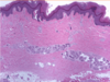
what is Systemic sclerosis?
Systemic scleroderma, or systemic sclerosis, is an autoimmune rheumatic disease characterised by excessive production and accumulation of collagen, called fibrosis, in the skin and internal organs and by injuries to small arteries
connective tissue disease characterised by fibrosis of the skin and internal organs

what are the different Classification of scleroderma?
Localised scleroderma
Systemic sclerosis (SSc):
- Limited cutaneous systemic sclerosis (CREST)
- Diffuse cutaneous systemic sclerosis
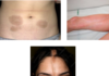
what si the difference between localised and systemic scleroderma?
Localised scleroderma (also known as morphoea or morphea) only affects the skin. In some cases it can spread to the tissues underneath the skin, such as muscles and bones. Systemic sclerosis affects the skin but may also involve the body’s internal organs
(pictures showing localised scleroderma)

what is the epidemiology of systmeic scleroderma?
UK Prevalence: 24/100,000
UK Incidence: 10/1,000,000
Onset: 30-50 years
Female : Male 3:1
what is the aetiology of systemic scleroderma?
Environmental:
- Silica
- Solvents
- Viral infections
Genetic predisposition
What is the pathogenesis of scleroderma?
Vascular damage (microcirculation)
Immune system activation/Inflammation
Fibrosis
what is Limited cutaneous SSc?
Limited cutaneous systemic sclerosis (lcSSc) is a subtype of systemic sclerosis (SSc; see this term) characterized by the association of Raynaud’s phenomenon with skin fibrosis limited to the hands, face, feet and forearms

What is Diffuse cutaneous SSc?
Diffuse cutaneous systemic sclerosis (dcSSc) is a subtype of Systemic Sclerosis (SSc; see this term) characterized by truncal and acral skin fibrosis with an early and significant incidence of diffuse involvement (interstitial lung disease, oliguric renal failure, diffuse gastrointestinal disease, and myocardial involvement).

SSc subsets - what may limited and diffuse cause?
Limited:
Anti centromere antibodies
Pulmonary hypertension
Gastrointestinal
Diffuse:
Anti Scl70 antibodies
Pulmonary fibrosis
Renal crisis
Small bowel bacterial overgrowth
what is Sjogren’s syndrome?
Systemic autoimmune disease
Characterised by lymphocytic infiltrates of the salivary and tear glands leading to oral and ocular dryness and autoantibody secretion
Sjögren’s (pronounced show-grins) syndrome is a condition that affects parts of the body that produce fluids, like tears and spit (saliva)
It usually starts in people aged 40 to 60 and is much more common in women than men
It’s a long-term condition that can affect your daily life, but there are treatments to help relieve the symptoms

What is the epidemiology of Sjorgens syndrome?
Prevalence: 1 in 100
Incidence: 4 in 100,000
Onset: 40-50yrs
Female : Male 9:1
what are the symptoms and sigsn of Sjogrens syndrome?
Dry eyes and mouth
Anti Ro (SSA), anti La (SSB) antibodies
Salivary gland biopsy
Parotid gland enlargement
1/3 have systemic upset - fatigue, fever, myalgia, arthralgia, dry skin

What are some complications of Sjogrens syndrome?
Lymphoma
Neuropathy
Cutaneous vasculitis
Interstitial lung disease
Renal tubular acidosis
what is Auto-immune myositis?
Myositis is a rare type of autoimmune disease that inflames and weakens muscle fibers
Autoimmune myositis causes inflammation and weakness in the muscles (polymyositis) or in the skin and muscles (dermatomyositis)
what is the epidemiology of Polymyositis and dermatomyositis?
Rare: 6/million incidence
More common in females (ratio 3:1)
Peak incidence is in 50 – 60 year olds
Increased risk of malignancy
what does myositis cause and how is it investigated?
Muscle weakness - symmetrical, proximal
Raised CK level
EMG, MRI, muscle biopsy
Interstitial lung disease – anti Jo1 antibodies
Cutaneous - Gottron’s papules (80%), Heliotrope rash (30-60%)

what is an overlap syndrome?
Mixed Connective Tissue Disease
what is mixed conncetive tissue disease?
Raynaud’s
Soft tissue swelling/sclerodactyly
Myositis
Arthralgia
Mixed connective tissue disease is a term used by some doctors to describe a disorder characterized by features of systemic lupus erythematosus, systemic sclerosis, and polymyositis. Raynaud syndrome, joint pains, various skin abnormalities, muscle weakness, and problems with internal organs can develop
The vasculitides - what are they and how are they classified?
The term vasculitides refers to heterogeneous disorders characterized by vascular inflammation affecting vessels of different sizes from large arteries to capillaries or tiny venules. Vasculitis leads to diminished blood flow or vessel occlusion resulting in ischemia, necrosis, and subsequent tissue damage
Inflammation of the blood vessels
Classified depending on size of vessel involved

what are different exmaples of the vasculitides?

what is Giant cell arteritis?
Most common type of vasculitis
Mainly effects white individuals
Incidence increases with age
More common in women
Temporal arteritis is a serious condition where the arteries at the side of your head (the temples) become inflamed
Almost all patients who develop giant cell arteritis are over the age of 50. GCA commonly causes headaches, joint pain, facial pain, fever, and difficulties with vision, and sometimes permanent visual loss in one or both eyes

what is the GCA classification criteria?
3 of the following (classic temporal arteritis):
Age at onset ≥50 years
New headache
Temporal artery tenderness/reduced pulsation
ESR≥50
Abnormal temporal biopsy
what is the gold standard investigaitong for GCA?
Temporal artery biopsy
Shows inflammatory infiltrate in the artery wall
Intimal hyperplasia
Luminal exclusion
Can also do Ultrasound doppler

what is shown here?

CT angiogram, MR angiogram
CT angiogram and MR angiogram good to investigate large vessel involvement
what is FDG PET good for diagnosing?
inflammation of the aorta

what are the complciations of GCA?
Irreversible visual loss
Aortic aneurysms
Arterial stenosis and limb ischaemia
Stroke
what is the treatment and management of GCA?
Urgent initiation of high dose Prednisolone
40-60mg per day, gradually tapered
PPI
Bone protection
Steroid sparing medication
what is ANCA associated vasculitis?
Antineutrophil cytoplasmic antibody (ANCA)-associated vasculitis (AAV) is a group of diseases (granulomatosis with polyangiitis, eosinophilic granulomatosis with polyangiitis and microscopic polyangiitis), characterized by destruction and inflammation of small vessels
Granulomatosis with polyangiitis (Wegener’s)
Microscopic polyangiitis
Eosiniphilic granulomatosis with polyangiitis
Overall incidence 15/million
Overall prevalence 150/million

what is Granulomatosis with Polyangiitis (Wegener’s) (GPA) and what does it cause?
Necrotising granulomatous inflammation
Usually involving the upper and lower respiratory tract
Hearing loss, sinusitis, hemoptysis
Necrotising glomerulonephritis is common
cANCA, anti PR3 antibodies
Collapse of nasal bridge
Can get investigated for cancer but biopsy shows it is this

what is Microscopic polyangiitis (MPA) and what does it cause?
Necrotising vasculitis, with few or no immune deposits, predominantly affecting small vessels
Granulomatous inflammation is absent
Renal and pulmonary involvement common
pANCA, anti MPO antibodies

what is Eosinophilic Granulomatosis with Polyangiitis (Churg Strauss) (EGPA) and what does it cause?
Eosinophil rich and necrotising granulomatous inflammation often involving the respiratory tract
Late onset asthma, nasal polyps and eosinophilia
Necrotising vasculitis predominantly affecting small to medium vessels
Neurological involvement
Cardiac, gastrointestinal – poor prognosis
40-60% anti MPO antibodies positive
Case scenario:
45 year old female patient
3-day history of breathlessness and right sided chest pain worse with deep inspiration
CXR confirms right pleural effusion
Treatment with antibiotics makes no difference
FBC showed persistently low WCC of 3.0 then 3.2 and low platelets of 100
Over the last year she has been experiencing intermittent pain and swelling in her joints and recurrent facial rash after sun exposure
What is the likely diagnosis:
A. Scleroderma
B. SLE
C. Giant Cell Arteritis
D. None of the above
Lupus in a women with evidence of cytopenia, arthritis, photosensitivity, pleuritic chest pain
So it is B
What bedside assessment would you prioritise?
A. Blood pressure
B. Fundoscopy
C. Auscultation for carotid bruits
D. Urine dipstick
D – make sure they don’t have any renal involvement
(3) Which of the following immunological tests would support your clinical suspicion
A. Positive anti dsDNA antibodies
B. Reduced complement levels
C. Positive anti Smith antibodies
D. Positive anti-nuclear antibodies
All answers are true here