Mini Symposium: Spine (Degeneration, Low Back Pain, Disc Prolapse) Flashcards

1
Q

what is the most common form of disc prolapses?

A

Normal spinal cord finishes at a level of above L1 and below that we just have spinal roots

Most common form of disc prolapses is lateral but occasionally central can be seen which causes different symptoms

2
Q

what type of joint are Intervertebral Discs?

A

Secondary Cartilaginous Joint

3
Q

what is the structure of the intervertebral discs?

A
  • Disc is largest avascular structure in the body
  • Annulus fibrosus - Tough outer layer
  • Nucleus pulposus - Gelatinous core
  • Annulus may tear and nucleus prolapse - Can cause cord/nerve root compression
4
Q
  • Cartilaginous end plate of each disc attaches to bony endplate of ________
  • The ___ and ___ connect discs with vertebral bodies
  • The fibres of the annulus fibrosis (collagen) run ________ and alternately between layers
  • They resist ________ movements
  • Discs fail with _______ movements
A

vertebra

ALL and PLL

obliquely

rotational

twisting

5
Q

what make sup the inververtebral disc?

A
  • The nucleus pulposus consists mainly of water - 88%
    • collagen & proteoglycans (very hydrophilic)
  • The disc is kidney bean shaped
6
Q

Disc prolapses are usually __________

A

postero-lateral

7
Q

what happens to the intervertebral discs as part of the normal ageing process?

A
  • Decreased water content of discs (see this on MRI)
  • Disc space narrowing
  • “Degenerative” changes on X-rays
  • Degenerative changes in the facet joints
  • Aggravated by smoking, etc.
8
Q

what are some different Pathological Processes?

A
  • Tearing of annulus fibrosis and protrusion of the nucleus
  • Nerve root compression by osteophytes
  • Central spinal stenosis
  • Abnormal movement (between vertebra)
  • Spondylolysis
  • Spondylolisthesis
9
Q

what is nerve root pain like?

A
  • Fairly common
  • Limb pain worse than back pain
  • Pain in a nerve root distribution (radicular)
  • Root tension signs (sciatic nerve stretch test)
  • Root compression signs
  • Dermatomes & myotomes
10
Q

what does radicular mean?

A

Pain in a nerve root distribution

11
Q

what is the management of nerve root pain?

A
  • Most will settle, about 90% in 3 months (without surgical intervention)
  • Physiotherapy
  • Strong analgesia
  • Referral after 12 weeks
  • Imaging - MRI (this is the modality of choice)
12
Q

MRI Scans showing disc prolapse in the second one and first is normal

Spinal canal boundaries:

Anterior – vertebral body

Posterior – laminar and facet joints posteriorly

A
13
Q

whata re the different kinds of disc problems?

A
  • Bulge (generalised) – common, majority asymptomatic, relevance?, nucleus contained, annulus slightly buldging
  • Protrusion (annulus weakened but still intact)
  • Extrusion (through annulus but in continuity)
  • Sequestration (dessicated disc material free in canal)
14
Q

what regions do disc prolapses most often occur?

A

Lumbar > cervical > thoracic

15
Q

wher eis a Cervical Disc Prolapse most common?

A

Most commonly C5/6

16
Q

how often and where do thoracic disc prolapses occur?

A
  • <1% of intervertebral disc prolapses
  • Mid to lower levels (75% T8-12)
  • Most at T11/12
  • Central, posterolateral and lateral herniations
17
Q

where do lumbar disc prolapses occur?

A
  • Usually L4/5 (45%), followed by L5/S1 (40%), then L3/4 (10%)
  • Most are posterolateral - (Posterior Longitudinal Lig weakest)

L3/4 – femoral neuralgia, pain on the anterior of the thigh

L5/S1 – symptoms are pain going right down the leg into the foot and

18
Q

what may central fisc prolapses in the lumbar region cause?

A

Central disc may give pain in both legs, or may be back pain only

19
Q

Prolapsed Lumbar Inter-Vertebral Disc - summary table

A

EHL - extensor hallucis longus

20
Q

what is Cauda Equina Syndrome?

A

Compression of cauda equina

sacral nerve roots compressed – can result in permanent bladder and anal sphincter dysfunction and incontinence

21
Q

what is the treatment of cauda equina syndrome?

A

Cant be treated conservatively

Need to treat rapidly

• Surgical emergency - admission, urgent MRI scan, emergency operation within 48h of onset; delay results in permanent dysfunction

22
Q

what is the aetiology of cauda equina syndrome?

A

central lumbar disc prolapse (commonest)

tumours

trauma (burst or Chance #, disc) or spinal stenosis

infection (epidural abscess – rare in this country)

iatrogenic (spinal surgery or manipulation, spinal epidural injection)

23
Q

whata re the clinica features of cauda equina syndrome?

A

Injury or precipitating event

Location of symptoms (bilat buttock & leg pain + varying dysaethesiae + weakness – beware)

Bowel or bladder dysfunction (urinary retention +/- incontinence overflow)

PR exam - saddle anaesthesia (perianal loss of sensation), loss of anal tone & anal reflex

High index of suspicion in spinal post-op patients with increasing leg pain in presence of urinary retention

24
Q

in cauds erquina syndrome is there a Delay in diagnosis?

A

•Shapiro (USA) – average delay 9 days

–4 patient related

–20 physician related!!

•Surgical emergency

25
Q

in cauda equina syndrome what is used for radiographic evaluation?

A

MRI

If contraindicated, then lumbar CT myelogram

Maybe if the patient has mental implants like pacemaker

26
Q

what is the treatment of cauda equina syndrome?

A

OPERATIVE!

Within 48h (of onset of symptoms)

(statistically significant improvement and difference if surgery < 48 hrs)

27
Q

what is the outcome of cauds equina syndrome?

A
  • 30% undergoing discectomy for cauda equina syndrome did NOT regain normal urinary function
  • 25% with motor deficits never regained full power (A motor deficit is a poor prognostic feature)
  • 33% with sensory deficits never regained normal sensation
  • 25% with perianal paraesthesiae did not return to normal
  • 26% had persitent sexual dysfunction
28
Q

what is Cervical and lumbar spondylosis (OA)?

A

Cervical spondylosis is also called cervical osteoarthritis. It is a condition involving changes to the bones, discs, and joints of the neck. These changes are caused by the normal wear-and-tear of aging. With age, the discs of the cervical spine gradually break down, lose fluid, and become stiffer

  • Common - Very common in cervical and lumbar areas of the spine
  • Degenerative change at - Facet joints, discs, ligaments, etc.
  • If severe, can compress whole cord (not just nerve roots) causing myelopathy - UMN signs in limbs (increased tone, brisk reflexes, etc.)

Degenerative changes cause osteophytes to form and calcification of the ligaments and leaves narrow space for nerves

In cervical spine the whole spinal cord can be compressed and cause a myelopathy

In lumbar they often don’t have upper motor neuron signs

29
Q

Lumbar Spine - what is the role of the facet joints and the intervertebral discs?

A
  • The facet joints (true synovial joints) allow mainly flexion and extension
  • The intervertebral discs (secondary cartilaginous joints) are specialised joints to allow movement between vertebrae
30
Q

whata re the ligaments of the spine?

A
  • Anterior Longitudinal Ligament (ALL – along the front of the vertebral bodies – broad, strong)
  • Posterior Longitudinal Ligament (PLL – along the backs of the vertebral bodies, i.e. front of the spinal canal; narrower)
  • Ligamentum Flavum (between laminae)
  • Interspinous and Supraspinous Ligaments (between spinous processes)
  • Intertransverse Ligament (between transverse processes)
31
Q

what is Lumbar spondylosis?

A

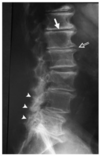
•OA of facet and disc joints (+ degeneration of ligaments, etc.)

Narrowing of disk space, osteophytes narrow the vertebral body, facet joints become arthritic so narrow, sclerotic and irregular

32
Q

what is spinal claudicaitona nd its presentation?

A

•Distinguish from vascular claudication

  • Usually bilateral
  • Sensory dysaesthesiae
  • Poss weakness (drop foot – tripping)
  • Takes several minutes to ease after stopping walking (unlike vascular)
  • Worse walking down hills because the spinal canal becomes smaller in extension, better walking uphill or riding bicycle
33
Q

Normal Canal

A

Stenotic Canal

34
Q

what ar ethe different types of Spinal Stenosis?

A
  • Lateral recess stenosis
  • Central stenosis (bilateral symptoms)
  • Foraminal stenosis
35
Q

what is hte treatment of Lateral Recess Stenosis?

A

Non-operative

Nerve root injection

Epidural injection (more general and doesn’t treat a specific area)

Surgery

36
Q

what is important in Central Stenosis?

A

•Canal shape important (congenital) - esp. trefoil canal

37
Q

what is the treatment of central stenosis?

A

Non-operative

Epidural steroid injection

Surgery (80% improve)

38
Q

what si the treament of Foraminal stenosis?

A

Non-operative

Nerve root injection

Epidural injection

Surgery (70% of patients respond to surgery)

When the nerve root is compressed just as it exits the spine

Most settle in 3 months (90%)

39
Q

Spondylolisthesis and Spondylolysis

A

When one vertebra is translated on another

Posterior elements are separated form the anterior elements

40
Q

what are the symptoms and treatment of Spondylolisthesis?

A

Often vary with type of spondylolisthesis

Treatment depends on symptoms:

  • Conservative with lifestyle changes
  • Surgery for persistent pain +/- nerve root entrapment