Glia Flashcards
(30 cards)
general glia facts
majority of cells in the brain, have receptors for NT and can release NT
serve many functions including: supporting neurotransmission, maintaining ionic balance, trophic support
different from neurons: lack axons, retain ability to survive, do not generate action potentials
types of glia
microglia
macroglia: oligodendrocytes, schwann cells, ependyml cells, astrocytes

microglia overview
macrophages of the nervous system, serve as the immune cells of the brain.
origin: myeloid lineage. Yolk Sac. collinate in the brain early in development
role: surveilence (constantly sampling environment), phagocytosis, synaptic maintencence
microglia response to tissue injury

microglia sculpt synapses
during development or after injury

Oligodendrocytes vs Schwann cells
Oligodendrocytes: Makemyelin, Found in CNS, Highly branching, Myelinate multiple axons/axon segments (up to 40 axons)
Schwann Cells: Make myelin, Found in PNS, One cell makes one myelin sheath

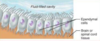
myelin
made of lipids and proteins, extension of cells, each process of oligodenrocytes or a single schwann cell wraps around a small portion of an axon to myelinate it, region that is myelinated is called internode, two internodes are separated by myelin free region called node of ranvier, myelin increases conduction velocity of nerve impulses (saltatory conduction)

disorders of myelin
multiple sclerosis: autoimmune attack on oligodendrocytes, loss of myelin loss of myelinated axons, unknown etiology, can be induced in rodents by injection of mylin resident proteins (MOG, PLP, MBP)

ependymal cells
form lining of ventricles, involved in creating CSF, have cilia (important for movement of CSF through ventricles
astrocytes
named for classic star shape, most common glial cell
fibrous astrocytes
found in white matter, orient parallel to neuronal axons, higher levels of GFAP, big/long fibers, role in K+ homeostasis

protoplasmic astrocyte
found in grey matter, very fine processes, little cytoplasm, very negative membrane potential (~-90 mV), prominent glutamate uptake, lower levels of GFAP

non-overlapping spacial domains
astrocytes have a tiling effect, single astrocyte ensheaths an average of 4 neronal cell bodies (can contact up to 100k sypases in mice and 2m in humans)

vasculature contact
astrocytes form perivascular end-feet around CNS capillaries and arteries
greater than 80% of capillary surface covered by astrocyte processes
helps form BBB
synapse contact
astrocytes are an important component of synapses, NT release activates astrocytes, glial resonse to NT is increase in Ca2+ and release of transmitters (allow astrocytes to modulate neuronal activity)

dynamic extension and retraction
astrocytes extend and retract their fine processes in response to stimuli

astrocyte number and complexity
increase in number and complexity evolutionarily


radial glia during development
scaffold for migrating cells, structural support, source of progenitor cells

synapse formation and maintenance
astrocytes regulate synapse formation (synaptogenesis), modulate synaptic strength (influence insertion of AMPA receptors into neurons), remove synapses (like microglia especially during early development)

control of cerebral blood flow
neuron activity influences blood flow via astrocyte (vessels expand after contact with NT, which increases flow of RBC), known as neurovascular coupling or functional hyperemia, basis of BOLD signal during fMRI
K+ spatial buffering
K+ goes out, hyperpolarization, need to remove K+ to facilitate further neuronal signaling -> astrocyte does passive K+- Cl- uptake, active transport via Na+K+ATPase,
AND K+ transfer by current flow through astrocyte syncytium via gap junctions called K+ spatial buffering
NT clearance
no extracellular digestion of glutamate- cleared by glutamate transporters on astrocyte (EAAT1/GLAST and EAAT2/GLT1)
ensures crisp synaptic transmission, limits amplitude or duration of synaptic signaling, limit excitotoxicity (spill over of NT), operate in reverse direction when Na+ gradients depleted
EAAT2/GLT1: predominant transporter, incredibly abundant, enriched in astrocyte areas near synapses, decreases in expression causes disease


