Dermatology Lecture 1 Flashcards

1
Q

What is the normal colour of a dogs skin?

A

Normal colour of a dogs skin is typically whitish-grey colour

2
Q

What is erythema?

A

Erythema = skin that appears to be redder than normal

3
Q

What is hyperpigmentation?

A

Skin that is darker than normal - leading to black/coloured skin

4
Q

Describe the change that is shown below:

A

Erythema

5
Q

Name the change that is shown below:

A

Erythema

6
Q

Name the skin condition that is shown below:

A

Erythema

7
Q

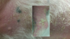
Name the skin condition that is shown in the image below:

A

Hyperpigmentation

8
Q

Name the condition that is shown in the skin below:

A

Hyperpigmentation

9
Q

Name the condition that is shown in the image below:

A

Hyperpigmentation

10
Q

Name the condition that is shown in the image below:

A

Hyperpigmentation

11
Q

Name the condition that is shown in the image below:

A

Hyperpigmentation

12
Q

Name the condition that is shown in the image below:

A

Loss of pigment

13
Q

Name the condition that is shown in the image below:

A

Loss of pigment

14
Q

Name the condition that is shown below:

A

Loss of pigment

15
Q

Name the condition that is shown below: (hint combination of three things)

A

Rash - collection of skin lesions - erythymatous macules, papules and pustules

16
Q

Name the conditions that are shown below and provide a definition of it:

A

Erythematous mascule: circular, flat area pf erythematous skin - up to 1 cm in diameter

17
Q

Name the condition that is shown below and define it:

A

Papules - small, red, raised, circular solid mass less than 1 cm in diameter

18
Q

Name the condition that is shown below and proivde a definition of it:

A

Pustule - red, circular spot containing a central, yellow sac of pus

19
Q

Name the condition that is shown below and provide a definition of it:

A

Staphylococcal ring

20
Q

Name the two ways in which allopecia can arise:

A
  1. Alopecia - common presenting sign - spontaneously/or secondary to pruritis
  2. Spontaneous allopecia - sign of disorder affecting hair follicle - follicular infections and endocrine disorders
21
Q

Name the condition that is shown below:

A

Focal alopecia

22
Q

Name the condition that is shown below:

A

Regional allopecia

23
Q

Name the condition that is shown below:

A

Multifocal allopecia

24
Q

Name the condition that is shown in the image below:

A

Symmetric diffuse truncal allopecia

25
Name the condition that is shown below and explain how it arises:
Excess scale - superficial layers of stratum corneum are being continously desquamated - any process that disrupts this leads to visible skin lesion of scale
26
Name the condition that is shown below:
Scale
27
Name the condition that is shown in the skin below:
Hyperkeratosis - pathological term meaning thickening of the stratum corneum \* note clinically is is reserved for thick adherent keratin on the nasal planum or footpads
28
Name the condition that is shown in the image below:
Comedone - dilated hair follicle filled with cornified cells and sebacceous material appears black/brown (black head)
29
Name the condition shown in the image below:
Follicular casts - adherent sheath of scale surrounding a hair shaft above the skins surface
30
Name the condition that is shown below:
Lichenification
31
Name the condition that is shown below:
Cutaneous atrophy
32
Name the condition that is shown in the image below:
Plaque: Localised patch of thickened skin - the one seen above is an example of a feline esoinophillic plaque
33
Define the term erosion:
A shallow defect in which epidermis is missing but basement membrane is intact
34
Define the term ulcer:
A deeper defect in which the epidermis is absent exposing the underlying dermis
35
What is a draining tract/sinus?
Opening between underlying dermis or subcutis and skin surface through which exudate is normally discharging
36
State the lesion that is shown in the skin below:
Erosion - shallow defect in which epidermis is missing but basement membrane is intact
37
Name the condition that is shown below:
Ulcer - Deeper defect in which epidermis absent exposing the underlying dermis
38
Name the condition that is shown below:
Draining tract/sinus - associated with deep infections
39
Name the condition that is shown below:
Nodule - raised, firm, palpable mass \> 1 cm
40
Name and define lesion that is shown below:
Wheal: Circumscribed, circular, raised lesion with a flat surface - caused by oedema - white to pink and occurs with urticaria
41
State the condition that is shown below:
Cyst - epithelial lined cavity filled with skin components - corneocytes, keratin, hait of sebum