Unit 9 - Speech and Language Flashcards
how does learning a 2nd language differ between children and adults
When learning a language when younger than 7 it’s easier - cortical area assigned overlaps with native language
Non overlapping cortex when an adult learns a new language
Small vascular lesion in adult means losing language aspect but not the other language
Stroke - may lose native but keep 2nd adult learned one
proportion of dominance in language
90% - left dominant
- 5% - right dominant
- 5% - share dominance
How is sound that enters outer ear transmitted
Amplified by ossicles in middle ear and is converted to pressure wave in cochlea - bends hair cells and creates signal in nerve branches
Define frequency of sound/pitch
no of times it passes a certain point per s = cycles per s = Hz
Corresponds the depth into spiral the pressure wave travels => specific axons transfer specific freq to auditory cortex
High pitch is detected at
Base of cochlea
Low pitch is detected at
apex of cochlea
Pathway of sound
Cochlea, cochlear nerve → pontine level brainstem → inferior colliculus of midbrain → medial geniculate nucleus of thalamus → acoustic radiations of internal capsule (sublenticular part) → primary auditory cortex → superior bank of sup. temporal gyrus and Heschl’s gyrus

Where is the primary auditory cortex
map preserved
Posterior superior bank of superior temporal gyrus
Heschl’s gyrus (BA 22) is included
tonotopic map

passively listening to tones involves
involves bilateral auditory cortex - wernickes - left - screening out non verbal material and frontal area 9 is supervisory role
Actively listening to words rather than tones involves
specific
- middle temporal lobe (BA21)
- posterior temporal lobe (BA37)
- angular gyrus (BA39)
these are phonemes
Speech met-analysis - areas involved
Actively listening to words
Monitor for slips of tongue
lacking in cases of receptive aphasia - impaired ability to comprehend speech
- Left dorsolateral prefrontal cortex (BA46)
- Broccas area (BA44 and 45)
Receptive aphasia
Impaired ability to comprehend speech
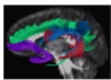
what does the arcuate fasciculus connect
Mediates communication between wernickes and brocas areas
concerned with phonological aspects of language
Runs posteriorly, ascends posteriorly to space occupied to space occupied by lateral fissure, connects to inferior frontal gyrus

pathway of direct segment
- direct from temporal lobe
- superior to angular gyrus
- goes to PT and PO
- PHONOLOGICAL
(red)

Posterior short/indirect
(yellow)
wernickes → angular gyrus (IPL)

Anterior short/indirect
from angular gyrus (IPL) → broca’s
(green)

What is the uncinate involved in
semantic processing
(purple)

Conduction aphasia
Lesion of anterior indirect segment of arcuate (angular → broca’s)
cannot repeat words or phrases
Where is the angular gyrus
Function
Overhangs superior temporal sulcus
Neural lexicon involved in word meaning retrieval and conversion of grapheme → phoneme
active during listening to spoken words
MCA stroke would have significant impact here

areas composing broca’s
Pars triangularis and opercularis
also involves cortex slightly posterior to these within inferior frontal operculum
Inferior parietal lobule =
Supramarginal and angular
where is supramarginal found
At the posterior most extent of lateral fissure
where is angular gyrus found
posterior part of superior temporal sulcus
posterior to supramarginal
Afferents and efferenta of angular gyrus
afferents = From inferior part of left lingual gyrus
efferents = to temporal plane (wernickes)
Function of Broca’s
Expressive aspects of spoken and written language
PT and POperc. in right vs left hemisphere
Tend to be larger in left hemisphere - connections to face, tongue and motor cortex within precentral gyrus
Homologous area in right is involved in non verbal communication including facial expression, gesticulation, modulation of rate rhythm and intonation of speech - prosity
Damage to non-dominant area - monotone robotic type of speech
CN V and speech
Trigeminal
Muscles of mastication
CN VII and speech
Facial
Muscles of facial expression
CN IX
glossopharyngeal
stylopharyngeus
deglutition
CN X and speech
Vagus
Larynx and pharynx
phonation
CN XI
Spinal accessory
laryngeal muscles
phonation
CN XII
hypoglossal
muscles of tongue
tongue movement
Where is speech initiated
In Broca’s

Areas of brain used in singers
singing requires integration of feedback of auditory systems - pitch precisely controlled
Superior temporal gyrus on both sides as well as left putamen in singing -> Anterior cingulate cortex - singers
aphasia
deficit affecting one’s ability to communicate
Different types of stroke
Stroke is an event affecting blood vessels that can be either a blockage or occlusion, which results in tissue death termed ischemia in those tissues that lost blood supply
A stroke can be haemorrhagic type where a weakness in the wall of a vessel is eventually breached and there is bleeding in brain tissue – this bleed can cause both loss of blood to the tissues that the vessel was supposed to supply and pressure on tissues due to the volume of the bleed
1 day after an acute infarction involving right middle cerebral territory
Left-most is a diffusion weighted image showing an area of infarct as a bright signal
To the right of that is a T1 weighted image which shows no evidence of blood in the area of infarct (blood would appear as white)
Next, moving right, is a post-contrast coronal image that shows vascular enhancement in the area of infarct
On the right is an MR angiography that show right MCA branches to be narrower in calibre as compared to left

receptive aphasia - where is there damage to
Wernickes
e.g. relevant MCA branch
inability to understand speech, deficit in auditory comprehension
If the angular gyrus is affected, then it can involve a compromised ability to read – alexia
Patients may be unaware of mistakes, they may have difficulty in understanding speech of others and retrieving correct descriptive terms (accessing lexicon areas) or word finding
how is speech comprehension facilitated
first arrival of a signal via the auditory branch of the vestibulocochlear cranial nerve in the superior posterior region of the superior temporal gyrus and Heschl’s gyrus (Wernicke’s area)
Expressive aphasia
Broca’s cortex (area of inferior frontal gyrus) may be affected resulting in impairment in the ability to produce speech
This patient may make sounds or words that are not what was intended, and they may or may not be aware of this depending on whether or not they have concurrent damage to the primary auditory cortex (Wernicke’s area)
Expressive aphasia can involve difficulty in expressing themselves or slow, laboured speech
It can often present with agraphia – inability to write thoughts
Larger areas of cortex affected can be accompanied with motor weakness in the right lower face and right arm, right lips and tongue and it can present with dysarthria (arthria refers to articulation or movement between joints) resulting in slurring of syllables
Conduction aphasia
When the arcuate fasciculus is affected (the white matter tracts subserving communication between Wernicke’s and Broca’s area)
This can occur independent of the other 2 types, and specifically involves the inability to repeat words or phrases
Global aphasia
when all these impairments are present, and all regions affected
receptive
expressive
conductive
prosody
Prosody is the pitch, intonation, cadence and musically or melodic aspects we use in speech to infer meaning beyond the words themselves
Most of language is subserved by the dominant hemisphere - left
Prosody appears to be associated with the non-dominant hemisphere (usually right hemisphere)
Aprosodia
a disturbance in melodic function, such as comprehension of the prosody in speech, especially in the right posterior temporo-parietal region, and for the production of melody in the right inferior frontal gyrus
speech changes to a dull monotone
dysphasia
acquired disorder of spoken and written language
It comes from the Greek -dys meaning disordered and -phasis meaning utterance
expressive dysphasia
Lesions involving Broca’s area, on the inferior frontal gyrus
Speech is hesitant, fragmented and telegraphic, with word finding difficulty and a paucity of grammatical elements such as verbs and prepositions
Since comprehension is relatively spared, patients tend to become frustrated as they struggle to express themselves
Receptive dysphasia
lesions in Wernicke’s area, on the superior posterior bank of the superior temporal gyrus and including Heschl’s gyrus as well
Speech is fluent but makes little sense, consisting of word fragments, substitutions and neologisms (a nonsense of words)
Since comprehension is affected, patients may be unaware of their own errors
global dysphasia/aphasia
combination of receptive and expressive aphasia
Conduction aphasia
specific problems repeating sentences
despite normal fluency and comprehension
attributed to a lesion in the arcuate fasciculus, which is said to disconnect the 2 primary language areas