Unit 6 - Cerebral White Matter Flashcards
What causes WM microstructure organisation
Damage to myelin, deficit in production by oligodendrocytes or change in genes that code for cell types (MAG and MOG)
What produces myelin
Oligodendrites
Concentric appearance
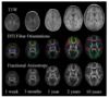
How does WM develop
At birth myelination is minimal
It increases dramatically during the 1st & 2nd year and continues to mature through life peaking at approx 40 yrs
then degenerates

What is the last to be derived
Association tracts (functionally speaking)
Striae
Thin bundles of fibres that pass longitudinally across the brain
Fascicles
Microscopically determined groups of fibres
Lamina
Relatively thin sheets of axons that proceed in a similar direction
Capsules
Curved sheets of fibres that partially enclose a grey matter structure
Tracts
Groups of axons subserving a similar or corresponding function
Radiations
Broad sheets of fibres that arch together to/from 1 target
Name for tracts that run together
Radiations
Where does the arcuate fasciculus lie
Within the longitudinal fasciculus
Commissural
Crossing the midline connecting cortical areas in 1 hemisphere to the other
Projection
Cortex to distant sites such as brainstem and spinal cord and vice versa
Association
Connecting cortical areas within the same hemisphere

Homotopic commissural fibres
Fibres that connect corresponding areas of cortex
Heterotopic
Fibres that connect a non-corresponding area in the contralateral hemisphere
main commissural fibres
Corpus callosum
Anterior commissure
Posterior commissure
Hippocampal commissure

Function of CC
Laterally the fibres of CC fan out into 2 wide cortical areas
Link those areas functionally related to midline - more relevant to trunk and to visual vertical meridian (than to the periphery of vision)
What does the rostrum of the CC continue as
Lamina terminalis
What is lamina terminalis embryonically
Closure point of anterior neural pore of neural tube during development
Synonym for genu of CC
what does it connect
Forceps minor
Interconnects the anterior frontal lobe, prefrontal cortices and anterior cingulate
Body of CC
What does it connect
roof of lateral ventricles between cingulum and laterally bounded by longitudinal fasciculus - interconnects precentral gyri - motor cortices, as well as insula more laterally and cingulate gyrus
Isthmus of CC is the location of
What does the isthmus denote
denotes a point of conversion during development between more posterior splenium (forceps major) - Fo
Location of commissure of fornices/hippocampal commissure

Function of CC fibres anterior VS posterior to the isthmus
CC fibres anterior to the isthmus connect frontal association cortices and those of the isthmus connect precentral motor and postcentral somatosensory and Heschls, those posterior to the isthmus connect visual and association fibres of the parietooccipital cortices
What does the splenium connect
Posterior parietal
Medial occipital
Medial temporal
Where is there more myelination
More myelinated when connecting primary motor and somatosensory areas -and smaller and poorly myelinated when connecting association cortices (NOT PRIMARY CORTICES)
Overall function of CC
Facilitates inter hemispheric interaction, underlying communicating and integrating perceptual learning and volitional information - higher order cognition and emotional functioning
Genu as a site of lesion
Diagnostic dyspraxia - alien hand syndrome - inter-manual conflict involving 1 hand (usually left) interfering with the correct execution of movement in the other hand
Isthmus/splenium region lesion
Main etrangere - patients inability to recognise his left hand as his own when its in his other hand and out of the visual field
Splenium as a site of lesion
Optic aphasia - failure to name objects present visually
Where is the ant. commissure located
What is its function
connections
Located on lamina terminalis
Reflex control of activity in the olfactory bulbs connecting them
Facilitates some inter hemispheric communication between the olfactory nuclei, primary cortices of the olfactory system and the amygdalae, as well as orbital cortices
?? Facilitates communcation between the 2 accumbens nuclei (reward and addiction)
Where does the ant. commissure run
Crosses midline in the anterior wall of the 3rd ventricle, just rostral to the ant. fornix leading to pre and post commissural columns
What structure is the post. commissure a part of
Epithalamus
Epithalamic commissure
interconnects superior posterior brainstem nuclei
bilateral pupillary reflexes in response to light
What does the hippocampal comissure connect
Crura of fornices
Where they converge towards the midline, immediately inferior to CC
Commissure connects the 2 medial temporal lobes (precebiculum, enteorhinal cortex and parahippocampal gyri but NOT hippocampal formation themselves - function is not well understood)
Where do most projection fibres pass through
Many fibres pass reciprocally through:
The crus cerebri or cerebral peduncles (superior to pons)
Pass in a compact area at the level of the basal ganglia (the IC)
Relay in thalamus
Reach cortex
Overview of fibre tracts

Overview of structures around the CC

What passes through the IC
Where does it exist
Inferior to IC, where do fibres pass through
Superior to IC, where do fibres pass through
Majority of structures between the cerebral cortex and subcortical structures travel through the IC
NOT a tract, but a compact area that tracts pass through - exists at the level of the basal ganglia
Inferior to it, the fibres are passing through the CPs
Superior to it, the fibres diverge to enter thalami nuclei - relay before emerging out through thalami peduncles - convey to cortex
Where do afferent fibres of IC arise from
thalamus and radiate to all parts of cortex - thalamic radiations, grouped into thalamic peduncles
Efferent fibres in IC arise from
cortical neurons and extend widely to specific portions of thalamus brainstem and spinal cord
Corticotegmental
Corticopontine
Corticobulbar (medulla)
Corticospinal
Ant. limb of IC connects
connects thalamus parts to cingulate and pons and thalamus to frontal cortices
What does the genu of the IC connect
What structure is it related to
IV foramen
Connects PONS and FRONTAL CORTEX - frontopontine tract
Corticobulbar tract
Nuclei in post. limb of IC
Thalami nuclei with motor and premotor cortex and with postcentral gyri
Corticospinal tract
Retrolenticular part of IC
Posterior to putamen/lenticular formation (putamen and GP)
Fibres connecting thalamus to cortex - optic radiations and parietopontine fibres
Sublenticular part of IC
Inferior to lenticular formation
Continuous with retrolenticular fibres and includes AUDITORY radiations
3 capsules
Internal - projection
External - association
Extreme - association
3 medullary lamina
Internal (within thalamus, projection)
Middle
Lateral
Define corona radiata
Collective term for all the fibre tracts connecting the brainstem to the thalamus (incl. the IC area) and those connecting thalamus and peduncles
4 thalamic peduncles
Anterior
Superior
Posterior
Inferior
Ant. thalamic peduncle connects
separate from anterior limb of IC to interconnect the thalamus with the prefrontal and cingulate cortices
Sup. thalamic peduncle
separate from posterior limb of IC
Corticospinal, corticopontine, corticotegmental - MOTOR FUNCTION - efferent
post. thalamic peduncle
separate from RETROLENTICULAR PART of the IC to interconnect the thalamus with occipital and posterior parietal and temporal lobe cortices - incorporate the geniculocalcarine tract (optic)
inferior thalamic peduncle
pass below the lentiform nucleus within the IC - sublenticular - to connect with the anterior temporal, orbitofrontal and insular cortices as well as the amygdala - also AUDITORY - geniculotemporal tracts
At what point is the transition from thalamic to cerebral peduncles
At tegmentum
what are the striae
Olfactory - CNI
Stria medullaris
Stria terminalis
Medial and lateral longitudinal striae
Where does stria medullaris run
What does it connect
Striae medullaris tracts along the medial surface of each thalami nucleus
Connects the septal area - cortical area just anterior to lamina terminalis and lateral pre-optic nucleus of the thalamus and GPi with the HABENULA
What is the habenula implicated in
Behaviour and sleep through connections with pineal gland
Pain
Stress
Reward
Depression and anxiety
Where does stria terminalis run
What does it connect
Courses along caudothalamic grooves where medial surface of caudate nucleus meets the thalamus on each side
Prominent outflow conguit of amygdala, beginning in temporal lobe and arching with the caudate in a path to terminate rostrally in nucleus of ST - lateral to anterior columns of the fornix and dorsal to ant commissure
Where do the medial and lateral longitudinal striae run
What do they connect
Connect dentate gyrus of the hippocampus in a path superifical to the CC with a GM - indusium grisium, terminating with the parahippocampal gyrus and continuing to diagonal band of Brocha and ultimately to amygdala
Where is the paraterminal gyrus
immediately anterior to lamina terminalis
Inferior posterior most part of medial surface of frontal lobe
What are the association fibres that connect diverse regions of the same hemisphere
Cingulum bundle
Superior longitudinal fasciculus (arcuate fasciculus)
Inferior longitudinal fasciculus
Inferior frontal longitudinal fasciculus
Uncinate fasciculus
REVIEW:
ILF
IFOF
UNCINATE