Unit 5 - Cerebellum (anatomy & physio lectures) Flashcards
How much of the mass of the brain does it comprise
Proportion of the brain’s neurons in the cerebellum
10% mass of brain
50% of its neurons
Functions of cerebellum
- Maintenance of equilibrium (vestibular input // the vestibulocerebellum)
- Adjusting the postural muscles of the body (spinal output - spinocerebellum)
- Programming and fine tuning movements (cortical input - cerebrocerebellum)
vestibulocerebellum function
Maintenance of equilibrium
Spinocerebellum Function
Adjusting the postural muscles of the body
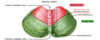
What can be seen on the superior surface of the cerebellum

Cerebrocerebellum function
Programming and fine tuning of movements
Name the dural folds related to the cerebellum
Falx cerebelli
Tentorium cerebelli
Name the 3 lobes of cerebellum
Anterior
Posterior
Flocculonodular
What lobes can be seen on the superior surface of the cerebellum
Anterior & posterior
What fissure separates the anterior lobe from the posterior/middle lobe
V-shaped primary fissure

What fissure separates the middle/posterior lobe from the flocculo-nodular lobe
Posterolateral fissure

What fissure does not mark the boundary between any lobes
Horizontal fissure

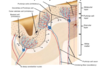
What structure is the roof of the 4th ventricle
Superior medullary velum
Name the structure found IN the 4th ventricle
Choroid plexus
(Surface can also be called the arbor vitae

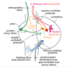
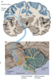
What are intracerebellar nuclei
Masses of grey matter embedded in white matter of cerebellum on each side of the midline
Name the 3 intracerebellar nuclei
Fastigial (medial)
Dentate (lateral)
Interposed (= emboliform + globose)

Name the 2 nuclei that compose the interposed nucleus
Emboliform + globose
Describe the structure of the dentate nucleus
Crumpled bag shape with opening facing medially

Where do the white matter fibres of the dentate nucleus exit
Superior cerebellar peduncle
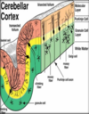
Name the 3 layers in the cerebellar cortex
Grey matter
White matter
Collection of nerve cells inside, from deep cerebellar nuclei

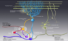
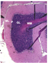
Distinguish between the outer, middle and inner layer of the cerebellar cortex
Outer - molecular layer (ML)
Middle - purkinje cell layer (P) - 1 cell thick
Inner - granular layer (GL)

What neurons & cells are present in the outer molecular layer
2 types of NEURONS:
Outer stellate cell
Inner basket cell
Also glial cells *

Describe the contents of the middle purkinje cell layer
Large Golgi type I neurons
Flask shaped
Arranged in a SINGLE layer
Dendrites branch profusely in molecular layer

Describe the synaptic relationship between the middle purkinje cell layer and the molecular layer
Branches of purkinje axons synapse with dendrites of basket cells and stellate cells in molecular layer

What is unique about middle purkinje cells
Form synaptic contact with parallel fibres from granule cell axons
They are the only cells that leave the cerebellum - the others remain in the molecular layer

Where do granule cell axons run
Run through the granule cell layer → white matter → synapses with cells in cerebellar nuclei
Contents of the inner granular layer
Small neurons with densely staining nuclei
Interneurons of the cerebellar cortex

Describe the path of the granule neuron
Where do the parallel fibres run
What cells are also present
Passes through the molecular layer → bifurcates at T junction → branches run parallel to long axons of folium → parallel fibres
Parallel fibres are at right angles to dendritic processes of purkinje cells
Glial cells are also present

Name the 2 cerebellar cortical mechanisms
What are they closely related to
Climbing fibres
Mossy fibres
Excitatory to PURKINJE cells
Origin & path of climbing fibres
Inferior olivary nucleus
Pass through granular layer
Number of synpases with dendrites of single Purkinje cell
Side branches synapse with stellate and basket cells

How do mossy fibres differ from climbing fibres
Where do they terminate
Stimulation produced by mossy fibres
Where do the fibres arise from (3)
- More diffuse excitatory effect
- Terminate in granular layer (NOT molecular) and synapse with granule cells
- Single fibre stimulates 1000s of purkinje cells through granule cells
- Fibres arise from vestibular, spinal, reticular, cerebral/pontine sources
Where do the efferent fibres of the purkinje cells end up
In deep cerebellar nuclei

Overview of cells in cerebellum

Describe the intrinsic fibres (white matter) of the cerebellum
They do not leave the cerebellum
Connect different regions of the cerebellum
Describe the afferent fibres (white matter) of the cerebellum
Form greater part of WM → proceed to cerebellar cortex → mainly enter through middle and inferior cerebellar peduncles
Where do the afferent fibres enter the cerebellar cortex
Through the middle and inferior cerebellar peduncles
Describe the path of the efferent fibres
Purkinje cell axons → cerebellar nuclei → superior cerebellar peduncle
Some fibres don’t synapse in nuclei

Name the 3 grps of white matter fibres
Intrinsic
Afferent
Efferent
Superior cerebellar peduncle links
Cerebellum with nuclei in midbrain
Middle cerebellar peduncle links
Cerebellum with pons
Inferior cerebellar peduncle links
Cerebellum with medulla
What passes through the inferior cerebellar peduncle
Largely afferent fibres passing to cerebellum
e.g. posterior spinocerebellar tract

What passes through the middle cerebellar peduncle (largest)
Transverse fibres of pons from pontocerebellar pathway

What passes through the superior cerebellar peduncle
Majority are efferent fibres from cerebellar nuclei

Role of the cerebellum
It continuously receives convergent input from various sensory pathways and from motor pathways
When the cerebellum detects a disparity between the intended and actual movement, it may generate an error-correcting signal
This signal is transmitted to the premotor and primary motor cortices via the thalamus and brainstem
Cerebellar pathway - voluntary movement
The primary motor cortex and the basal nuclei in the forebrain send impulses through the nuclei of the pons to the cerebellum
BLUE

Cerebellar pathway - assessment of voluntary movements
Proprioceptors in skeletal muscles and joints report degree of movement in the cerebellum
PURPLE

Cerebellar pathway - integration & analysis
The cerebellum compares the planned movements (motor signals) against the result of the actual movements (sensory signals)
ORANGE

Cerebellar pathway - corrective feedback
The cerebellum sends impulses through the thalamus to the primary motor cortex and to motor nuclei in the brainstem
ORANGE

FLOCCULONODULAR LOBE
- Anatomically
- Phylogenetically
- Functionally
- Oldest portion of cerebellum
- Archicerebellum
- Vestibulocerebellum - associated with vestibular system
ANTERIOR LOBE, UVULA, PYRAMID
- Phylogenetically
- Functionally
- Paleocerebellum
- Spinocerebellum - gross movements of head and body
POSTERIOR LOBE (EXCEPT UVULA & PYRAMID)
- Phylogenetically
- Functionally
- Neocerebellum
- Cerebral cerebellum (pontocerebellum) - fine voluntary movements
Overview of archicerebellum, paleocerebellum and neocerebellum

Midline lesion - flocculonodular lobe (vestibulocerebellum)
Responsible for co-ordination of the paraxial muscles associated with equilibrium
Truncal ataxia
Tumours in children

Anterior lobe syndrome (spinocerebellum)
Chiefly maintain co-ordination of limb movements
Chronic alcoholics (also Vit B deficiency)
Damage to Purkinje neurons
Gait ataxia

Posterior lobe syndrome (cerebrocerebellum)
Learning and storage of all of the sequential components of skilled movements
CVA, tumour, trauma, degenerative diseases
Loss of co-ordination of voluntary movement (appendicular ataxia), intention tremor, decreased muscle tone

Functions of cerebellum
What side of the body does it influence
What does damage result in
- Indirectly regulates movement and posture by adjusting motor fibre output
- Improves accuracy of movement
- Motor learning - motor adjustments
- Ipsilateral side
- Damage disrupts co-ordination of movement, motor learning, balance, causes flaccidity and active tremors
Where does input to cerebellum come from (w/ regard to cortex)
Mainly from cerebral cortex
- Primary motor and premotor
- Sensory cortices and visual areas
- Also spinal cord and brainstem
Where does input to cerebellum come from (w/ regard to nuclei)
Input mainly via nuclei of pons and medulla
- Pontine nuclei - vestibular nuclei
- Inferior olive - receives info from descending spinal circuits
- External cuneate/dorsal nucleus of Clarke - motor & sensory input

Name the cells of the granule cell layer
Granule cells
Golgi cells

Where do mossy fibres receive inputs from
Where do they originate
Where do they synapse
Receive inputs from sensory and motor pathways
Axons of cell bodies from BRAINSTEM nuclei (mostly pontine)
Synapse onto granule cells which then synapse onto purkinje cells
Where do climbing fibres originate
Where do they receive inputs from
Where do they synapse
Originate in inferior olivary nucleus
Receive inputs from RED nucleus, cerebral cortex & spinal cord
Climbing fibres synapse directly onto purkinje cells
Afferents of cerebellar cortex

What do interneurons influence
The firing of the purkinje fibres
CEREBROCEREBELLUM
What is the input output particular with
What is its function
Consequence of lesions
- Input-output particular with premotor cortex
- Motor planning (motor cortex), timing of sequential movements
- Lesions result in delays in initiation of movement, disturbances in coordination and sequence of muscle movements, slurring of speech

SPINOCEREBELLUM
Receives input from
What is it responsible for
Consequences of damage to the area
- Input from spinal cord & motor areas - output to motor cortex and brainstem motor pathways
- Motor execution and controls muscle tone - compares sensory signals with ingoing movement & corrects for deviation in movement
- Danage results in ACTIVE TREMOR particularly arms, past pointing, reduced muscle tone
VESTIBULOCEREBELLUM
What is the input-output related to
Function
What does damage result in
- Main input-output with vestibular nuclei in medulla
- Balance, motor execution (medial descending tracts), facial (eye) movements
- Damage results in balance problems, slow speech, ataxia in legs
Deep cerebellar nuclei

Overview of cerebellar circuits

What do lesions in the cerebellum cause
A movement incoordination syndrome
→ ATAXIA
Other than movement, what else does the cerebellum influence
Involved in cognition and executive control - impact on pathologies like dyslexia and autism
Autonomic control via hypothalamus regulation