Brainstem Flashcards
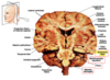
Sagittal section through the brainstem

Name the structures of the prosencephalon (forebrain)
Telencephalon - cerebral hemispheres/cerebrum
Diencephalon - between brain
Name the 3 structures of the rhombencephalon (hindbrain)
Metencephalon - pons
Myelencephalon - medulla
Metencephalon - roof portion (cerebellum)
*** NOTE: hindbrain does not include midbrain
What structures surround the brainstem
Brainstem lies anterior to cerebellum
Superiorly, continuous with the diencephalon
Medulla is continuous with the spinal cord
What structure is bulbar palsy associated with
Medulla
Function of brainstem
- Vital role in basic attention, arousal & consciousness
- Passes information between the spinal-cord and the cerebrum
- located in an area near bony protrusions, making it vulnerable to damage during trauma
- cranial nerve exit points
What cranial nerves arise from the brainstem
CN 3-12

Sagittal section through the brain

Coronal section through brain & brainstem

What feature tells you that you’re in the brainstem specifically the midbrain
Substantia nigra

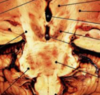
Name the structures on the posterior aspect of the brainstem

Posterior aspect of the brainstem

Where does the tegmentum lie

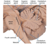
Locations of the pyramid, tegmentum and tectum of the
- medulla
- pons
- midbrain

What features define the anterior surface of the midbrain
- Interpeduncular fossa - deep depression in the midline
- Cerebral peduncles
- Posterior perforated substance - small BVs perforate floor
- Oculomotor nerve emerges from groove on medial side

What features define the posterior surface of the midbrain
Superior & inferior colliculi (corpora quadrigemina)
Trochlear nerve emerges below inferior colliculi

What are superior colliculi responsible for
Visual centres for visual reflexes
What are inferior colliculi responsible for
Lower auditory centres
What are the internal structures of the midbrain
Cerebral peduncles
Crus cerebri - descending axonal tracts
Cerebral aqueduct

How is the cerebral peduncle divided
By pigmented substantia nigra
ANTERIOR - crus cerebri
POSTERIOR - tegmentum

What are crus cerebri
Descending axonal tracts
Structure of cerebral aqueduct
What lies posterior to the cerebral aqueduct
Lined by ependymal cells
Surrounded by central grey matter - periaqueductal grey
TECTUM lies posterior to cerebral aqueduct
What is the superior colliculi
What is the function of the SC
- Large nucleus of grey matter
- Part of visual system
- Afferent corticotectal fibres from visual cortex - occipital lobe and frontal eye field of frontal lobe
- Controls eye movement

What defining feature lies at the level of the superior colliculi
RED NUCLEUS
- Grey matter mass between cerebral aqueduct and substantia nigra
- Redish hue due to iron containing pigment in cytoplasm of neurons
- Fibres pass to spinal cord via rubrospinal tract
- Motor control

Function of the inferior colliculi
- Nucleus of grey matter
- Receives auditory info
- Afferent auditory fibre → inferior colliculus
- Efferent fibres → thalamus → auditory cortex of temporal lobe

What is the substantia nigra
- Dopaminergic neurons between crus cerebri & tegmentum
- Melanin pigment within cytoplasm of neurons
- Muscle tone - Parkinsons (degeneration of dopaminergic neurons)
- In all midbrain levels

What structures can be found throughout the entire length of the midbrain
Cerebral aqueduct
Substantia nigra
What cranial nerves can be seen emerging from the pons

What do the nuclei and tracts in the pons do
Relay info to and from cerebellum
What cranial nerve nuclei lie in the pons
Function of nuclei in pons
Sensory and motor nuclei for CN V, VI, VII, VIII
Nuclei involved in control of respiration
Structure of pons

Features of the anterior surface of the pons
- Many transverse fibres converge on each side - middle cerebellar peduncle
- Midline - shallow basilar groove
- Anterolateral surface - trigeminal nerve
- Pontomedullary junction - abducent, facial, vestibulocochlear nerve

Features of the posterior surface of the pons
- Triangular shape - forms upper half of floor of 4th ventricle
- Limited laterally by superior cerebellar peduncles
- Divided symmetrically by median sulcus
- Inferior medial eminence expanded to form the FACIAL COLLICULUS

What is the sulcus limitans
Divided the alar from basilar plate in embryology
=> divided sensory from motor in posterior and anterior grey matter
What is the facial colliculus
Expansion of inferior medial eminence
Nucleus of CN VI, but axons of facial nerve sweep around it
Describe the internal structure of the pons (anterior and posterior)
What pathway is the pons a part of
Posterior part - tegmentum
Anterior part - basal part - pontine nuclei with transverse pontocerebellar fibres
Part of corticopontocerebellar pathway

Where does the corticopontocerebellar pathway travel

Function of medulla oblongata
Autonomic nuclei controlled visceral activities (reflex centres)
CV centres - cardiac centres & vasomotor centres
Respiratory rhythmicity centres

Describe the anterior surface of the medulla
- Anterior median fissure - continuous with fissure of spinal cord
- Pyramid - swelling on each side of the fissure (Corticospinal fibres)
- inferior pyramid - decussation of pyramids
- Olives - posterolateral to pyramids
- underlying olivary nucleus
- Hypoglossal nerve between olive and pyramid
- Inferior cerebellar peduncles posterior to olives
- connecting groove site of glossopharyngeal, vagus, accessory nerves

What nerve lies between the olive and pyramid
Hypoglossal nerve
Where do the inferior cerebellar peduncles lie
What connects to them
Posterior to olives
Connecting groove site of glossopharyngeal, vagus and accessory nerves

Describe the features of the posterior surface of the medulla
SUPERIOR - lower part of floor of 4th ventricle
INFERIOR - continuous with spinal cord
Posterior median sulcus - gracile tubercle on each side of the sulcus, contains the gracile nucleus
What structure lies lateral to the gracile tubercle
Cuneate tubercle

Superior part of medulla
Pyramids formed by pyramidal tracts
Olive formed by the inferior olivary nucleus - associated with muscle movement

Inferior part of medulla
Decussation of _____
Central canal
Decussation of pyramidal/corticospinal tract
- most corticospinal fibres cross the median plane and continue down the spinal cord as the lateral corticospinal tract
Fasciculus gracilis & cuneatus
- continue to ascend superiorly posterior to central grey matter

Overview of decussation at inferior medulla

Lateral & posterior view of brainstem

Name the centres in the pons and medulla

Arterial supply of midbrain
Posterior cerebral a.
Posterior communicating a.
Arterial supply of pons
Anterior inferior cerebellar a.
Superior cerebellar a.
Pontine arteries
Arterial supply of medulla
Posterior & inferior cerebellar a.
Anterior and posterior spinal arteries - branches of vertebral
