Unit 9 - Hearing Anatomy Flashcards
Auditory system
Exteroceptive system concerned with perception of sound
Vestibular system
Proprioceptive
Concerned with maintenance of equilibrium
Where does the vestibulocochlear nerve (CN VIII) emerge
SENSORY
Emerges from brainstem at cerebellopontine angle - junction between cerebellum, pons and medulla
Lateral to facial nerve

Skull opening that transmits the vestibulocochlear nerve
Internal auditory meatus
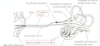
Structures of ear

Separation between external and middle ear
Tympanic membrane (ear drum)
Contents and boundaries of middle ear
Air filled space
Bounded by tympanic membrane and inner ear
Contents of inner ear/lebyrinth (in petrous portion of temporal bone)
Cochlea
Semicircular canals
Vestibule (utricule & saccule)
Transmission through outer ear
Air pressure fluctuations move tympanic membrane (ear drum) back and forth
Attached to tensor tympani muscle - which it pulls inwards
Cochlea in inner ear conducts sound through fluid instead of air
Before sound passes into inner ear it must be amplified - OSSICLES
ossicle bones

How do the ossicles amplify sound
Stapes rests against cochlea - through oval window
Air pressure pushes on tympanic membrane - vibrates - ossicles move - stapes pushes against cochlea and displaces fluid within SCC and in cochlea to move fibres on hair cells
Tensor tympani muscle attached to malleus
stapedius attached to stapes
Tensor tympani muscle attaches to
Malleus

Stapedius attaches to
Stapes
Receptors of auditory system
Function
Where are they found
Responsible for converting mechanical energy (fluid displacement) into electrochemical energy - travels along cochlear nerve
Hair cells enclosed within inner ear in tubular system are filled with fluid
Auditory hair cells in spiral organ of corti in cochlea
What is the cochlea spiral shell composed of
3 fluid filled spaces
Scala vestibuli
Scala tympani
Cochlear duct

Scala vestibuli & scala tympani
Partially enclosed in bone
Bony labyrinth - contains perilymph
continuous with each other
Cochlear duct
Part of membranous labyrinth (contains endolymph) suspended between bony labyrinth
membranous labyrinth also includes
utricle
saccule
semicircular canals
Outer spirals =
pitch of noise
= base
Low pitch of noise
Centre spirals =
pitch of noise
Apex
High pitch of noise
Where are the hair cells located
Basilar membrane (blue)

Oval vs round window
Relationship between movement of stapes and round window
Oval window - opens into scala vestibuli
- Stapes footplate occupies oval window
Round window - opens into scala tympani
- flexible tympanic membrane at round window
As the stapes moves inward, the round window moves outward

What happens when vibrations reach cochlear duct
Cochlear duct and basilar membrane are set in motion - hair cells are activated (mechanoreceptor cilia)

Categories of auditory hair cells
Organ of Corti - 2 hair cell types
Flask shaped inner hair cells
Rectangular shaped outer hair cells

Where are stereocilia found
What is the kinocilium in contact with
At apical end of each hair cell
Kinocilium located at tallest row of stereocilia
Tips of longest stereocilia in contact with overlying membrane
Basilar membrane moved by fluid movement - stereocilia bend and it changes MP of hair cells