Neurons & Glia Flashcards
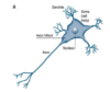
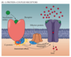
Structure of neurons
Unipolar => 1 main projection from it
How many neurons are in the cerebrum
1011 neurons
In 99% of cases how do neurons receive info
Through their dendrites
How do neurons function
Using bioelectricity (like muscle cells)
Nerve and muscle cells are ________ __________
electrically excitable
Transmembrane potential
voltage difference across a cell membrane
resting membrane potential
- -60 -> -80 mV
- Unequal distribution of ions (Na+, K+, Cl-) across cell membrane
- Greater permeability to K+ than Na+
What does the Na+/K+ electrogenic pump
Pumps 3 Na+ out and 2 K+ in
hence inside is made negative relative to outside
Distribution of ions within the cell and outside of it
Lots of K+ inside

EC and IC concentrations of:
- Na+
- Cl-
- K+

What also pushes against the movement of K+
K+ is a +ve ion and outside is more positively charged than inside => more difficult for K+ to move due to electrical gradient
equilibrium potential for K+
When concentration gradient for K+ = electrical gradient pulling K+ in, the result is the equilibrium potential for K+
Nernst equation

Equilibrium potential for:
- Cl-
- K+
- Na+
- -70 mV
- -80 mV
- +50 mV (cell membrane is relatively impermeable to Na+ when the cell is at rest
How many protein subunits compose an ion channel
4/5 subunits

how do mechanosensitive ion channels open
Sense sound - prise channel open - something PHYSICAL makes the ion channel open
How do ligand gated ion channels work
Shape of pore is altered by the binding of ligand to receptor on the surface of the channel
e.g. Na+ receptor binds ACh (nicotinic colinergic receptor) - binding site for ACh - Na+ passes through - when it binds to its receptor on the surface it changes the shape of the protein subunit and changes the lumen shape or size it becomes wide enough to allow Na+ to get through
How do VG ion channels work
The cause of the change in shape of protein subunit is the change in MP (ie change in proportion of +ve and -ve charges across the membrane) in vicinity of protein subunits
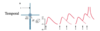
Action potential graph

4 principles that define an AP
- Threshold
- All or nothing
- Self-propagating
- Refractory period
* After an AP has occured there will be a period of time during which a) an AP cannot happen b) it would be very difficult for an AP to happen
Explain threshold and its relation to an AP being ‘all or nothing’

What does it mean when the MP is hyperpolarised
It is less likely that the neuron will become activated
Name an inhibitory NT
GABA
- many anti-epileptic (abnormal electrical discharge within neurons) medicine mimics GABA
- Hyperpolarises cell
- Moves MP away from threshold
- Reduces likelihood that the neurons are electrically active
Name an excitatory NT
Glutamate (most common)
What channels are responsible for depolarisation
VG Na+ channels
threshold in relation to eq potential for Na+
+ 15 mV
Absolute vs relative refractory period
- ABSOLUTE - impossible to reactivate that neuron
- RELATIVE - physiologically very difficult to reactivate the neuron
Quantify the increase in conductance of Na+ at AP
x5000 increase in Na+ conductance
What channels are responsible for repolarisation
VG K+ channels
Explain the refractory period of an AP
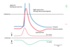
What happens at threshold
VG Na+ channels are opened
15 mV more +ve than the RMP in neurons
When does the inward flow of Na+ stop & how
When MP reaches the positive values
Related to a voltage sensitive change in the shape of the ion channels
=> inactivation of the VG Na+ channels
When MP is raised, what remedies the situation
The Na+/K+ ATPase
- when the MP gets up to +ve values there will also be activation of the VG K+ channels which can then facilitate the repolarisation event
How are APs self-propagating
Due to local circuits
- Na+ influx depolarises the cell for up to 3mm along the axon
- Adjacent areas reach threshold
- Propagation of AP
- Refractory period facilitates AP propagation in 1 direction only
Where do APs usually happen
Within the axon hillock (cell body/dendrite area)
It then propagates down the axon in 1 direction due to refractory period
What do you find in the dendrite region
Ligand sensitive channels, where the stimulus will occur
What cells produce myelin
Glial cells
Name the glial cells found in the PNS
Schwann cells
Name the glial cells found in the CNS
Oligodendrocytes
What is contained in the layers that the glial cells create
What does myelin do to ion channels
Sphingomyelin (lipid rich)
Creates a distance between active ion channels
What is the size of glial cells
3mm in diameter
Where are nodes of ranvier found
In between glial cells - ion channels are voltage sensitive
What part of the brain makes us consciously aware of things
Only when it reaches our cerebral cortex, outer mantel of cerebrum
Impact of diameter on nerve conduction
Wider the diameter, the greater the conduction
General classification of nerves
- A (α, β, γ, δ) - largest // I
- B // I
- C (smallest) // II, III, IV
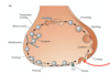
What is activated the AP is propagated to the axon terminal
VG Ca2+ channels
What does Ca2+ (greater conc outside, enters through ion channel) release activate
Ca2+ activates Ca2+-calmodulin dependent protein kinase II
What does CAM kinase II do
Primes vesicles for mobilisation, docking to release sites on presynaptic membrane asnd membrane fusion
Precipitates exocytosis
Name the scaffolding within the axon terminal
What does it do
Cytoskeleton (network of tubules and filaments) gives structure
It anchors NTs (vesicles) in the nerve terminal region for control
What substance holds vesicles in place, as well as cytoskeleton
Synapsin
What does Ca2+ phosphorylate
Synapsin to then release vesicle to docking site and hence release of NT
What is a vesicle
A bubble of liquid bilayer
What happens to NT left in the synaptic cleft
Vesicles reformed and NT taken back in
explain synaptic transmission
There are receptors on the post-synaptic surface
Interactions of NTs with the receptors precipitates changes in the next cell

How do you characterise most synaptic transmission
Axodendritic
how do you characterise a small amount of transmission
Axosomatic => binding sites are on the cell body
Describe ionotropic receptors
- Protein subunits arranged around a pore
- Fast activation
- Short duration of action
- ligand gated
Describe metabotropic receptors
- G-protein linked (inside surface)
- Slow activation
- Long duration of action
- 7 transmembrane regions
- NT dissociates from surface of the receptor
Example of ionotropic receptor
Nicotinic
Example of metabotropic receptor
Muscarinic
What sort of effect does ACh have
Short sharp effect & can also have prolonged effect - subject to modulation by drugs
What receptors does glutamate (most common excitatory NT in the brain) have
Ionotropic and metabotropic
G protein coupled receptors
What do excitatory NTs produce
Depolarisation of the postsynaptic membrane
Is an EPSP an AP
It may favour an AP but it is not in itself one
- no propagation
- Graded responses obtainable
What do inhibitory NTs produce
Hyperpolarisation of the post synaptic membrane
Makes it more difficult to trigger an AP
Where is an AP often triggered
Axon hillock
What is the role of summation
Key in occurence of AP

What is spatial summation
Simultaneous activation of several dendrites because of the NT that’s released => AP triggered

What is temporal summation
Zapped before it’s back to rest fully so it builds from there
Repetitive firing of a neuron can also result in threshold being reached
Define glia
Protectors and support cells of neurons
Heighten the functional capacity of neurons
Name the 6 different types of glia
- Schwann cells
- Oligodendrocytes
- Astrocytes
- Ependymal cells
- Microglia
- Radial glia
What are the most common of all the glial cells
Astrocytes are the most common
What glia are found when we’re developing as babies
Radial glia - facilitate the proper development of the brain
What myelin-producing cells are found in the PNS
Schwann cells
What myelin-producing cells are found in the CNS
Oligodendrocytes
What glia control the EC environment around neurons
Astrocytes
Ependymal cells
What glia have an immune function
Microglia
Proportion of glia vs neurons
10-50x more glia than neurons
Where are microglia derived from
Macrophages outside of CNS
- Phagocytes - activated by infection and injury
Where are macroglia derived from
Neural stem cells
What are the 7 functions of macroglia
- Structural support
- Insulate axons
- BBB (CSF is basically BBB)
- Promote efficient signalling between neurons (e.g. clear NT from synapses, such as glutamate - taken into astrocytes)
- Release growth factors to nourish neurons
- Guide migrating neurons and axon outgrowth
- Synaptogenesis
Astrocytes actively control _____________
What cells perform a similar function
synaptogenesis
- Regulate synapse number
- Regulate synapse function
- Regulate synapse stability
(schwann cells can perform similar functions)
Explain how macroglia have a crucial role in the anatomical development of the brain
A macroglial cell in the vicinity releases chemicals that can interact with neuron in the region
This communication results in neuron growing towards glial cell
Determines where the axon of a neuron actually terminates
What do MACROglia do in synaptogenesis
- EC protein signals from astrocytes trigger synapse formation in CNS
- Neurons migrate during development but synapse formation only occurs when astrocytes (or other macroglia) are present
- Microglia cannot perform this function
- adult hippocampal sten cells display similar dependence on astrocytes for synapse formation
- Schwann cells in the periphery trigger neuromuscular junction formation
What are macroglia also needed for
Synapse maintenance
How do glia sense synaptic activity
What do they do in response
Through increasing IC Ca2+ levels (calcium transient currents)
- Respond by releasing gliotransmitters
- Release transmitters in response to neuronal activity
