Cardio - ECG Flashcards
What are ECGs great at telling us?
HR, arrhythmias, conduction abnormalities
What are ECGs good at telling us?
Chamber enlargement (specific but not sensitive)
What are ECGs bad at telling us?
Mechanical activity of the heart
What is an arrhythmia?
General term to describe electrical activity that has an irregular rhythm and/or an abnormal HR
What is an electrocardiogram?
ECG - graph of the electrical activity of the heart
How wide are the “small” boxes on an ECG strip?
1 mm
How wide are the “big” boxes on an ECG strip?
5 mm
What is the amplitude of an ECG strip?
10 mm/mV
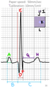
What are the 2 common paper speeds for ECGs?
25 mm/sec and 50 mm/sec
What is the normal electrical activity of the heart?
- The SA node initiates normal electrical events (“pacemaker”)
- Signals –> AV node that slowly conducts electricity to allow atria to contract and empty blood into ventricles before depolarizing the ventricles
- Signals –> L bundle branch/LV and R bundle branch/RV
Mechanical activity of the heart follows _____ _____.
electrical activity
What is highlighted and what occurs here?
P wave - atrial depolarization
What is highlighted and what occurs here?
PR (or PQ) interval - conduction slowly passes through AV node
What is highlighted and what occurs here?
QRS complex - ventricular depolarization
What is highlighted and what occurs here?
T wave - ventricular repolarization
What is A depicting?
Atrial depolarization (SA node)
What is B depicting?
Conduction of signals slowly through the AV node
What is C depicting?
Ventricular depolarization (L and R bundle branches to the respective chambers)
What is D depicting?
Ventricular repolarization
How is HR calculated on a 25 mm/sec strip?
QRS in 15 big boxes (3 sec) x 20
How is HR calculated on a 50 mm/sec strip?
QRS in 30 big boxes (3 seconds) x 20
What “trick” can be used to measure 3 seconds in a 50 mm/sec strip?
Use a BIC pen
What is the average HR here?
160 bpm
What is the average HR here?
80 bpm
What is the average HR here?
240 bpm
What are potential causes for bradyarrhythmia that can be seen on an ECG?
Sinus bradycardia, AV block, atrial standstill, sinus pause/arrest
What are some ECG findings that will still have a normal HR?
Normal sinus rhythm, 1st degree AV block, intermittent ventricular arrhythmia, intermittent supraventricular arrhythmia
What is a normal HR for dogs?
60-160 bpm
What is a normal HR for cats?
140-240 bpm
What is a normal HR for horses?
24-45 bpm
What are potential findings on an ECG that shows tachyarrhythmia?
Sinus tachycardia, ventricular tachycardia, supraventricular tachycardia (SVT), atrial fibrillation (a type of SVT)
What is a regularly irregular arrhythmia?
Irregular rhythm with a discernable pattern to the irregularity.
(This is a characteristic of sinus arrhythmias)
What is an irregularly irregular arrhythmia?
Irregular rhythm without a discernable pattern to the irregularity.
(This is characteristic of atrial fibrillation)
How can you tell if a beat is of sinus origin?
Every P has a QRS and every QRS has a P (QRS usually is narrow)
How can you tell if a beat is of supraventricular ectopic origin?
Narrow QRS complex that looks like sinus origin;
P’ waves may have different morphology than sinus P waves (taller/small, wider/narrower) or be absent
How can you tell if a beat is of ventricular ectopic origin?
Wide and bizarre QRS complex, no P waves
This is a _____ rhythm.
regular
This is a _____ _____ rhythm.
regularly irregular
This is a _____ _____ rhythm.
Irregularly irregular
What is the origin of this rhythm?
sinus
What is the origin of this rhythm?
supraventricular
What is the origin of this rhythm?
ventricular
Every 10 mm is ___mV
1
1 small box = ___ sec
0.02
1 small box = ___ mV
0.1
What is the P wave duration in this ECG?
0.04 sec
What is the P wave height in this ECG?
0.3 mV
What is the PR/PQ interval in this ECG?
0.14 sec
What is the QRS duration in this ECG?
0.04 sec
What is the R wave height in this ECG?
1.54 mV
What is the ST segment in this ECG?
0.02 sec
What is the QT interval inthis ECG?
0.2 sec
A
P wave
B
PQ interval
C
QT interval
D
Q
E
R
F
S
G
ST segment
H
T wave
I
0.1 mV
J
0.02 sec
K
0.5 mV
L
0.1 sec
This is an ECG from a 2y FS mixed breed dog. What is the diagnosis for this ECG?
Atrial standstill (no P wave, low rate, supraventricular) with RV enlargement (deep S wave)