The Thigh & Knee Anatomy Flashcards

1
Q

What surrounds the muscles of the thigh?

A

a layer of fascia called the fascia lata

2
Q

What is formed from the fascia lata and how does it divide the muscles of the thigh?

A

fascia lata forms three intermuscular septa that run deep into the thigh and attach to the linea aspera

the three intermuscular septa divide the muscles of the thigh into 3 compartments:

anterior, medial and posterior

3
Q

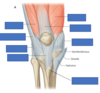
label the components/compartments of the thigh

A
4
Q

What is the iliotibial tract and which muscles insert into it?

A

a thickening of the fascia lata on the lateral side of the thigh

the gluteus maximus and tensor fascia lata insert into the iliotibial tract

they help to stabilise the knee through the iliotibial tract

5
Q

What is the pathway of the iliotibial tract and where does it insert?

A

it runs down the lateral aspect of the thigh, over the lateral part of the knee joint

it inserts onto the lateral aspect of the tibia

6
Q
A
7
Q

What are the muscles of the anterior thigh?

A
  1. quadriceps femoris (4 muscles)
  2. sartorius
  3. pectineus
  4. iliopsoas
8
Q

label the muscles of the anterior thigh

A
9
Q

What are the 4 muscles that compromise the quadriceps femoris?

A
  1. rectus femoris
  2. vastus lateralis
  3. vastus intermedius
  4. vastus medialis
10
Q

What is the innervation and common insertion of the quadriceps muscles?

A

common insertion - patella

common innervation - femoral nerve (L2, L3, L4)

11
Q

What is the common action of the quadriceps muscles?

What is different about rectus femoris and why?

A

they extend the leg at the knee joint

the rectus femoris also spans the hip joint so can flex the thigh at the hip joint

12
Q

What is the origin of rectus femoris?

A

anterior inferior iliac spine (AIIS)

13
Q

What is the origin of vastus lateralis?

A

greater trochanter and linea aspera

14
Q

What is the origin of vastus intermedius?

A

anterior surface of femur

15
Q

What is the origin of vastus medialis?

A

intertrochanteric line and linea aspera

16
Q

What do all 4 quadriceps muscles converge on?

A

a common tendon called the quadriceps tendon

this runs over the knee joint onto the patella and becomes the patellar ligament

17
Q

What is the role of the patellar ligament?

A

it connects the patella to the tibial tuberosity on the anterior tibia

18
Q
A
19
Q

How should the patient be positioned when testing the knee jerk reflex?

A

sat on a couch with their leg dangling

OR

their knee is flexed with the examiner supporting the weight of the leg with their arm

20
Q

What are the steps involved in testing the knee jerk reflex?

A
  1. the examiner palpates for the patella and the tibial tuberosity
  2. they find the halfway point between them - this is the patellar ligament
  3. they tap the patellar ligament with a tendon hammer
21
Q

What does the knee jerk reflex test for?

A

it tests the femoral nerve and the spinal nerves it contains (L2, L3, L4)

sometimes it is described as only testing L3 and L4 as the contribution from L2 is less

22
Q
A
23
Q

What will be seen in a normal knee jerk reflex?

A

the leg is extended once and then comes to rest

absence or decrease of this reflex is Westphal’s sign

24
Q

What is the function of pectineus?

What is its innervation?

A

it adducts and flexes the thigh at the hip joint

it is innervated by the femoral nerve (L2, L3)

25
What is the insertion and origin of pectineus?
**origin:** pectineal line and adjacent bone of the pelvis **insertion:** the oblique line, extending from the base of the lesser trochanter to the linea aspera
26
What are the origins and insertion of iliopsoas?
**origins:** * psoas major - lumbar transverse processes of posterior abdominal wall * iliacus - iliac fossa of posterior abdominal wall **insertion:** * lesser trochanter of femur
27
What is unusual about iliopsoas? What is its function?
iliacus and psoas major are separate muscles that converge on a common tendon iliopsoas is a primary **flexor** of the hip
28
What is the innervation of iliopsoas?
psoas major is innervated by anterior rami (L1, L2, L3) innervation from femoral nerve (L2, L3)
29
label the components of iliopsoas and the pelvis
30
What is the origin and insertion of sartorius? What is its innervation?
**origin:** anterior superior iliac spine (ASIS) **insertion:** medial aspect of proximal tibia **innervation:** femoral nerve (L2, L3)
31
What are the actions of sartorius?
1. flexes the thigh at the hip joint 2. flexes the leg at the knee joint
32
What are the borders of the femoral triangle?
**superior border:** inguinal ligament **lateral border:** sartorius muscle **medial border:** adductor longus muscle
33
What forms the roof and floor of the femoral triangle?
**roof:** * fascia lata **floor:** * pectineus * iliopsoas * adductor longus
34
What are the contents of the femoral triangle?
1. femoral nerve 2. femoral artery 3. femoral vein 4. femoral canal - containing deep lymph nodes and vessels
35
What structure drains into the femoral vein within the femoral triangle?
the great saphenous vein
36
what are the following components of the femoral triangle?
37
What is contained within the femoral canal? What is the femoral ring?
1. lymphatic vessels - draining the deep inguinal lymph nodes 2. deep lymph node - the lacunar node 3. empty space 4. loose connective tissue the femoral ring is the opening to the femoral canal
38
what is the purpose of the empty space within the femoral canal?
it allows for distension of the adjacent femoral vein this allows it to cope with increased venous return or increased intra-abdominal pressure
39
What is a femoral hernia? How does it present?
part of the small intestine protruding through the femoral ring it presents as a lump situated inferolateral to the pubic tubercle
40
what is a problem associated with femoral hernias?
the borders of the femoral canal are tough and not particularly extensible this can compress the hernia and interfere with its blood supply this forms a **strangulated hernia**
41
What is the origin of the femoral artery?
it is a continuation of the external iliac artery it becomes the femoral artery when it passes underneath the inguinal ligament
42
Where is the pulsation of the femoral artery palpable?
in the region of the femoral triangle this is half-way between the ASIS and the pubic tubercle
43
What is a major branch of the femoral artery?
the femoral artery runs down the anterior thigh and gives off branches that supply the anterior compartment a major branch is the **profunda femoris**
44
label the muscles and components of the adductor canal
45
Where does the adductor canal run to and from?
it extends from the apex of the femoral triangle to the adductor hiatus of the adductor magnus
46
what are the boundaries of the adductor canal?
**anteromedial border:** sartorius muscle **lateral border:** vastus medialis **posterior border:** adductor longus and adductor magnus
47
What is the role of the femoral vein? What happens when it passes through the femoral triangle?
all the veins in the lower limb drain back towards the femoral vein it continues proximally as the external iliac vein it receives the great saphenous vein as it passes through the femoral triangle
48
label the veins
49
Where do the muscles in the medial compartment of the thigh arise from?
the external surface of the pubis and the ischiopubic ramus
50
What is the main function of the medial thigh muscles?
**adduction** of the thigh at the hip joint (except obturator internus) the adductor muscles also **medially rotate** the thigh obturator internus is a **lateral rotator** of the thigh
51
label the medial thigh muscles
52
What are the 5 medial thigh muscles?
1. adductor longus 2. adductor brevis 3. adductor magnus 4. gracilis 5. obturator externus
53
What is the insertion, origin and innervation of adductor longus?
**origin:** body of pubis **insertion:** linea aspera **innervation:** obturator nerve (anterior division) (L2, L3, L4)
54
What are the actions of adductor longus?
it adducts and medially rotates the thigh at the hip joint
55
What is the origin, insertion and innervation of adductor brevis?
**origin:** body of pubis and inferior pubic ramus **insertion:** linea aspera **innervation** obturator nerve (L2, L3)
56
What are the actions of adductor brevis?
it adducts and medially rotates the thigh at the hip joint
57
What is the origin, insertion and innervation of adductor magnus?
**origin:** ischiopubic ramus **insertion:** linea aspera, medial supracondylar line **innervation:** obturator nerve (L2, L3, L4)
58
What are the actions of adductor magnus?
it adducts and medially rotates the thigh at the hip joint
59
What is the origin, insertion and innervation of gracilis?
**origin:** body of pubis and inferior pubic ramus **insertion:** medial surface of proximal tibia **innervation:** obturator nerve (L2, L3)
60
What are the actions of gracilis?
1. adduction of thigh at hip joint 2. flexion of leg at knee joint
61
What is the origin, insertion and innervation of obturator externus?
**origin:** external surface of obturator membrane **insertion:** trochanteric fossa **innervation:** obturator nerve (posterior division) (L3, L4)
62
What is the function of the obturator externus?
it laterally rotates the thigh at the hip joint
63
Which arteries supply the medial compartment of the thigh?
the medial compartment is supplied by the **obturator artery** the **profunda femoris** gives off perforating branches that perforate the adductor magnus and supply the muscles of the medial thigh
64
Which muscles are supplied by the anterior branch of the obturator artery?
1. pectineus 2. obturator externus 3. adductor muscles 4. gracilis
65
What is the adductor hiatus?
a gap between the adductor magnus and the femur it allows for the passage of femoral vessels from the anterior thigh to the posterior thigh and then the popliteal fossa
66
What is the nerve of the medial compartment?
**obturator nerve** this is composed of nerve roots L2 - L4
67
How does the obturator nerve reach the medial aspect of the thigh to supply the adductors?
it passes through a defect in the obturator internus muscle (obturator canal), through the obturator foramen and emerges into the medial aspect of the thigh
68
What does the location of the obturator nerve render it vulnerable to?
it is on the lateral wall of the pelvis it is vulnerable to compression by pelvic masses if a uterine or ovarian mass grows large enough to compress the obturator nerve, symptoms are felt in the medial thigh