The Knee Joint Flashcards
What are the 3 broad categories of reason for knee injury?
acute injuries:
- present to A&E
- sports, falls in the elderly
- fractures and tears/sprain of soft tissues
chronic knee pain/swelling:
- often seen by GP
- osteoarthritis, bursitis
atraumatic acute swelling/pain:
- present to A&E
acute gout, flare of osteoarthritis, septic joint
What are the 2 different components of the knee that could be injured?
bony injuries:
fractures of the patella, tibia or distal femur
dislocations
soft tissue injuries:
meniscal tears or ligament tears/rupture
Where do fractures of the patella, tibia or distal femur tend to occur?
- traumatic
- osteoporotic bone - low energy forces can cause fracture
- peri-prosthetic (around a prosthesis)
dislocation of the whole knee joint is uncommon and catastrophic
What are the 2 types of meniscal tears?
- due to acute injury/trauma
- due to chronic wear & tear and degeneration
What type of joint is the knee joint?
Which bones make up the knee joint?
It is a synovial hinge joint
It consists of:
- distal femur
- proximal tibia
- patella
The fibula is NOT associated with the knee joint
What are the 3 articulations involved with the knee joint?
2 femorotibial
this is the articulation between the femoral condyles and tibial condyles
1 femoropatellar
this is the articulation between the posterior patella and anterior surface of the femur
What is the fit like at the knee joint?
Why?
The tibial plateau is a poor fit for the rounded femoral condyles
This is because the tibial plateau is flat
Label the components of the knee joint


What structure helps to make the fit of the knee joint better?
menisci
these are wedge-shaped plates of fibrocartilage that sit on top of the tibial plateaus
they deepen the tibial articular surface to receive the rounded femoral condyles
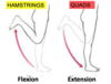
When is the knee joint most stable and least stable?
Why?
Most stable during extension:
this is when there is the best possible fit between the femur and tibia
Least stable during flexion:
this is when there is the least congruence between the tibia and the femur

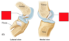
Label the diagram as if you were looking down onto the inside of a left knee


Where do the menisci attach to?
Where are they thicker?
The external edges attach to the fibrous capsule of the joint
They are crescent shaped discs of fibrocartilage that are ticker at the external margins
What are the 4 functions of the menisci?
- increase joint congruency
- distribute weight evenly throughout the joint
- shock absorption
- assist in locking mechanism
Label the 6 types of meniscal tear


What tends to cause meniscal tears?
What is the damage associated with?
caused by injury or degenerative changes
damage associated with development of osteoarthritis in later life
What can meniscal tears cause?
What is the treatment?
displaced cartilage can become trapped during knee movements
this causes pain or locking
treatment is by repair or resection
What components involved in stability are represented by the red, green and blue lines?

red:
intra-articular ligaments (inside the joint)
green:
extra-articular ligaments (outside the joint)
blue:
surrounding muscles
What are the ‘intra-articular’ ligaments of the knee?
cruciate ligaments
there is an anterior and a posterior cruciate ligament
What are the ‘extra-articular’ ligaments?
collateral ligaments
there is a fibular and a tibial collateral ligament
Label the 2 ligaments
What is their main function?

they sit in a crossed fashion inside the joint and prevent lateral displacement of the femur and tibia

What is the origin and attachment of the posterior cruciate ligament?
it attaches to the posterior intercondylar region of the tibia
it travels supero-anteriorly to insert onto the medial femoral condyle
What is the attachment and insertion of the anterior cruciate ligament?
it attaches to the anterior intercondylar region of the tibia
it travels supero-posteriorly to attach to the lateral femoral condyle
Which cruciate ligament is stronger?
the posterior cruciate ligament is STRONGER than the anterior cruciate ligament
Label the cruciate ligaments as if looking down inside a left knee


Label the anterior and posterior cruciate ligaments


What are the 3 main functions of the posterior cruciate ligament?
- prevents posterior displacement of the tibia on the femur
- prevents hyperflexion
- main stabiliser of the flexed knee when weight bearing e.g. walking downhill
When does the posterior cruciate ligament tend to be damaged?
when landing on the tibial tuberosity with the knee flexed
What are the 2 main functions of the anterior cruciate ligament?
- prevents anterior displacement of the tibia on the femur
- prevents hyperextension
When does the anterior cruciate ligament tend to be damaged?
when the knee is hyperextended or force is applied anteriorly
Label the ligaments of the knee joint


What is an alternative name for the lateral collateral ligament?
What is its function?
fibular collateral ligament
it prevents adduction of the leg at the knee
What is another name for the medial collateral ligament?
What is its function?
tibial collateral ligament
it prevents abduction of the leg at the knee
How do the fibular and tibial collateral ligaments differ?
fibular collateral ligament:
this is a cord-like band that is NOT attached to the meniscus
tibial collateral ligament:
this is a flat band that is attached to the meniscus
What is the main consequence of the tibial collateral ligament being attached to the meniscus?
if the ligament tears, this often also leads to a tear in the meniscus
What is meant by the “unhappy triad” of combined knee injuries?

- torn anterior cruciate ligament
- torn tibial collateral ligament
- torn medial meniscus

When does the “unhappy triad” combined knee injury tend to occur?
it is caused by excessive lateral twisting of the flexed knee
or
blow to the lateral side of the extended knee
What movements are shown?


How does the femur move in flexion and extension of the hip?
the femur comes towards the trunk in flexion of the hip
the femur moves away from the trunk in extension of the hip
What are the 4 muscles that make up quadriceps femoris in the anterior compartment of the thigh?
- vastus medialis
- vastus lateralis
- vastus intermedius
- rectus femoris
Where to the vastus intermedius originate from and run to?
What is significant about its tendon?
it originates from the anterior part of the femur
its tendon runs over the patella and inserts on the tibial tuberosity
the tendon that runs over the patella is the quadriceps tendon (connects muscle to bone)
it becomes the patellar ligament after passing over the patella (connects bone to bone)
Where do vastus medialis and vastus lateralis originate and insert?
they arise from the linea aspera on the posterior aspect of the femur
they converge on the quadriceps tendon
Where does rectus femoris originate and insert?
How is it positioned relative to the vasti?
it originates from the anterior superior iliac spine (ASIS)
it inserts onto the quadriceps tendon
it runs superficial to the vastus intermedius
What is the function of quadriceps femoris?
all 4 muscles extend the knee joint when they contract
they pull the tibial tuberosity towards the femur
What is the additional action of rectus femoris?
it extends the knee joint and contributes to flexion of the hip
Why is quadriceps femoris important in stabilising the knee joint?
the quads are active any time when standing as the knee is extended
label the quadriceps muscles


Other than the quadriceps muscles, what other muscles form part of the anterior compartment?
- iliopsoas
- pectineus
- sartorius
What is the origin and insertion of iliopsoas?
psoas major:
originates at the lumbar and lower thoracic vertebrae
iliacus:
originates at the iliac fossa
They converge on a common tendon (iliopsoas) that inserts on the lesser trochanter
What is the function of iliopsoas?
it is the main flexor of the hip
it pulls the lesser trochanter towards the trunk when it contracts
What is the insertion and origin of pectineus?
it originates at the superior pubic ramus
it inserts close to the lesser trochanter
What is the function of pectineus?
it helps with flexion of the hip
What is the insertion and origin of sartorius?
Why is it easily recognisable?
it originates at the ASIS, comes across the thigh and inserts on the medial tibia
it is easily recognisable as it runs on a diagonal across the thigh
What is the function of sartorius?
flexion of the hip joint and flexion of the knee joitn
What is the nerve supply (and spinal roots) of the anterior compartment muscles?
femoral nerve
this is formed from the ventral rami of spinal nerves from L2 - L4
label the muscles of the anterior thigh


What are the 4 medial compartment muscles?
What is their function?
- adductor brevis
- adductor longus
- adductor magnus
- gracilis
they are adductors of the thigh
What is the insertion and origin of the 3 adductor muscles?
(brevis, longus, magnus)
they originate from the pubic bone and inferior pubic ramus
they come across in a diagonal oblique direction to insert on the femur
this allows them to adduct the hip when they contract
How are the 3 adductor muscles arranged?
adductor brevis and longus are smaller than magnus and lie on top of each other nearer to the hip
longus is more superficial than brevis
What is the innervation of the medial compartment muscles?
obturator nerve (L2 - L4)
what is the insertion and origin of gracilis?
how is it identified and what is its function?
it originates at the medial part of the pelvis and inserts onto the tibia
it is long and thin and the most medial muscle in the leg
it is a weak adductor
label the medial thigh muscles


What are the borders of the femoral triangle?
superior border
inguinal ligament
medial border
medial border of the adductor longus muscle
lateral border
medial border of the sartorius muscle
Label the borders of the femoral triangle


What structures form the floor of the femoral triangle?
- iliopsoas
- pectineus
- adductor longus
What important structures run through the femoral triangle?
Why are they susceptible to injury?
- femoral nerve
- femoral artery
- femoral vein
the femoral artery and vein are superficial so are vulnerable to injury and stab wounds
What is meant by ‘locking’ of the knee?
as the joint approaches full extension, the femur undergoes a few degrees of medial rotation on the tibia
this is a stable position that allows the thigh muscles to relax

How is the knee “unlocked”?
the popliteus muscle contracts to laterally rotate the femur
What injury is shown?

patellar dislocation
Label the bursae of the knee


What is meant by bursitis?
inflammation of the bursa around the knee
these are fluid-filled sacs located between points of friction
What is meant by ‘bowing of the legs’ or being ‘knock-kneed’?

What are following changes observed in osteoarthritis?

