LL Ultrasound and Cases Flashcards
Someone has slipped whilst fell-running and has a deformed and painful right ankle
What is the diagnosis?

dislocation of the ankle
In a dislocated ankle, how would you assess the vascular status (perfusion) of the foot?
Which arteries should be palpated and which is easiest to palpate?
try to palpate pulses
try to palpate posterior tibial artery and dorsalis pedis
DP is easier to palpate as it is superficial and easy to compress against the bones of the foot
PT is harder to palpate as it is close to the tendons behind the medial malleolus
Why can it be difficult to palpate pulses after dislocation of the ankle?
the normal bony landmarks are now deformed or lost
especially the medial malleolus - the landmark for posterior tibial artery
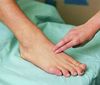
where is the dorsalis pedis pulse palpated?
lateral to the extensor hallucis longus tendon on the dorsal surface of the foot
If distal pulses are not palpable, how else could you assess perfusion?
looking at warmth, colour and capillary refill time
In general, how would you assess the neurological status of the foot after ankle dislocation?
you examine the regions of the skin of the foot innervated by major peripheral nerves
compare this to the other foot
Why would you examine the sole of the foot when assessing neurological status?
this assesses the function of:
- medial and lateral plantar nerves
- saphenous nerve
- tibial nerve
- sural nerve
Which nerve supply these areas of the sole of the foot?


Which nerves supply the skin of the foot?


How might a patient experience compromise of nerves?
numbness or reduced sensation
or an abnormal sensation and tingling
How can this fracture be described?

comminuted intra-articular tibial plateau fracture
the distal portion of the fragment (tibial shaft) is anteriorly displaced

What feature is visible in this X-ray?

lipohaemarthrosis
What is meant by a comminuted fracture?
a break or splinter of the bone into more than two fragments
What is acute compartment sydrome?
increased pressure inside a muscle compartment that most often happens in response to trauma
the fascia does not expand to accomodate swelling (oedema, bleeding, etc.)
the pressure increases inside the compartment and causes pain
What can acute compartment syndrome lead to if it is not treated?
How is it treated?
it can lead to vascular occlusion, nerve compression and muscle necrosis
it is a surgical emergence and requires immediate fasciotomy
What is the likely diagnosis when:
a patient has a swollen, warm and tender leg but their thigh looks normal
deep vein thrombosis
if a patient has had a fracture, their leg is immobile
if they are not eating or drinking much, they may be dehydrated
Should a fracture patient with a suspected DVT be anticoagulated?
YES
Unless the patient has a contraindication to being anticoagulated
If the thigh is not swollen in a DVT, which vessel is most likely to be affected?
popliteal vein
if the thigh is not swollen, it is unlikely to be more proximal than this
How should a patient with a suspected DVT be managed initially?
by calculating the Wells score and anticoagulating as per DVT treatment guidelines
(treatment dose - not prophylactic dose)
What questions should be asked when a patient has a suspected DVT?
- chest pain - check sats, resp rate
if there are any chest symptoms, need to consider PE
- tell the patient to inform staff if they experience any chest symptoms (pain, tightness, breathlessness)
What is the diagnosis?
What structures may be damaged by this injury?

posterior dislocation of right hip
potential injury to the acetabulum, joint capsule, hip ligaments and sciatic nerve
What nerve could be damaged in a posterior dislocation of the hip?
the sciatic nerve
i.e. tibial and common fibular nerves
How would you assess the motor function of the sciatic nerve?
Which muscle groups and associated movements would need to be examined?

In general, how would you assess the sensory function of the sciatic nerve?
test sensation in regions of the skin innervated by the tibial and common fibular nerves
compare this to the opposite leg
