The Foot Flashcards
What types of joints are present in the foot?
the small joints in the foot are synovial joints
this means they may be affected by both rheumatoid arthritis and osteoarthritis
Label the bones of the foot


Label the tarsals, metatarsals and phalanges


What are the two inter-tarsal joints?
- subtalar joint
- transverse tarsal joint
Whar structures articulate at the subtalar joint?
What movements occur here?
the talus and calcaneus
it is involved with inversion and eversion of the foot
When do the phalanges and metatarsals become fractured?
when heavy objects are dropped onto or run over them
people may fracture or dislocate their toes by tripping over or kicking something hard
What are two conditions that commonly affect the first MTP joint?
bunions:
- known as hallux valgus
- it is a painful bony bump that develops on the metatarsophalangeal joint
gout:
- form of arthritis caused by excess uric acid in the bloodstream
- uric acid crystals form in the joints
What causes calcaneum fracture?
when a person falls from height and lands directly onto their feet (or foot)
What are the 3 arches of the foot?
- medial longitudinal arch
- lateral longitudinal arch
- transverse arch
Label the arches of the foot


What are the important functions of the transverse and longitudinal arches of the foot?
they act as a spring and bear the weight of the body
they absorb the shock produced by locomotion
the flexibility conferred to the foot by these arches facilities functions such as walking and running
What structures maintain and support the arches of the foot?
they are supported by ligaments and tendons in the foot
(plantar ligaments)
What is pes planus?
“flat foot”
the longitudinal arches of the foot are lost
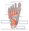
Where are the long extensor tendons of the toes visible?
Extensor digitorum longus and extensor hallucis longus tendons travel over the dorsum of the foot to their insertions
the tendons are visible under the skin of the foot
What are the 3 intrinsic muscles on the dorsum of the foot?
- extensor digitorum brevis
- extensor hallucis brevis
- 4 dorsal interossei
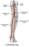
Which artery can be palpated on the dorsum of the foot?
dorsalis pedis artery
it arises at the anterior aspect of the ankle joint, as a continuation of the anterior tibial artery
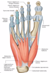
label the tendons and muscles of the foot


what is the function of extensor digitorum brevis?
it extends the first four digits at the metatarsophalangeal joint
it assists in extending the second, third and fourth digits at the interphalangeal joint
it has no action on the fifth digit
What is the function of extensor hallucis brevis?
it assists in extension of the big toe
What is the function of the dorsal interossei?
abduction of the toes
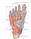
What lies deep to the tough skin on the sole of the foot?
a sheet of tough fibrous connective tissue - the plantar aponeurosis (or fascia)
this is thick and strong centrally but weaker in its medial and lateral parts
What is the role of the plantar aponeurosis?
it supports the arches of the foot and protects the deeper structures within the sole
What helps to cushion the heel of the foot?
there is a fat pad that lies between the skin and the calcaneum
How is the plantar fascia related to the flexor tendons of FDL and FHL?
fibrous bands project from the plantar fascia to merge with the fibrous sheaths that surround the long flexor tendons
What is plantar fasciitis?
inflammation of the plantar fascia along the bottom of the foot that connects the heel bone to the toes
this causes intense heel pain
What usually happens when someone presents to A&E having stepped on something sharp?
this causes a laceration or puncture wound to the sole
the foot is usually X-rayed to make sure that there is no foreign body embedded in the soul before the wound is closed
deeper wounds to the sole can potentially damage muscles, tendons and/or blood vessels
What muscles make up the intrinsic muscle layers of the sole of the foot?
layer 1 - most superficial:
- flexor digitorum brevis
- abductor hallucis
- abductor digiti minimi
layer 2:
- tendons of FHL and FDL (extrinsic)
- lumbricals
- quadratus plantae
layer 3:
- flexor hallucis brevis
- flexor digiti minimi brevis
- adductor hallucis
layer 4 - deepest:
- four dorsal and three plantar interossei
Label the muscles of layer 1 of the intrinsic muscles of the sole

- flexor digitorum brevis
- abductor hallucis
- abductor digiti minimi

Label the muscles in layer 2 of the intrinsic muscles of the sole

- tendons of FHL and FDL (extrinsic muscles)
- lumbrical muscles
- quadratus plantae

What is the bifurcation of the posterior tibial artery and tibial nerve?
posterior tibial artery:
bifurcates into the medial and lateral plantar arteries
tibial nerve:
bifurcates into the medial and lateral plantar nerves
Label the muscles of layer 3 of the intrinsic muscles of the sole

- medial and lateral heads of flexor hallucis brevis
- flexor digiti minimi
- adductor hallucis (transverse and oblique head)

Label the muscles of layer 4 of the intrinsic muscles of the sole

4 dorsal and 3 plantar interossei

Why are patient’s feet often assessed in clinic?
to assess sensation in the skin over the feet during a peripheral nervous system examination
this is often conducted in diabetic patients
Why are diabetic patient’s feet assessed?
diabetes can lead to peripheral neuropathy, in which sensation in the feet is impaired
this can lead to foot ulcers
What are the dermatomes of the foot?

How is the plantar reflex examined?
an item with a firm, blunt tip is used to stroke the sole of the foot in one swift continuous movement from the heel, up the lateral side of the sole and then across the ball of the foot towards the big toe

What is the normal response to the plantar reflex?
plantarflexion (curling) of the toes - normal Babinski sign

Which spinal nerves/segments are tested in the plantar reflex?
L5, S1
What is an abnormal response to the plantar reflex?
What does it indicate?
extension of the big toe - positive Babinski sign
this indicates the presence of an upper motor neurone lesion
Which peripheral nerves and spinal segments are being tested when a patient is asked to extend and flex the big toe?
extend the big toe:
- deep fibular (peroneal) nerve - L4-L5
flex the big toe:
- tibial nerve (L5-S2)
Label the arteries on the sole of the foot

the deep plantar arch lies in the sole of the foot

Why is clinically assessing the perfusion of the foot important?
What condition can be detected through this assessment?
it indicates whether the arteries of the lower limb are patent
peripheral vascular disease affecting the LL carries significant morbidity and can result in limb amputations
signs of PVD are first detected in the most distal parts of the limb (the foot)
Label the superficial venous drainage of the foot


Into which larger veins to the great saphenous and small saphenous veins drain?
great saphenous vein:
- drains into the deep femoral vein at the femoral triangle
small saphenous vein:
- drains into the popliteal vein
What is the pathway of the popliteal artery before it bifurcates?
it descends down the posterior thigh, giving rise to genicular branches that supply the knee joint
it moves through the popliteal fossa, exiting between the gastrocnemius and popliteus muscles
What are the bifurcations of the popliteal artery?
at the lower border of popliteus it divides into:
- anterior tibial artery
- tibioperoneal trunk
the tibioperoneal trunk then divides into:
- posterior tibial artery
- fibular (peroneal) artery
What is the passage of the posterior tibial artery like?
it continues inferiorly along the surface of the deep posterior leg muscles
it enters the sole of the foot via the tarsal tunnel, accompanying the tibial nerve
What is the passage of the fibular (peroneal) artery like?
it descends posteriorly to the fibula, within the posterior compartment of the leg
it gives rise to perforating branches, which penetrate the intermuscular septum to supply muscles in the lateral compartment of the leg
What is the pathway of the anterior tibial artery like?
What vessel does it become?
it passes anteriorly between the tibia and the fibula, through a gap in the interosseous membrane
it moves inferiorly down the leg and into the foot, where it becomes the dorsalis pedis artery
Which artery and nerve are shown?


Which arteries and nerves are shown?


What are the 2 arteries which supply blood to the foot?
- dorsalis pedis (continuation of anterior tibial artery)
- posterior tibial artery
What is the passage of the dorsalis pedis artery in the foot like?
it begins as the anterior tibial artery enters the foot
it passes over the dorsal aspect of the tarsal bones, then moves inferiorly towards the sole of the foot
it anastomoses with the lateral plantar artery to form the deep plantar arch
Which structures are supplied by the dorsalis pedis artery?
it supplies the tarsal bones and dorsal aspect of the metatarsals
via the deep plantar arch it contributes to the blood supply of the toes
What are the bifurcations of the posterior tibial artery?
it enters the sole of the foot through the tarsal tunnel
it splits into:
- lateral plantar artery
- medial plantar artery
What structures are supplied by the medial and lateral plantar arteries?
the plantar side of the foot
they contribute to the supply of the toes via the deep plantar arch
Where can the femoral pulse be palpated?
it can be palpated as it enters the femoral triangle - midway between the ASIS and the pubic symphysis
Where can the popliteal artery be palpated?
it lies deep in the popliteal fossa, so the pulse requires deep palpation to feel
ask the patient to slightly flex their leg as this relaxes the fascia around the popliteal fossa
How is the dorsalis pedis pulse palpated?
by palpating on the dorsum of the foot, just lateral to the extensor hallucis longus tendon
How can the posterior tibial pulse be palpated?
inferoposteriorly to the medial malleolus, where the artery turns to enter the foot