SDL 3 - Brain, Meninges & Blood Supply Flashcards

1
Q

What is meant by ‘rostral’ and ‘caudal’?

A

Rostral:

  • this is used to describe the position of a structure with reference to the nose
  • Occurring near the front end of the body

Caudal:

  • towards the tail end or the posterior part of the body
2
Q

What is meant by the cephalic flexure?

A

The axis of the adult brain bends at an angle of 100 degrees between the midbrain and the diencephalon

this is the cephalic flexure

3
Q

What terms are used to describe structures in the brain, relating to the cephalic flexure?

What does this mean?

A

The terms dorsal and ventral are used as though the flexure did not exist and the CNS was still a straight tube (as in the early embryo)

In the spinal cord and brainstem, dorsal means posterior

In the forebrain, dorsal means superior

4
Q

Label the aspects of the forebrain and spinal cord

A
5
Q

Label the diagram of the neurone

A
6
Q

What is the difference between grey matter and white matter?

A

Grey matter:

  • contains neuronal cell bodies
  • also contains neuropil, glial cells, synapses and capillaries

White matter:

  • contains axons coated in a myelin sheath
7
Q

What is meant by ‘neuropil’?

A

Dendrites, as well as myelinated and non-myelinated axons

8
Q

What is meant by the cortex?

A

Cortex is the outermost (or superficial) layer of an organ

The cerebral cortex is the outer portion of the cerebrum that is covered by the meninges, often referred to as grey matter

9
Q

What is meant by nucleus?

A

A nucleus is a cluster of neurones within the CNS

they are located deep within the cerebral hemispheres and the brainstem

10
Q

What is meant by ‘tract’ in neuroanatomy?

A

A bundle of nerve fibres (axons) connecting nuclei of the CNS

11
Q

What is meant by ganglion?

A

A group of neurone cell bodies within the PNS

12
Q

Label the ventral view of the brain

A
13
Q

Label the midsagittal view of the brain

A
14
Q

Which subdivisions of the brain make up the forebrain (or cerebrum)?

A

Telencephalon:

  • contains cerebral cortex (2 cerebral hemispheres)

Diencephalon:

  • contains the thalamus and hypothalamus
15
Q

Which subdivisions of the brain make up the brainstem?

A
  1. Medulla oblongata
  2. Pons
  3. Midbrain
16
Q

Label the lobes and structures of the brain

A
17
Q

Are the cerebral lobes functional or descriptive subdivisions?

A

Functional

the cerebellum has four functionally and spatially defined lobes:

  1. Parietal
  2. Occipital
  3. Temporal
  4. Frontal
18
Q

What is the formation of the dura mater like?

What structures does it form periodically?

A

It is formed from the periosteum lining the inside of the skull, together with a fibrous inner layer

throughout most of the cranial cavity, the two layers are tightly fused together

at some sites, the fibrous inner layer of dura separates from the periosteum to enclose blood-filled spaces - dural venous sinuses

19
Q

What is contained within the dural venous sinuses?

A

Venous blood flows into them from the brain

20
Q

At two sites of separation of the 2 layers of dura, what is formed?

A

The fibrous inner layer of dura projects into the cranial cavity to form a dural reflection (fold/septum)

this extends into the fissures between the major components of the brain

21
Q

What is the name of the dural septum that extends down between the two cerebral hemispheres?

A

Falx cerebri

22
Q

What is the name of the dural septum that extends between the occipital lobes of the hemispheres and the cerebellum?

A

Tentorium cerebelli

23
Q

What is the function of the two dural septa?

A

They restrict rotary displacement of the brain

24
Q

Label the structures indicated on the diagram

25
What occupies space A, and what is its function?
**Subarachnoid space** it contains cerebrospinal fluid (CSF) that acts as a cushion/shock absorber it provides a passage for blood vessels and nerves in the brain
26
What are the arachnoid villi and what is their function?
They are small protrusions of arachnoid mater in the outer layer of the dura mater They reduce pressure on the CNS they allow CSF to pass from the subarachnoid space into the venous system
27
Which vessel provides the arterial supply to the dura mater?
**Middle meningeal artery** This is a branch of the maxillary artery An injured middle meningeal artery is the most common cause of an epidural haematoma
28
Which two pairs of blood vessels provide the arterial supply to the brain?
* Vertebral arteries * internal carotid arteries
29
What is the course of the vertebral artery as it ascends to enter the skull?
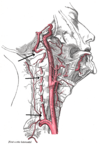
* Arises from the **subclavian artery** * Passes through the transverse foramina of the cervical vertebrae * Passes through the **foramen magnum** and into the skull * The vertebral arteries then fuse to form the **basilar artery**
30
What is the course of the internal carotid artery like as it passes into the skull?
* Arises at the level of C4, where the **common carotid artery** bifurcates * it passes through the **carotid canal** to enter the skull * it arises on the floor of the skull, on either side of the **optic chiasma**
31
Label the circle of Willis in the brain
32
Which vessels are A and B? Which brain regions are supplied by each pair of arrows?
33
Label the distribution of the anterior, middle and posterior cerebral arteries
34
Label the distributions of the anterior, middle and posterior cerebral arteries
Blue = anterior cerebral artery Orange = posterior cerebral artery Pink = middle cerebral artery
35
Which brain regions are supplied by which pairs of cerebral arteries?
**Posterior cerebral artery:** * occipital lobe * inferior & medial surface of temporal lobe **Anterior cerebral artery:** * corpus callosum * superior & medial surfaces of frontal & parietal lobes **Middle cerebral artery:** * lateral areas of frontal, temporal and parietal lobes
36
What is meant by 'perforating arteries'? What is an alternative name for them?
Penetrating arterial branches that arise from around the arterial circle and pass into the substance of the brain Those arising from the **MCA** are also called **striate arteries**
37
What do striate arteries supply?
* Arise from middle and anterior cerebral arteries * supply deep structures in the cerebrum including basal ganglia and internal capsule
38
What are the 2 sets of veins that drain blood from the brain?
Superficial and deep veins
39
What is the passage of superficial veins like? Where do they drain into?
They run over the surface of the hemispheres the majority of superficial veins drain into the superior sagittal sinus
40
What is the passage of the deep veins like?
They converge on the internal cerebral veins these eventually unite to form the **great cerebral vein**
41
Complete the venous drainage of the brain
42
What are the 3 main types of intracranial haemorrhages?
1. Extradural 2. Subdural 3. Subarachnoid
43
Rupture of which vessels leads to an extradural, subdural and subarachnoid haemorrhage?
**Extradural haemorrhage:** * also known as epidural haematoma * Usually caused by torn **middle meningeal artery** **Subdural haemorrhage:** * usually caused by tearing of the **bridging veins** that cross the subdural space **Subarachnoid haemorrhage:** * usually caused by ruptured berry aneurysm
44
What is the proper name for a stroke? Why do they occur?
**Cerebrovascular accident** they occur when a blood vessel is occluded and brain tissue becomes infarcted the symptoms depend on which artery has been occluded and which area of the brain has been damaged