L1 - Anatomy of the Vertebral Column Flashcards
What are the more sinister causes of back pain?
Malignancy and infections can present as back pain It is important not to delay a diagnosis and treatment
What is the biggest risk associated with damage to the vertebrae?
the spinal cord and the nerves are potentially at risk
How many vertebrae are there?
How are they divided into sections?
- 7 cervical vertebrae
- 12 thoracic vertebrae
- 5 lumbar vertebrae
- 5 sacral vertebrae (fused)
- 4 fused vertebrae of the coccyx

How and why do the vertebrae change in size as you go down the spine?
they get larger
this is because they act to support the weight of the body
What is the shape, strength and stability of the vertebral column dependent on?
ligaments, muscles and joints surrounding the vertebral column
muscles help with movement of the back - flexion, extension and lateral flexion
What is the main function of the vertebral column?
to protect the spinal cord
the spinal cord sits in a foramen between the vertebral body and the vertebral arch

What is the function of the natural curvatures of the spine?
they give additional flexibility and provide shock absoprtion
What are the primary curvatures?
What is their proper name and when are they formed?
kyphoses
these are convexities seen in the foetus
there are thoracic and sacral kyphoses
What are the secondary curvatures?
What is their proper name and when are they formed?
lordoses
these are concavities which develop in later life
they develop as a child starts to lift its head up and walk
they are the cervical and lumbar lordoses
Which curvatures of the spine are primary and which are secondary?
Primary - thoracic and sacral
secondary - cervical and lumbar

Label the curvatures of the spine

What is kyphosis?
a spinal disorder in which an excessive outward curve of the spine results in an abnormal rounding of the upper back
often caused by osteoporosis in the vertebral bodies

What is scoliosis?
sideways curvature of the spine
vertebral bodies rotate leading to the spine being curved laterally

What is lordosis?
an excessive inward curvature of the lower back

What are the 5 main components of a vertebra?

Red - Body:
This is the weight-bearing portion of the vertebra
Blue - Vertebral arch:
this is the whole region behind the body
it is split into the pedicles (dark blue) and the laminae
Green - Articular facets:
there are 2 superior and 2 inferior articular facets
each vertebra articulates with the vertebra above and the vertebra below to form an articulated column
Pink - Processes:
There are 2 transverse processes and one spinous process
Yellow - Vertebral foramen:
this is the hole between the body and the arch, where the spinal cord passes through

Why is the alignment of the articular facets crucial?
it determines movement
What condition occurs if parts of the vertebral arch fail to form/fuse properly?
spina bifida
in most severe cases, there is no vertebral arch, and the spinal cord herniates out the back of the spine
this leads to neurological conditions
How is the vertebral column formed?
What types of structures are involved in its formation?
the vertebrae stack up on top of each other
the column is held together by:
- facet joints
- intervertebral discs
- ligaments

Between which structures do facet joints form?
between superior and inferior articular processes on adjacent vertebrae
What is the function of the intervertebral foramen?
spinal nerves pass through the intervertebral foramen as they leave the spinal cord
How is the body of a cervical vertebra different?
the body is smaller and more oval-shaped
it has an elevated uncus laterally
How is the transverse process and pedicle different in a cervical vertebra?
there isn’t really a transverse process - there are anterior and posterior tubercles instead
there is a foramen in this region that is not present in other vertebrae
How is the shape of the transverse foramina different in cervical vertebrae?
it is more triangular shaped
it is much larger in relation to the size of the spinal cord running through it than in other regions
How does the size of the transverse foramina in the C-spine affect injuries in this region?
the spinal cord isn’t necessarily going to be injured in this region
this is due to the size of the foramen being large compared to the spinal cord
How are the articular facets directed in the cervical spine?
What does this mean for possible likelihood of injury?
- superior facets directed superoposterioly
- inferior facets directed inferoanteriorly
They come more onto the horizontal plane, making it easier for cervical vertebrae to slide on top of each other (subluxation_
What is different about the cervical spinous process?
it is bifid

What is the function of the transverse foramina?
It allows for the passage of the vertebral artery, which supplies the brain with blood

What are the alternative names for vertebrae C1 and C2?
C1 - atlas
C2 - axis
They are highly atypical
How does the C1 vertebra (atlas) differ?
It is a ring
It has anterior and posterior arches with lateral masses, which have a large articular surface for the occiput
It has no body or spinous processes

How is the body of vertebra C2 modified?
It has an upward projection called the dens

How do the atlas and the axis fit together?
What movement does this allow for?
the dens of C2 articulates with the anterior arch of C1 to make a pivot joint
This allows for rotation of the head from side to side
The dens is held in place by the transverse ligament

What is significant about the joint between the dens and the atlas being synovial?
it can be affected by rheumatoid arthritis
What supports the joint between the dens and the atlas?
the transverse ligament
this helps to maintain the stability of the joint when the head is being pivoted
What is the risk if the dens is fractured?
the dens can be driven into the upper cervical spinal cord - this is catastrophic
if the transverse ligament is still intact, it prevents this from happening
What are the most common injuries/pathologies that affect the cervical spine?
- fractures and dislocations
- dislocations can cause fractures as the joints are interlocking
- rupture of ligaments
What can ligament rupture in the cervical spine lead to?
spinal cord/nerve injuries due to fracture fragments or dislocation causing compression or severing
stability is compromised
What are the most flexible parts of the spine?
What contributes towards this?
The cervical spine and lumbar spine
having thick intervertebral discs increases flexibility
Why is the dens more prone to fracture?
What tends to cause fracture of the dens?
the bone of the dens is less dense, making it more prone to fracture
this is caused by hyperextension or hyperflexion

What is the main risk associated with fracture of the dens?
the spinal cord is not usually affected as the dens is held in place by the transverse ligament of the atlas
30-50% of fractures result in non-union
How is an X-ray usually taken to show fracture of the dens?
The X-ray is taken with the patient’s mouth open wide
This allows you to see the articulation between the atlas and the axis
A black line shows fracture of the dens

What does this X-ray show?

fracture of the dens
Why is subluxation more likely to occur in the cervical spine?
the facets are more horizontally orientated
this means the C-spine is more prone to dislocations and subluxation (slipping)
What does this X-ray show?
Why is it often not a serious problem?

subluxation (slipping) of the cervical vertebrae
as the vertebral foramen is realtively wide, the subluxation may not compress the spinal cord
Why is C-spine immobilisation necessary when there is a C-spine injury?
it keeps the C-spine in-line so that a suspected fracture or dislocation cannot be made worse through movement

How is the vertebral body different in a thoracic vertebra?
it is heart-shaped with superior and inferior costal demi-facets for articulation with the head of the rib

How are the spinous and transverse processes different in a thoracic vertebra?
the spinous process is long and extends postero-inferiorly
the transverse process has a costal facet for articulation with the tubercle of the rib

Which direction do the articular facets of a thoracic vertebra face?
superior facet faces posteriorly
inferior facet faces anteriorly
dislocation of the thoracic vertebrae is unlikely
Which structures on the thoracic vertebrae does the head of the rib articulate with?
- superior demifacet of the corresponding (same number) vertebra
- inferior demifacet of the vertebra above

What structure does the tubercle of the rib articulate with?
the costal facet on the transverse process (purple)

After the C-spine vertebrae, which are the most commonly fractured vertebrae and why?
T11 and T12
the lumbar spine is much more flexible than the thoracic spine and T11 and T12 form the joint between them
In which group of people is osteoporosis most common?
postmenopausal females
What is involved in osteoporosis?
How can it lead to vertebral fracture?
there is thinning of the bones and loss of bone density
the weight of the body and the vertebrae above cause crush fractures of the vertebrae
the vertebrae lose height anteriorly more than they do posteriorly , creating a compression “wedge” fracture

What can osteoporosis lead to in the long-term?
- excessive kyphosis (due to there being more height at the back of the spine than the front)
- loss of height
- pain
- reduced mobility
What does a compression/wedge fracture look like?
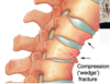
What is shown in the X-ray?

the vertebra has lost height anteriorly due to osteoporosis
How does ageing affect the vertebrae?
loss of bone density with age leads to concave vertebral bodies
this puts increased force on the rims of the vertebrae and oestophytes develop in response
What is shown in the X-ray?

osteophyte development
How does the vertebral body and foramen of the lumbar vertebrae differ?
the vertebral body is much larger as it is carrying more weight of the body
the triangular foramen is much smaller in relation to the spinal cord

What is the appearance of the transverse and spinous processes like in the lumbar vertebrae?
the spinous process is short and sturdy
the transverse processes are shorter as there is no articulation with the ribs
Which direction do the articular facets of the lumbar vertebrae face?
the superior facets face medially
the inferior facets face laterally
What tends to cause fracture of the lumbar vertebrae?
Large forces are needed to fracture the lumbar vertebrae in young people
it is usually caused by forced sudden flexion (e.g. car accident)
What is the risk associated with a lumbar spine fracture?
there is a risk to the spinal cord and spinal nerves as the vertebral foramen is relatively narrow in these vertebrae
What fracture is shown in the X-ray?

fracture of the lumbar transverse process

Which fractures are shown in the CT scan?

Fracture of L3 spinous process
Burst fracture of L5 (in many fragments)
Fragments may fall posteriorly into the region of the vertebral canal and cause neurological damage

What is meant by spondylolithesis?
Where is it most common?
a slipping of vertebra that often occurs at the base of the spine
the lumbar spine is most commonly affected
What condition is shown in the X-ray?
how can you tell?

spondylolithesis
L4 is just slipping off of L5 and L5 is just slipping off of the sacrum
Which parts of the sacrum are marked in pink and green?

the ala and the sacral promontory
the sacrum articulates with bones in the pelvis, allowing the weight to be transmitted either side onto the pelvis

What determines which movements are present in the vertebral column?
movements vary in different regions depending on the orientation of facet joints and the thickness of intervertebral discs
What movements are present in the vertebral column?
- extension
- flexion
- lateral flexion
- rotation

What 3 muscles make up erector spinae?
- longissimus muscle
- iliocostalis muscle
- spinalis muscle

What is the role of erector spinae?
it is the main extensor of the back
it brings you back to the upright position after bending over
Where are the vertebral discs the thickest?
What does this mean?
in the C and L spine
the thickness determines flexibility - these regions are more flexible
What are the 2 components of an intervertebral disc?
annulus fibrosis:
this is the peripheral fibrocartilage ring that is attached to the rim of the vertebral body
nucleus pulposus:
this is the central gelatinous “shock absorber”

What happens to the intervertebral discs with increasing age?
they dehydrate with age and the water content is reduced
this makes them become less spongy

How does dehydration of the intervertebral discs affect their function?
they lose their ability to deform when pressure is applied to the back
this means that forces in the vertebral column are more likely to tear the discs
What is meant by a ‘slipped’/’prolapsed’ disc?
the centre of the intervertebral disc herniates out of the surrounding fibrous ring
this can impinge on the nerves of the spinal cord
Which vertebrae are most commonly implicated in IV disc herniation (slipped disc)?
L4/L5 or L5/S1
What happens if spinal nerve roots are compressed in IV disc herniation?
it can cause pain/paraesthesia in the distribution of that nerve
When does IV disc herniation become a surgical emergency?
if the disc prolapses immediately posteriorly and compresses the spinal cord itselt
in cauda equina syndrome where the disc compresses the chain of spinal nerves in the cauda equina
Which procedure is carried out to decompress the spinal nerves?
laminectomy
this involves removing the lamina to decompress the contents of the vertebral canal
the incision passes through both the lamina (removing the spinous process) or the pedicles (taking away the whole vertebral arch)

How could an X-ray for IV disc prolapse be identified?

What are the 5 ligaments of the vertebral column?

- anterior longitudinal ligament
- posterior longitudinal ligament
- ligamentum flavum
- interspinal ligament
- supraspinous ligament

What are the roles of the ligaments in the vertebral column?
they are important for resisting hyperextension and hyperflexion, particularly in the C-spine
Where is the anterior longitudinal ligament found and what is its role?
runs from occipital bone and C1 to the sacrum
it is the only ligament to resist hyperextension of the vertebral column
Where does the posterior longitudinal ligament run to and from?
What is its function?
it runs from C2 to the sacrum
it prevents posterior herniation of the IV discs
What is the role of the ligamentum flavum?
it binds the lamina of adjacent vertebrae
What is the clinical significance of ligament injury?
it destabilises the vertebral column, increasing risk of injury if there are already fractures/dislocations present
can injure/tear/rupture IV discs
In a car accident, what is the motion of the neck like and how does this cause injury?
the neck firstly goes backwards and then forwards
this is hyperextension followed by hyperflexion
this can tear the anterior longitudinal ligament and sometimes it can take some of the bone with it

What is the term for a flexion or extension injury of the cervical spine?
whiplash
What procedure is shown?

lumbar puncture
