SM 268: Connective Tissue Disease in Skin Flashcards

You may prefer our related Brainscape-certified flashcards:
1
Q

What is Lupus Erythematous?

A

A chronic mulltiorgan disease with specific cutaneous presentations

2
Q

What leads to the development of Lupus Erythematous?

A

Genetic susceptibility + UV light induced Keratinocyte damage + environmental stressors = autoantigens

3
Q

What are the 3 ways Lupus can present cutaneously?

A

Chronic Cutaneous: Discoid Lupus Erythematous Subacute Cutaneous Lupus Erythematous Acute Cutaneous Lupus Erythematous

4
Q

What is the chronic mucocutaneous form of Lupus Erythematous?

A

Discoid Lupus Erythematous

5
Q

Who does DLE effect and how does it present?

A

Discoid Lupus Erythematous is common among young african american women

Presents as scaly pink or brown plaques with an annular appearance on the head and upper body

Heals with dispigmentation
6
Q

How does DLE Present and how does it heal?

A

Discoid Lupus Erythematous presents as scaly pink or brown plaques with an annular appearance on the head and upper body

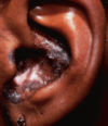
Heals with light Dispigmentation and Alopecia

7
Q

What is this and why?

A

Discoid Lupus Erythematous:

Young AA woman

Pink/brown Annular lesions on face/scalp

Alopecia and dispigmentation

8
Q

How does Subacute Cutaneous Lupus Erythematous present?

A

Subacute Cutaneous Lupus Erythematous presents as Scaled, Annular pink papules and plaques in the face, upper trunk, and upper extensor extremities

9
Q

What is this and why?

A

Subacute Cutaneous Lupus Erythematous:

Annular Pink Lesion

Upper Extremity

Plaques

10
Q

What antibody is associated with Subacute Cutaneous Lupus?

A

Anti-SSA (Ro)

11
Q

What antibody would you expect in this patient’s blood?

A

Subacute Cutaneous Lupus:

Upper trunk

Pink Annular Papules/Plaques

Minor Scaling

= Anti-SSA

12
Q

What tends to cause Subacute Cutaneous Lupus Erythematous and how does this effect diagnosis?

A

Drugs, such as Thiazides and CCB’s

Get a detailed drug history!

13
Q

How does Acute Cutaneous Lupus Erythematous present?

A

Classic Malar Rash that spares the Nasolabial folds

+
Painless Oral Ulcerations

Variable: erythema, edema, telangiectasia, erosions

14
Q

What caues a classic malar rash?

A

Sun exposure

15
Q

If you see this, what antibodies do you expect and what should you evaluate for?

A

Expect anti-dsDNA

Evaluate for Systemic Lupus: PFT’s, LFT’s, UA, etc.

16
Q

How does Neonatal Lupus Erythematous arise, and how does it present?

A

Maternal Anti-SSA antibodies cross the placental barriers, causing a neonatal rash and potentially complete heart block

17
Q

Does Neonatal Lupus Erythematous require sun exposure?

A

There is no sunlight in the womb, so no

18
Q

What is Dermatomyositis and who gets it?

A

Effects women more than men, causes proximal extensor inflammatory myopathy

19
Q

How does Dermatomyositis present and what shows up on labs?

A

Proximal extensor weakness that impede’s ADL’s

Heliotrope Rash

Shawl Sign

Dilated Fingernail Capillary Loops

Gottron’s Papules

Elevated CK

EMG/MRI abnormalities

20
Q

What is this and what does it suggest?

A

Heliotrope Rash around the eyes that spares the nose

= Dermatomyositis

21
Q

What are Gottron’s Papules and what do they suggest?

A

Gottron’s Papules = rash on extensor joints: MCP/IP/Elbow

Suggests Dermatomyositis

22
Q

What is this and what does it suggest?

A

Rash on IP + MCP = Gottron’s Papules = Dermatomyositis

23
Q

What’s shown here and what does it suggest?

A

Dilated Capillary Loops at the Proximal nail bed

Gottron’s Papules on the IP

= Dermatomyositis

24
Q

What is this and what does it suggest?

A

Rash on sun-exposed areas that spares clothing covered areas = Shawl Sign

Suggests Dermatomyositis

25
Q

If you see this, what antibodies should you expect and what disease?

A

Shawl Sign = Anti Mi2 Antibodies + Dermatomyositis

26
Q

What’s this guy have and why?

A

Dermatomyositis:

Malar Rash that does NOT spare the area under the nose

Extensor surface rash on elbow

27
Q

What is Calcinosis Cutis and what conditions is it associated with?

A

Calcinosis Cutis is a painful, swollen finger with Calcium deposits that show up as white spots/nodules

Associated with Dermatomyositis and Systemic Sclerosis (CREST)

28
Q

Does Calcinosis Cutis predict a good outcome in Dermatomyositis?

A

Nope - expect alopecia, and high mortality due to ILD

29
Q

What’s shown here?

A

Nail bed erythema/capillary changes

30
Q

What antibody predicts malignancy in Dermatomyositis, and in what population?

A

Anti-TIFgamma antibodies predict malignancy in adults

Dermatomyositis does not cause malignany in kids

31
Q

Does Dermatomyositis predict cancer in adults or children?

A

Dermatomyositis predicts cancer in adults, not children

Evaluate adults for malignancy

32
Q

How does the facial rash in Dermatomyositis differ from Lupus?

A

Lupus spares the nasolabial folds under the nose, while Dermatomyositis does not

Also, Dermatomyositis specifically involves extensor surfaces (IP/MCP/Elbow) with Gottron’s Papules as well as the Shawl sign

33
Q

What differences between SLE and DM can be found on skin biopsy?

A

None - check the rash for nasolabial folds, extensor surfaces

34
Q

What is Relapsing Polychondritis, who does it effect, and where?

A

Relapsing Polychondritis is repeated inflammation of Cartilaginous structures in 20 - 60 year olds, including the Respiratory Tract and Ear

Spares the Earlobe

35
Q

What is this?

A

Relapsing Polychondritis, a process that involves antibody-mediated autoimmunity

Note: it spared the earlobe

36
Q

What are the two types of Systemic Sclerosis, and which one involves internal organs?

A

Limited Cutaneous SSc

Diffuse Cutaneous SSc

Both involve internal organs with Inflammation, Vasculitis, and Fibrosis

37
Q

What type of antibodies are found in dcSSc?

A

Anti-Scl70 antibodies = bad outcome

38
Q

What type of antibodies are found in lcSSc?

A

Anti-centromere antibodies = good outcome

39
Q

What is CREST syndrome?

A

The presentation of lcSSC

Calcinosis Cutis

Reynauds

Esophageal involvement

Sclerodactyly

Telangiectasia

40
Q

What is Sclerodactyly and what is it associated with?

A

Sclerodactyly is fibrosis of the skin on the hands, leading to contractures and nail changes

41
Q

What is this and why?

A

Relapsing Polychodritis, because the inflammation of the ear spares the earlobe

42
Q

How does Systemic Sclerosis effect the face?

A

Leads to difficulty opening the mouth and loss of wrinkles due to extensive fibrosis/fiber deposition

43
Q

What is this and what does it suggest?

A

Salt and Pepper Dyschromia: loss of pigment around follicle due to fibrosis

Systemic Sclerosis

44
Q

What is Morphea and when does it turn into Systemic Sclerosis?

A

Morphea is an erythematous/violaceous expanding indurated plaque that can lead to loss of range of motion

Morphea does not turn into Systemic Sclerosis

45
Q

What is En Coup de Sabre?

A

A loss of Hair in a sabre pattern due to Morphea

46
Q

What is Livedo Reticularis?

A

A netlike appearance that shows up on the skin after cold exposure and resolves with warming

47
Q

What is Livedo Racemosa?

A

A fixed version of “livedo vascularis” that causes irregular networks to appear

Does not vary with temperature

Indicates sytemic disease

48
Q

How do Livedo Reticularis and Livedo Racemosa differ?

A

Livedo Reticularis is temperature dependent, a regular pattern, and comes and goes with cold/warmth

Livedo Racemosa is fixed, irregular, and indicates systemic disease

49
Q

What is anti-phospholipid antibody syndrome and how does it present?

A

Anti-phospholipid antibody syndrome is a thrombotic process seen in autoimmune diseases that causes venous and arterial thrombosis

Leads to abortions and skin necrosis, as well as thrombocytopenia

50
Q

How does anti-phospholipid antibody syndrome present and why?

A

Anti-phospholipid antibody syndrome causes both arterial and venous thrombi, extensively depelteing thrombocytes

Leads to thrombocytopenia, as well as infarcts/necrosis of the skin and spontaneous abortions

51
Q

What antibodies are involved in anti-phospholipid antibody syndrome?

A

Anticardiolipin and lupus anticoagulant antibodies are involved

52
Q

How is anti-phospholipid antibody syndrome treated?

A

Treat the autoimmune disease causing it and use anticoagulants to break up the venous and arterial clots it induces

53
Q

How does HSP present?

A

AKA IgA Vasculitis:

Preceding URI

Acute onset Palpaple Purpura + Colicky Abdominal Pain

IgA deposits in vessel walls

54
Q

How does GPA present?

A

Inflammation of the Upper and Lower respiratory tract

Hemoptyis + Glomerulonephritis

55
Q

How does EGPA present?

A

Allergic Rhinitis into Peripheral Eosinophelia ending with systemic vasculitis/inflammation

Commonly seen as an adult onset asthma