Protozoa - Flagellates Flashcards
Flagellate cyst
Transmissive stage
Comes from asexual reproduction
Is a vegetative form resistant to unfavourable environment conditions outside of the host
Flagellate trophozoite
Parasitic stage
- active, feeding and motile
Flagellate Promastigote
Has a single anterior flagellum
Flagellate Amastigote
non-flagellated intracellular stage
Where does replication of Giardia occur?
In the intestine -> diarrhoea
How Giardia causes diarrhoea?
Disrupts absorption of nutrients -> malabsorption
- causes immune response
- extracellular parasite - sits on top of the hosts cells and absorbs glucose from the host
Significance of Giardia
Malabsorption and diarrhoea -> reduced growth and reduced cognitive development
Wheres vets come across Giardia?
In high populations desities - kennels
Day care centre
Farms (intensive)
Giardia zoonosis?
In waterborne outbreaks - Giardia cysts more susceptible to chlorination than Cryptosporidium oocysts
particularly from cattle
Lifecycle of Giardia
Cyst shed in faeces -> 24-48 hr for trophozoite to mature -> ingested -> into stomach -> pH changes, CO2 stimulates hatching -> trophozoite ready to go with flagella (suction cup to hold onto intestinal wall) -> asexual replication -> pushed further down GI tract OR immune system attacks it -> encystation process -> forms a cyst -> passed in faeces
Pathogenesis of Giardia
Villous atrophy and malabsorption via sheer bulk of numbers
immune response is to completely flush out GI tract -> Gi tract also loses mucous layer
-> IBS post Giardia
Clinical presentation with Giardia
Acute/chronic diarrhoea
flatulence
vomiting
Bali belly
OR asymptomatic
Diagnosis of Giardia
Faecal smear -> look for cysts
In acute stage may see trophozoite with 2 nuclei and a central spine
- Zinc sulphate floatation of cysts
- Giardia SNAP (cysts shedding is intermittent so may need to take a few samples over time)
- ELISA
- PCR
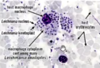
What is this?

Giardia cyst
Treatment of Giardia
Supportive care
Metronidazole (resistance) needs multiple doses for 5-7 days (can cause encephalopathy)
- Febantel for 3 days
- Fenbendazole for 5 days
Control of Giradia
Hygiene, reduce overcrowding and stress
- wash diarrhoea with ammonia
- wash bedding in hot soapy water - dry in sunlight
Trichomonads features
Faecal-oral transmission (fresh faeces)
Can be transmitted during sex
No cysts! Trophozoites only
Single nucleus
3-6 anterior fagella with one that forms undulating membrane
Bovine - Tritrichomonas foetus significance
Transmission?
Epidemiology?
Infertility and reduced pregnancy
- sexually transmitted through preseminal fluid (no transmission by AI)
- Bulls are asymptomatic carriers - survives in folds of penis
- extensive cattle farms (North QLD) with uncontrolled mating
Tritrichomonas blagurni infects what species?
Feline
Significance of Tritrichonomas blagburni
- Large bowl disease with chronic diarrhoea
- outbreaks in catteries
- asymptomatic carriers with chronnic shedding
Diagnosis of felin tritrichonomas
Fresh faecal smear and look for motile trophozoite
Faecal culture - grow trophozoites in anaerobic bag
PCR
Treatment of feline tritrichomoniasis
- off label use of ronidazole (30mg/kg) once daily for 14 days
- borad spectrum antibiotic -> use probiotic after treatment
- beware of neurotoxicity
Bovine tritrichomonas replication?
Uterine mucosa and causes infertility by disrupting the uterine membrane
Tritrichomonas foetus presentation in a cow
mild vaginitis +- discharge, metritis, salpingitis
- embryonic death and absorption -> return to service
- early foetus abortion
- retained foetus -> pyometra
- abnormal calving
- cow susceptible to repeat infection with poor immunity
Diagnosis of bovine tritrichomonas
Bull sheath wash sample - once a week (3 neg in succession after last service
- Cow cervical wash following abortion
- direct microscopy
- Culture
- PCR
Control of Bovine Tritrichomoniasis
- Test and cull bulls
- Replacement virgin heifers and bulls
- test bulls before coming in
- good fencing
- Vaccine reduces severity of infection in cows
Avian Trichomonas causes
Canker in pigeons
Frounce in falcons
Causes of trichomoniasis in birds
Trichomonas gallinae
Site of Trichomonas gallinae
Upper digestive tract
nasal cavity -> resp tract
Transmission of Trichomonas gallinae
Epidemiology
- through feeding of young
- contaminated drinking water
Pigeons are asymptomatic carriers
Young, immuncompromised birds susceptible
(poor hygiene, overcrowding and stress all factors)
Presentation of trichomonas is birds
SQUABS
Stop feeding and lose weight, ruffled and dull, dyspnoea, difficulty swallowing
- circumscribed caseous plaques on oro-pharynx, oesophagus, crop, proventriculus. Ca form ulcers and abcesses
- can be localised or systemic
Diagnosis of trichomonas in birds
- detect trichomonads
- scrape/crop fluch
- Microscopy
- PCR
- culture
Treatment of trichomonas in birds
- Surgical removal of caseous material
- Quarantine
- Ronidazole, metronidazole
- eliminate carries
Cause of enterohepatitis or Blackhead
Histomonas melaegridis + E. coli
Histomonas melaegridis is most pathogenic in which species?
TURKEYS>>>> chickens >> pheasants, ducks and geese
Outbreaks of M. melaegridis mortality and morbidity rate
80-100%
Difference of H. meleagridis to Eimeria in birds
- this spreads out of the GI system and up the bile duct and into the liver -> target lesions
Lifecycle of H. meleagridis
no cyst stage
Have amastigote that go into the Heterakis worm and passed in the Heterakis egg in the faeces -> egg ingested by paretenic hosts (earthworm) -> bird eats the worm -> infected with Heterakis and Histomonas trophozoites-> replicates in caecum and intestine -> up bile duct -> goes between the liver cells and replicates here to-> causing destruction

Presentation of Histomonas is Turkeys
- yellow diarrhoea
- Necrotising typhlitis
- thinkening and caseous exudate on caeca
+- peritonitis
Target lesions in liver - maybe also spleen and lungs
Diagnosis of Histomonas
- Necropsy - lesions on liver and caecal infection
- lesion scraping / microscopy Look for trophozoites feeding outside of the cells (to differentiate from Eimeria where you look for intracellular replicating stages)
- Lesions on histopath
Control of Histomonas
Control the Nematodes (Heterakis)
Separate chickens and turkeys
Turkeys do cloacal drinking where they squat over another birds faeces and and suck it up their cloaca. Spread the disease.
- quarantine
- Management (high stocking density they pass to each other via cloacal sucking (so clean up faeces) but in extensive system have worms -> control worms)
- strict biosecurity
- No registered drug. Only use Nitroimidazoles eg ronidazole if not being used for food production
What is a Trypomastigote?
- motile, extracellular, flagellum attached via undulating membrane. Kinetoplast posterior to nucleus

What is the Amastogote?
Kinetoplastid stage that is intracellular, has no flagella, has asexual binary fission. Kinetoplast anterior to nucleus

What is the Epimastigote
extracellular, motile. Kinetoplast centrally located just anterior to nucleus, flagellum

What is a promastigote?
Extracellular motile. Kinetoplast anterior to nucleus, flagellum

Trypanosoma Features
Not in Aus
Old world - African sleeping sickness (anaemia->chronic fatigue)
New world - Chagas disease
100s of millions of people infected
Trypanosomiasis 3 Groups
disease caused and vector/transmission
Salivaria (sleeping sickness: Tsetse fly)
Stercoraria (chagas disease: Triatoma)
Mechanica (Dourine via coitus)
Salivaria pathogenesis
Replicate in blood -> anaemia feeding intercellularly on the serum
Taking glucose from the bloodstream
Systemic disease, fever, lethargy
- wasting and organ failure
Pathogenesis of Stercoraria
Transmitted in the faeces of the vector. Tratoma come out at night and bite the corners of peoples eyes and feed on blood and are also daefecating into to eye -> inflammation (intracellular)
Mechanica pathogenesis
Sexually transmited
Important Trypanosomes
T. brucei brucei (domestic animals)
T. brucei rhodesiense (humans)
T. b gambiense (humans + pigs)
T. evansi (bovine, equine, camels, dogs)
Salivary tryanosomiasis life cycle

African trypanosomiasia features
Sleeping sickness in humans
Tse tse fly
increased decreased 70%
causes Nagana in animals
Presentation of Salivarian trypanosomiasis in animals
- fever
- can evade immune system by changing its surface antigens
- lymph node enlargement
- splenomegaly
- anaemia and lethargy
- oedema
- wasting - starvation of glucose
Neurological due to sugar starvation
Diagnosis of African trypanosomiasis
Blood smear - see spindly flagella tail and wavy curtain with dark staining nucleus
smear of CSF
- agglutination tests
- ELISA
- PCR

Treatment of salivarian trypanosomiasis
Humans:
Pentamidine
Nifurtimox and eflornithine
Melarsoprol (arsenic - 10% die from the drug)
Animals:
Diminazine, Homidium bromide
Control of salivary trypanosomiasis
Release of sterile males
Trypano-tolarant cattle
Test and treat
Drugs
Stercoraria trypanosomiases life cycle
In blood and into muscles as amastogote still - replicating (in heart muscle)

Trypanosoma cruzi (chagas disease) problems
Have affinity for cardiac muscle -> heart failure
Hard to get to to treat
Leishmania epidemiology
- not endemic in Aus
- Mediterranean region
affects humans and animals
100s of millions infected
Three main forms of leishmaniasis
Cutaneous
Muci-cutaneous
Visceral (hepato-splenomegaly, anaemia, oedema)
Lifecycle of Leishmania
Replicate in white blood cells. Which cause WBCs to rupture -> massive inflammation.

Leishmania species
Many many species
Old and new world species
L. infantum (dogs)
L. donovani
L. major
Features of visceral leishmaniasis
Healthy person: asymptomatic or mild infections
Young, old or immunocompromised: 85% fatality rate withour treatment
0-50% with treatment
Replication in and around the viscera. Mostly white blood cells.
- > fever, hepato-splenomegaly and pancytopenia
- liver failure, bleeding, anaemia, secondary infections
Cutaneous leishmaniasis
Localised nodulo-ulcerative lesion
- 1.5 mill new cases annually
- if left untreated may become diffuse with invasion of mucosa and cartilages
Infantile leishmaniasis
Leishmania infantum
Visceral and cutaneous leishmaniosis.
Dogs are primary reservoir but it is zoonotic
- non specific dermatitis
- signs of visceraldisease
Leishmania diagnosis
Blood smear-amastigotes in WBCs
lymph node aspiration
impression smear of lesion
PCR
Serology
Control of leishmania
Vector and reservoir control
- repellents
- Commercial canine vaccines (80% efficacious)
Hard to treat dogs - Drugs need lots of doses. Drugs are nasty Antimonials.
all you are doing in decreasing the clinical signs
