Platyhelminths - Paramphistomatidae, Dicrocoeliidae Flashcards
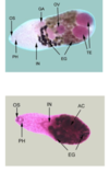
Paramphistomatidae shape
pear shaped, 5-12mm long and 2-4mm in diameter
Juvenile fluke length
1-2 mm
Position of ovaries
Behind testes
Pathogenicity of adult
Do next to nothing
Planorbid vectors of paramphistomatidae
Orthocoelium, Paramphistomum, Gyraulus
Lifecycle of paramphistomatidae

Difference between fasciola and paramphistomatidae
paramphistomidae don’t migrate to the liver. They stay in the duodenum before going into the rumen.
Pathogenesis with a moderate infection with paramphistomatidae
Ill thrift, reduced weight gain, loss milk production
Clinical signs of paramphistomatidae (heavy infection - 72 000 worms)
juvanile migration delayed: in duodenum for up to 4 months
- watery diarrhoea, dehydration and death
Exposure increases immunity
Weaners or immuno-naive animals most susceptible
- drought, movement, stress
Diagnosis of paramphistomatidae
Good history
- temp, rainfall, grazing conditions, age group affected, drench history
Clinical signs
Post-mortem (look for larvae in duodenum
Eggs in faeces? (not if only larvae -> clinical signs)
- response to treatment
- keep on differential list if weaners affected
Treatment of paramphistomatidae
Off label use of closantel and oxyclozanide
Control of paramphistomatidae
- Fence/drain affected areas
- Antihelmintics (off label)
- Get adults in rumen in late winter to reduce pasture contamination
- Juvanile flukes in summer to autumn
Features of Dicrocoelium dentriticum
- lancet fluke
- small, elongated 0.5cm (scalpel blade)
- genital porw in front of ventral sucker
- unbranched caeca
- NOT IN AUSTRALIA
Eggs of Dicrocoelium dentriticum
40-25um
smaller eggs with a thcker shell

Lifecycle of Dicrocoelium dentriticum

Pathogenicity of Dicrocoelium
- Juvaniles migrate up bile ducts to become adults
- mildly pathogenic except heavy infections (50 000+)
- calcification of bile ducts -> liver condemnation
Treatment of Dicrocoelium
Tough!
Benzimidazoles at a high dose 20mg/kg
Praziquantel
Fish borne liver flukes name?
Opisthorchis and Clonorchis
Signs of Opisthorchis and clonorchis
Aymptomatic
Gall bladder stones
Cholangitis, jaundice
Cholagiocarcinoma (bile duct tumour results from infection)
Fish borne intestinal flukes?
Heterophyidae, Echnostatidae
Signs of intestinal fish borne flukes
Asymptomatic or enteritis/gastritis, ulceration
- diarrhoea, upper gastric pain (peptic ulcers)
SE Asia and China
Fish borne liver and intestinal fluke lifecycles

Risk factors for humans with fish borne liver and intestinal flukes
- traditional eating of raw, fermented or pickled fish
- poor sanitation (use of animal or human effluent as fish food
- uncontrolled reservoir host population