MSK - Histology - Muscular Tissue Flashcards
Which of these types of muscle is striated?
Cardiac
Smooth
Skeletal
Cardiac,
skeletal
What type of tissue is shown in this micrograph?
What are some of its defining characteristics?

Skeletal muscle;
striations
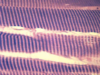
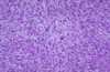
What type of tissue is shown in this micrograph?
What are some of its defining characteristics?

Cardiac muscle;
striations, intercalated discs
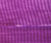
What type of tissue is shown in this micrograph?
What are some of its defining characteristics?

Smooth muscle;
non-striated
When we say some muscle tissue is striated, what does that mean?
The tissue has alternating dark and white lines that are perpendicular to the direction of the muscle fibers
State the proper terminology for the following in relation to a myocyte:
Plasma membrane
Smooth endoplasmic reticulum
Cytoplasm
Sarcolemma;
sarcoplasmic reticulum;
sarcoplasm
What type(s) of muscle is(are) striated?
Skeletal;
cardiac
What type of muscle is striated and involuntary?
What type of muscle is striated and voluntary?
What type of muscle is non-striated and involuntary?
What type of muscle is non-striated and voluntary?
Cardiac;
skeletal;
smooth;
none (does not exist)
Dystrophin connects what filament type to protein complexes of the plasma membrane?
These plasma membrane complexes connect to what extracellular structure?
F-actin;
laminin

What are the two main myofilaments?
Myosin (thick);
actin (thin)
What proteins are associated with myosin?
Titin (anchors thick filaments to Z lines)
What proteins are associated with actin?
Tropomyosin (covers myosin-binding sites);
troponin (binds calcium, moves tropomyosin);
nebulin (stabilizes and aligns actin polymers);
dystrophin (anchors thin filaments to plasma membrane protein complexes)
What protein runs alongside myosin, anchoring it to the Z-lines and running the entire length of a sarcomere?
Titin
What type of protein gives muscle its elasticity?
Titin
What are two unique facts about skeletal muscle nuclei?
Skeletal myocytes are multinucleated;
the nuclei are peripherally located
Skeletal myocytes are basically elongated plasma membranes filled with:
and peripherally lined by:
myofibrils;
many nuclei
In striated muscle, the dark bands are called __ bands.
The light bands are called __ bands.

A
(dArk bands);
I
(lIght bands)

What type of cell has a very limited role in providing regenerative effects in damaged skeletal muscle?
Satellite cells
Many __________ fuse into a single elongated ___________ that runs with other similar cells to make up a muscle fascicle.
Myoblasts;
myofiber (myocyte)
What type of cell junction unites myocytes?
None!
They are held together by layers of connective tissue (endomysium, perimysium, epimysium)
Name 1, 4, 5, 7, 9, 10, and 11.

1 - Z-line
4 - M-line
5 - titin
7 - sarcomere (Z-to-Z)
9 - H-zone
10 - A-band
11 - I-band
What proteins are found in the I band?
Actin,
tropomyosin,
troponin,
nebulin,
z-line proteins,
titin

What proteins are found in the A band?
Myosin, M-line proteins
+ I band proteins
(actin, tropomyosin, troponin, nebulin, titin)

What proteins are found in the H-zone?
Myosin, myomesin (M-line protein), titin

What layer of muscle connective tissue carries most of the vasculature and nervous innervation for the muscle?
Epimysium
Epimysium surrounds bunches of muscle fascicles. It is made of what type of connective tissue?
This is associated with what type of connective tissue fiber in particular?
Dense irregular connective tissue;
type I collagen
In close examination of striated muscle, a thin line can be seen bisecting each I band (lIght band). What is that line?
Z lines
In striated muscle, what dark line bisects each I band?
In striated muscle, what light area bisects each A band?
The Z-line;
the H band

A sarcomere is defined as the space from one __ line to the next.
Z
In striated muscle, which are the A bands and why are they called this?
In striated muscle, which are the I bands and why are they called this?
Dark bands (dArk), they are anisotropic;
light bands (lIght) they are isotropic
How is myocyte contraction linked to surrounding connective tissue?
Dystrophin (intracellular) connects actin to plasma membrane protein complexes;
these complexes connect to laminin in the endomysium basement membrane (extracellular)

Name as many proteins of the sarcomere as you can.
Myosin, titin, myomesin (and C-proteins);
actin, tropomyosin, troponin, nebulin, alpha-actinin
(peripherally associated: dystrophin, desmin)

Which striation bands shorten during muscle contraction?
Which striation band remains the same length during muscle contraction?
The I and H bands;
the A band
What are the two binding sites of a myosin head?
Actin-binding;
ATP-binding
When activated, protein 1 pulls protein 2 off the myosin-binding site on F-actin.
What is protein 1 and what is protein 2?
Troponin;
tropomyosin
What type of myofilament is found in the A band but not in the H band?
Light myofilaments (actin + associated proteins)
What band of the sarcomere is thick filaments only?
What band of the sarcomere is both thick and thin filaments?
What band of the sarcomere is thin filaments only?
H band;
A band (length of thick filaments);
I band
What structure connects two adjacent thick filaments within one sarcomere?
Myomesin (M-line protein)
1st step - myosin bound to actin
What is the 2nd step of the actin-myosin crossbridge cycle?
What is the 3rd step of the actin-myosin crossbridge cycle?
What is the 4th step of the actin-myosin crossbridge cycle?
What is the 5th step of the actin-myosin crossbridge cycle?
ATP binds myosin (myosin releases actin);
ATP hydrolysis (myosin bending);
force generation (contraction, phosphate release);
reattachment (myosin inactive, bound to actin)

What are the triads extending down into each myocyte?

One T-tubule + two sarcoplasmic reticula
(i.e. a long, narrow sarcolemma invagination with two long, narrow smooth endoplasmic reticula on either side)

What protein binds F-actin to Z-lines?
Alpha-actinin
The specific protein found in the M-line of a sarcomere is called:
Myomesin
Identify the muscle spindle apparatus in this cross-section of skeletal muscle.

(Blue circle)

What type of tissue is shown here?
Outline one single cell in this cross-section.

Skeletal muscle
(blue line)

Abnormalities in the dystrophin protein are liked to what two disorders?
Duchenne muscular dystrophy (dystrophin nearly completely absent);
Becker muscular dystrophy (dystrophin partially absent)
What structure is shown in this slide?

A myotendinous junction
(skeletal muscle attaching to tendon)
Duchenne muscular dystrophy is linked to mutations in what protein?
Becker muscular dystrophy is linked to mutations in what protein?
Dystrophin;
dystrophin
What role does desmin play in myocytes?
Does it assist in contraction?
It anchors Z-lines to the sarcolemma;
no (it is structural, maintaining shape)
What protein anchors actin to Z-lines?
What protein anchors myosin to Z-lines?
What protein anchors Z-lines to the sarcolemma?
What protein anchors F-actin to the sarcolemma?
Alpha-actinin;
titin;
desmin;
dystrophin
What protein anchors actin to Z-lines?
Alpha-actinin
What protein anchors myosin to Z-lines?
Titin
What protein anchors Z-lines to the sarcolemma?
Desmin
What protein anchors F-actin to the sarcolemma?
Dystrophin
What causes rigor mortis?
The lack of ATP prevents actin-myosin dissociation
Why are smooth myocytes not striated?
The sarcomeres are randomly arranged in various directions
(i.e. not in myofibrils)

Instead of Z-lines, smooth myocytes have:
Dense bodies

What structure stores calcium within myocytes?
Sarcoplasmic reticula
Myasthenia gravis involves antibodies to cholinergic receptors in what location?
The sarcolemma (at the neuromuscular junction)
What is the difference in myosin between cardiac, skeletal, and smooth muscle?
Skeletal/cardiac muscle = bipolar;
smooth muscle = side polar

Dense bodies in smooth muscle are analogous to what structure in skeletal and cardiac muscle?
The bodies are attached to what proteins and what other structure?
Z-lines;
intermediate filaments (vimentin, desmin), the sarcolemma

What two special sensory organs relate information back to the CNS about the degree of stretch and tension in skeletal muscle?
Muscle spindles (within the muscle fibers);
Golgi tendon apparatus (within the myotendinous junctions)
Tendons attach to what bony structure?
Periosteum
(both are types of dense connective tissue)
What are the three main types of skeletal muscle according to function?
Type I;
type IIa;
type IIb
What type of skeletal muscle fiber is slow oxidative and fatigue-resistant?
Type I (red)
What type of skeletal muscle fiber is fast oxidative glycolytic?
Type IIa (intermediate)
What type of skeletal muscle fiber is fast glycolytic and fatigue-prone?
Type IIb (white)
What type of skeletal muscle fiber is ‘red?’
What type of skeletal muscle fiber is ‘intermediate?’
What type of skeletal muscle fiber is ‘white?’
Type I;
type IIa;
type IIb
Which of the following is more prevalent in each type of skeletal muscle (red or white):
1. Mitochondria
2. Vascularity
3. Myoglobin
4. Sarcoplasmic reticulum
1 - Red
2 - Red
3 - Red
4 - White
What type(s) of muscle is(are) characterized by a high quantity of gap junctions?
Cardiac;
smooth
Which type of muscle is characterized by branching and anastomosing between fibers?
Cardiac
What type(s) of muscle is(are) characterized by centrally located nuclei?
What type(s) of muscle is(are) characterized by peripherally located nuclei?
Smooth, cardiac;
skeletal
Intercalated disc are made up of what type of cellular junction(s)?
Gap;
desmosomes
What type of muscle is not characterized by the T-tubule system?
What does it have instead?
Smooth myocytes;
caveolae (smaller invaginations)
Are any muscle types able to easily regenerate?
Only smooth muscle
What proteins anchor smooth muscle dense bodies in place?
Vimentin, desmin
(intermediate filaments)
Dense bodies are analogous to __________ in skeletal muscle.
Two proteins (which?) and what other structure anchor these bodies in place?
Z-lines;
vimentin, desmin
(intermediate filaments),
the sarcolemma

Name the differences in myosin orientation between smooth, cardiac, and skeletal muscle.
Skeletal/cardiac = bipolar myosin;
smooth = sidepolar myosin

Starting at the initial influx of calcium into the myocyte, what happens next in smooth muscle activation?
Ca2+ binds/activates calmodulin;
this complex than activates myosin light-chain kinase (MLCK);
the kinase phosphorylates the circular (self-bound) myosin to unbind/activate it

Skeletal myocytes gets calcium from:
Smooth myocytes gets calcium from:
Cardiac myocytes gets calcium from:
The sarcoplasmic reticulum
The extracellular fluid; adjacent smooth muscle cells
The sarcoplasmic reticulum; adjacent cardiac muscle cells
What shape are skeletal myocytes?
What shape are smooth myocytes?
Polygonal;
fusiform
Where are skeletal myocyte nuclei?
Where is a cardiac myocyte nucleus?
Where is a smooth myocyte nucleus?
Peripheral;
central;
central
What types of tissue are the three with boxes obscuring them in this slide?

Smooth muscle cross-section;
smooth muscle longitudinal section;
dense irregular connective tissue


Cardiac muscle

Smooth muscle

Cardiac muscle
(Bs indicate intercalated discs)


Skeletal muscle fibers

Skeletal muscle

Cardiac muscle

Smooth muscle (cross-section)


A skeletal muscle fiber (myofiber) is just one, long:
Myocyte
One single myofiber (myocyte) is filled with many, many chains of:
myofibrils
Where are muscle satellite cells found?
On the surface of skeletal muscle cells
Are muscle striations parallel lines or perpendicular lines to the direction of the myocyte and its myofibrils?
Perpendicular lines
What is here described:
“fluid-filled capsules enclosing a few small muscle cells and nerve fibers”
Muscle spindles
How many nuclei are typically present in cardiac myocytes?
1
(but may be 2)
Do any types of muscle cell exhibit branching?
Yes, only cardiac myocytes
What type of tissue is shown in this slide?
What is the yellowish substance seen in some cells?

Cardiac muscle;
lipofuscin
What are the three types of cellular junction found in cardiac myocyte intercalated discs?
Gap;
macula adherens (desmosome);
fascia adherens (specialized Z-lines)
Some cardiac myocytes stain an intense magenta if treated with periodic Acid-Schiff stain (PAS). What are these cells and why do they stain this way?

Purkinje cells
(modifed myocytes that conduct electrical impulses);
abundant glycogen inclusions
How does a smooth myocyte nucleus appear when the myocyte is relaxed?
How does a smooth myocyte nucleus appear when the myocyte is contracted?
Cigar-shaped;
corkscrew-shaped
What type of tissue is indicated by the circles in this slide?
(Orange, red, green, purple)

Orange - dense irregular CT fibers
Red - smooth muscle (longitudinal)
Green - a parasympathetic ganglion
Purple - smooth muscle (cross-section)
What disorder is characterized by antibodies (IgG) to the cholinergic receptors on the motor end plate?
Myasthenia gravis
Is there any type of muscle that is able to actively regenerate?
Smooth muscle
How does smooth muscle regenerate?
Simple mitosis of smooth myocytes
Upon injury to skeletal muscle, what normally quiescent cells are responsible for some very limited regeneration?
Where are they found?
Satellite cells;
the external lamina
Which type of muscle shows no regenerative capacity beyond simple scar formation?
Which type of muscle shows some very limited regenerative capacity through satellite cells?
Which type of muscle shows active regeneration through simple mitosis?
Cardiac;
skeletal;
smooth
Does cardiac muscle contain satellite cells?
How is damage addressed?
No;
fibroblast proliferation and scar tissue formation
What term refers to an increase in cell number?
Hyperplasia
What term refers to an increase in cell size?
Hypertrophy
During pregnancy, does the uterus undergo hyperplasia or hypertrophy?
Both
What type of tissue is shown here?
How can you tell?

A leiomyoma (benign tumor of smooth muscle);
whorled (fascicular) pattern of smooth muscle bundles separated by well-vascularized connective tissue
< 5 mitotic figures per HPF, no significant atypia
What is the most common tumor found in females?
What is the most common tumor found in males?
Leiomyomas (fibroids);
lipomas
What type of tissue is this?
How can you tell?

Leiomyosarcoma;
atypia + either mitotic activity, tumor cell necrosis, or size > 10cm
How does weightlifting result in skeletal muscle hypertrophy?
Through repair of microtraumas
What effect can each of the following have on skeletal muscle?
- Inactivity, bedrest, cancer, congestive heart failure,*
- COPD, burns, liver failure, glucocorticoids -*
Atrophy
Cardiac ventricular hypertrophy can result from anything that causes what?
Increases in afterload
What are the changes in color seen in myocardium as a result of ischemia?
(Start with normal myocardium and end with a scar)
No change –>
dark mottling –>
hyperemia –>
yellow-brown softening –>
gray-white scar
During what time period following infarction will myocardial histology show no color changes?
1 - 4 hours
During what time period following infarction will myocardial histology show dark mottling?
4 - 24 hours
During what time period following infarction will myocardial histology show hyperemia?
1 - 5 days
During what time period following infarction will myocardial histology show hyperemia?
5 - 14 days
During what time period following infarction will myocardial histology show gray-white scar formation?
2 - 8 weeks
How does skeletal muscle regenerate?
How does cardiac muscle regenerate?
How does smooth muscle regenerate?
Satellite cells (VERY limited);
it doesn’t (no satellite cells);
simple mitosis
