Cardio - Histology - Vascular Tissue; Lymphoid Tissue Flashcards

1
Q

What are the three layers (tunicas) of any blood vessel?

A

Tunica intima

Tunica media

Tunica adventitia (externa)

2
Q

The lumen is wider in which, arteries or veins of comparable size?

A

Veins

3
Q

The wall is thicker in which, arteries or veins of comparable size?

A

Arteries

4
Q

What fiber is found in uniquely high concentrations in the aorta and other large arteries?

A

Elastin

5
Q

What are the three types of artery?

A

Elastic;

muscular;

arterioles

6
Q

Which arteries are elastic?

A

The aorta, carotids, and subclavians

7
Q

What does the elastin in the aorta do?

What does the collagen in the aorta do?

A

Propel blood (rebound effect);

provides strength to control distension

8
Q

What transition occurs in the tissues from elastic to muscular arteries?

A

A shift from elastic tissue to smooth muscle

9
Q

What subendothelial layer is especially prominent in muscular arteries?

A

The internal elastic membrane

10
Q

What is a normal blood pressure?

A

< 120 / < 80

11
Q

What blood pressure is in the elevated category?

A

120 - 129 / < 80

12
Q

What blood pressure defines hypertension stage 1?

A

130 - 139 systolic OR 80 - 89 diastolic

13
Q

What blood pressure defines hypertension stage 2?

A

≥ 140 systolic OR ≥ 90 diastolic

14
Q

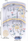
What structure connects arterioles and capillaries?

A

Metarterioles

15
Q

Is capillary blood flow continuous or pulsatile?

A

Pulsatile

(for maximum nutrient/waste exchange)

16
Q

Where are precapillary sphincters located?

What are they?

A

Metarterioles;

bands of smooth muscle

17
Q

What intermediate filaments lend structural support to capillaries?

A

Desmin and vimentin

18
Q

What type of tissue is shown in this micrograph?

A

A capillary bed

19
Q

What molecule promotes tight junction leakage in capillary beds?

A

Histamine

20
Q

How do large molecules leave capillary beds?

A

Transcytosis

(endocytosis and then exocytosis on the other side)

21
Q

What type of well-developed mesenchymal cell surrounds capillary endothelial cells and have the ability to differentiate into smooth muscle?

A

Pericytes

22
Q

What contractile filaments are present in pericytes?

A

Tropomyosin;

isomyosin

23
Q

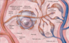
What are the three types of capillary?

A

Continuous;

fenestrated;

sinusoidal (discontinuous)

24
Q

What types of vessel in the body contain valves?

A

Veins;

lymphatics

25
Where do Purkinje cells travel?
The subendocardium
26
Purkinje cells are large cells containing large amounts of what substance?
Glycogen
27
Identify the structure with an 'A' in the lumen. Identify the structure with a 'B' in the lumen.
A = Medium vein B = Muscular/medium artery
28
Identify the areas indicated by green bars and yellow bars. Identify the structure at the tip of the blue arrows.
Yellow bar = Tunica media Green bar = Tunica adventita Arrows indicate internal elastic lamina
29
Identify the structures labeled 'A', 'B', and 'C'. All three structures are components of what structure?
A = Peripheral nerve B = Muscular artery C = Medium vein Neurovascular bundle
30
What structure is surrounded by arrows in this micrograph?
Vasa vasorum
31
Identify the entire structure. Identify the area indicated by the black bar. Identify the area indicated by the blue bar. Identify the structure at the tip of the arrows.
Large/elastic artery Black bar = Tunica media Blue bar = Tunica adventita Arrows = Vasa vasorum
32
The bar indicates the thickness of the wall of this structure. Identify the structure.
Large vein/vena cava
33
Identify A and B.
A = Muscular artery B = Small vein
34
Identify the structure at the tips of the black arrows. Identify the structure at the tips of the blue arrows.
Black arrows = Arteriole Blue arrows = Venule
35
Identify the blood vessel in this micrograph.
A fenestrated capillary
36
Identify the blood vessel in this micrograph.
Sinusoid
37
Identify this structure.
Medium/small vein
38
Identify the vessel. Identify the structure indicated by black arrows. Identify the structure indicated by black bar.
Muscular artery Arrows = Internal elastic lamina in tunica intima Bar = Tunica media
39
As you move from arteries to veins, what change would you expect to see in the relative sizes of the tunica intima, media, and adventitia?
**Intima** - remains the same **media** - decreases in size **adventitia** - increases in size
40
The internal elastic lamina is part of what layer of the blood vessel wall? The external elastic lamina is part of what layer of the blood vessel wall?
The tunica intima; the tunica media
41
Which blood vessels are 'resistance' vessels? Which blood vessels are 'capacitance' vessels? Which blood vessels are 'conducting' vessels? Which blood vessels are 'distributing' vessels? Which blood vessels are 'exchange' vessels?
Resistance - **a****rterioles** Capacitance - **veins** Conducting - **elastic arteries** Distributing - **muscular arteries** Exchange - **capillaries**
42
The tunica adventitia (externa) is thicker (relative to wall thickness) in which type of vessel, arteries or veins?
Veins
43
The tunica media is thicker (relative to wall thickness) in which type of vessel, arteries or veins?
Arteries
44
Describe the changes in blood pressure as blood travels out through arteries and back through veins.
45
Where are capillaries with extensive tight junctions especially common?
The blood-brain barrier
46
In what example tissues are fenestrated capillaries likely to be found?
Endocrine glands, the intestines, the kidneys
47
Organs such as endocrine glands, the intestines, and the kidneys are likely to be supplied by what type of capillary?
Fenestrated
48
What type of capillary is characterized by an abundance of small pores covered by a thin diaphragm?
Fenestrated
49
What type of capillary is found in the adrenal medulla?
Fenestrated
50
What is another term for sinusoidal capillaries?
Discontinuous capillaries
51
Which type of capillary allows for quickest exchange of material between tissues and the bloodstream?
Sinusoidal (discontinuous) capillaries
52
Which of the following capillary types are characterized by fenestra covered by a thin diaphragm? Continuous Fenestrated Discontinuous (sinusoidal)
Fenestrated only
53
Which of the following capillary types are characterized by fenestra with no diaphramagtic barrier? Continuous Fenestrated Discontinuous (sinusoidal)
Discontinuous (sinusoidal)
54
**True/False.** Sinusoidal (discontinuous) capillaries are characterized by fenestra and no diaphragm, yet they have an intact basal lamina.
**False.** The basal lamina is also discontinuous
55
In what example tissues are sinusoidal (discontinuous) capillaries likely to be found?
Liver, spleen, gallbladder
56
What are the two terminal vessels of the lymphatic system?
The thoracic duct; the right lymphatic duct
57
What two structures can often be found acompanying a muscular artery?
A vein and peripheral nerve of proportional sizes
58
What structures are indicated by the arrows in this micrograph? Red - Orange - Blue - Green - Gray -
Red - **Nerve** (shows perineurium) Orange - **Lymph cap** Blue - **Artery** (shows 2 layers of smooth muscle + a pericyte) Green - **Vein** Gray - **Vein**
59
How much of total blood volume can be held in the veins at one time? How much of this blood can remain stationary without negative effects?
70%; 0% (venous blood is not stationary)
60
From smallest to largest, what are the names of vessels returning blood to the heart?
Venules, small veins, medium veins, large veins
61
**True/False.** Many large and medium veins are muscular.
True
62
Does cardiac contraction begin at the apex or the base of the heart? Does cardiac contraction begin in the deep layers or superficial layers of the heart?
Apex; deep (endocardium --\> epicardium)
63
During microscopic examination of a tissue specimen, the pathologist notices numerous capillaries with continuous endothelium, lacking fenestrations but with many pinocytotic vesicles. Based on these observations, the tissue being examined most likely is: A. muscle B. liver C. spleen D. adrenal medulla
**A. muscle** * B. liver (sinusoidal)* * C. spleen (sinusoidal)* * D. adrenal medulla (fenestrated)*
64
What are some examples of large veins?
Vena cava, portal v., splenic v., renal v.
65
What are some examples of medium veins?
Typically, named deep veins | (e.g. popliteal, radial, tibial)
66
What type of vein is the great saphenous?
A muscular (medium) vein
67
What vessels are typically most likely to be affected by atherosclerosis?
Large and medium arteries
68
Which gender is more at risk for atherosclerotic development?
Males
69
Describe the (very basic) components of an atheroma.
A soft lipid core + a fibrous cap
70
What is the 'response to injury' theory of atherosclerosis?
Atherosclerosis is a chronic inflammatory reaction to endothelial damage ## Footnote **1) I**njury to the endothelium of the arterial wall **2)** Tissue response of the vascular wall to the injury
71
Describe the basic pathophysiology of atherosclerosis.
72
What are three negative outcomes that may occur at a site of severe atheroma development?
Vessel stenosis; plaque aneurysm and rupture; vessel occlusion by thrombus
73
What is the difference between stable and vulnerable atheromas?
Thickness of the fibrous cap; degree of inflammation
74
The collagen and ECM in atheromatous plaques are synthesized largely by what type of cell?
Smooth muscle cells
75
Although they decrease the risk of plaque rupture, what risk do stable atheromas have when compared to vulnerable plaques?
Increased risk of vessel stenosis
76
Why would rupture of an atheromatous plaque lead to clot development? What are two potential adverse outcomes for these clots?
Exposed thrombogenic factors in the underlying ECM; thrombosis, embolus development
77
What is a 'true' aneurysm of the cardiovascular system? What is a 'false' aneurysm?
One that involves all three layers of the vessel wall; rupture of the intima --\> hematoma between the media and externa (adventitia)
78
What type of aneurysm shape is a balloon-like direct outpouching of the vessel wall? What type of aneurysm is a more football-shaped expansion in all directions?
Saccular, fusiform
79
What is the most common location for an atherosclerotic aneurysm?
The abdominal aorta
80
**True/False.** A false aneurysm of the aorta is an aortic dissection.
**False.** False aneurysm --\> between the media and externa (adventitia) Dissection --\> between the intima and media
81
Dissections take place between what layers of the vessel wall?
The intima dissecting off the media
82
What is congestive heart failure in terms of bodily metabolic need?
Cardiac output is insufficient to match bodily metabolic requirements
83
Are most cases of congestive heart failure due to systolic or diastolic function?
Systolic | (inadequate contractile function)
84
What are the most common causes of left-sided heart failure?
Ischemic heart disease; hypertension; aortic/mitral valve diseases; 1° myocardial diseases
85
What is the most common causes of right-sided heart failure?
Left-sided heart failure
86
What immediate effect(s) does left-sided heart failure have on associated organs?
Pulmonary congestion/edema; poor perfusion of systemic organs
87
What pulmonary immune histology is expected in cases of left-sided heart failure?
Wet, congested lung tissue with **hemosiderin-laden macrophages**
88
Hemosiderin-laden macrophages are a sign of:
Left-sided heart failure
89
What is cor pulmonale?
Isolated right-sided heart failure | (i.e. not caused by left-sided HF)
90
Cor pulmonale typically occurs in patients with:
Chronic disorders affecting the lungs ## Footnote *(e.g. COPD, pulmonary fibrosis, sleep apnea, PE, poliomyelitis, myasthenia gravis, etc.)*
91
What are some major signs or symptoms of left-sided heart failure?
**Pulmonary congestion** --\> cough with frothy sputum, orthopnea, paroxysmal nocturnal dyspnea, shortness of breath; ## Footnote **cyanosis**
92
What are some major signs or symptoms of right-sided heart failure?
Hepatosplenomegaly, JVD, dependent edema; anorexia, GI distress, weight loss
93
Cardiac ventricular hypertrophy can result from anything that causes what?
Increases in afterload
94
What are the changes in color seen in myocardium as a result of ischemia? *(Start with normal myocardium and end with a scar)*
No change --\> dark mottling --\> hyperemia --\> yellow-brown softening --\> gray-white scar
95
During what time period following infarction will myocardial histology show no color changes?
1 - 4 hours
96
During what time period following infarction will myocardial histology show dark mottling?
4 - 24 hours
97
During what time period following infarction will myocardial histology show a yellow-brown softening with a hyperemic border?
3 - 10 days
98
During what time period following infarction will myocardial histology show gray-white scar formation?
2 - 8 weeks | (scar complete after ~2 months)
99
What proportion of patients with a STEMI typically die within the first hour before receiving medical care? Of what?
1/3; fatal arrythmias
100
What is the in-hospital MI death rate?
7%
101
Cardiac rupture is a complication of 1 - 5% of MIs. In what time frame is it most likely to occur?
3 - 7 days following the infarction
102
What percentage of PEs arise from DVTs?
\> 95%
103
How do peri-mortem clots appear? Are they attached to the underlying wall? How do post-mortem clots appear? Are they attached to the underlying wall?
Grayish-red; yes. Gelatinous, yellow, 'chicken fat' clots; no
104
Via activation of what proteins do macrophages know that bacterium are present for phagocytosing?
Toll-like receptors | (common invader patterns)
105
How long do neutrophils typically live? Do they have APC abilities?
5 days; no
106
What type of substance do natural killer cells phagocytose?
Nothing; they are not phagocytes
107
Via what two mechanisms do natural killer cells exert any effect?
1. Release of cytokines 2. Inducing suicide in tumor cells, virus-infected cells, etc.
108
Memory cell activation by an antigen leads to a rapid increase in plasma cells specific to that antigen via what process?
Clonal selection
109
Via what two methods do immunoglobins exert an effect on pathogens?
Opsonization; neutralization
110
Macrophages have receptors for what part of the immunoglobin?
The Fc region
111
T cell MHCs are useful for identifying what two types of problem?
Problems within the cell (MHC I); APCs presenting problems outside the cell (MHC II)
112
What type of connective tissue fiber is found in high concentrations in lymphoid tissues? What cells produce this tissue?
Type III collagen (reticulin); reticular cells
113
What type of cell provides the support framework of the thymus?
Thymic epithelial cells | (epithelioreticular cells)
114
What are the primary lymphoid organs?
Bone marrow; thymus
115
**True/False.** Secondary lymphoid tissues can be found in _each_ of the following: nasopharynx oropharynx bronchus lung tissue small intestine urogenital tissues
True.
116
Are reticular fibers found in the thymus?
No
117
**True/False.** The thymus is histologically homogenous all the way through, but it is divided by septa into various lobules.
**False.** The thymus has a cortex and a medulla that are histologically distinct.
118
Based on structure and location, how many types of epithelioreticular (thymic epithelial cells) are there throughout the thymus?
6
119
**True/False.** Many macrophages can be found in the thymus.
True
120
What types of T cell selection occur in the thymic cortex and medulla, respectively?
Positive selection, negative selection
121
What is the purpose of thymic positive T cell selection? Where does it occur?
To select for T cells with the correct surface proteins (e.g. CD4 or CD8, TCR, CD3, etc.); the thymic cortex
122
What is the purpose of thymic negative T cell selection? Where does it occur?
To ensure certain T cells don't have the incorrect surface proteins (e.g. just CD4 or just CD8); the thymic medulla
123
Positive and negative T cell selection in the thymus is all contained and controlled by what type of cell? How many types of this cell are there (based on location and structure)?
Thymic epithelial cells (epithelioreticular cells); 6
124
What are the six types of epithelioreticular (thymic epithelial) cell found in the thymus?
Type I - outer boundary of cortex Type II - positive selection Type III - inner boundary of cortex Type IV - outer boundary of medulla Type V - negative selection Type VI - Hassall's corpuscle *(express thymic stromal lymphopoietin - TSLP)*
125
What two types of epithelioreticular (thymic epithelial) cell are the boundaries of the thymic cortex?
I and III
126
What two types of epithelioreticular (thymic epithelial) cell are responsible for positive and negative T cell selection, respectively?
Types II and V
127
What type of epithelioreticular (thymic epithelial) cell makes up Hassall's corpuscles in the thymic medulla? What do they secrete?
Type VI; thymic stromal lymphopoeitin (TSLP)
128
Which is more basophilic, the thymic cortex or medulla?
Cortex
129
What specialized structure lines thymic capillaries? What type of capillaries are these?
The blood-thymus barrier; continuous
130
Between the endothelial cells / pericytes of thymic capillaries and the epithelioreticular (thymic epithelial) cells of the thymic medulla, what type of cell is found in the connective tissue space?
Macrophages
131
How many basal lamina are found surrounding thymic capillaries?
Two | (see image)
132
Where are most B cells found in a lymph node? Where are most T cells found in a lymph node? Where are most plasma cells found in a lymph node?
**Cortex** (follicles, nodules) **Paracortex** (deep cortex) **Medulla** (medullary cords)
133
Name the predominant type of cell in the following locations: **Cortex** (follicles, nodules) **Paracortex** (deep cortex) **Medulla** (medullary cords)
Cortex (follicles, nodules) - **B lymphocytes** Paracortex (deep cortex) - **T lymphocytes** Medulla (medullary cords) - **Plasma cells**
134
What type of cell is the predominant type found in a lymph node cortex (follicles)?
B lymphocytes
135
What type of cell is the predominant type found in a lymph node paracortex (deep cortex)?
T lymphocytes
136
What type of cell is the predominant type found in a lymph node medulla (medullary cords)?
Plasma cells
137
What type of cell and fiber makes up much of the lymph node stroma?
Reticular cells / reticular fibers (type III collagen)
138
**True/False.** Lymph nodes are non-encapsulated lymphatic tissue.
False.
139
The lymph node paracortex has a large concentration of what vessel type?
High endothelial venules
140
What is in the center of a lymphatic follicle (with all the B cells)?
A germinal center
141
Afferent lymphatic vessels pierce lymph node capsules and drain into what space?
The subcapsular space
142
Describe the flow of lymph through a lymph node. (Note: from afferent lymphatic vessel to efferent lymphatic vessel.)
**Afferent lymphatic vessel** --\> Subcapsular space --\> Paratrabecular sinuses --\> Through cortex --\> Through medulla --\> **Efferent lymphatic vessel**
143
Where does blood flow enter the lymph node? Where does it exit?
The hilum; the hilum
144
Describe the route taken by the fluid that enters the lymph node as blood but leaves as lymph. ## Footnote *(Note: obviously, this is not all the blood. Most of it leaves through lymph node venules.)*
Enters through arteries --\> Leaves circulation through high endothelial venules (perfuses paracortex) --\> Enters lymphatic sinusoids --\> Exits lymph node through efferent vessels --\> Returns to circulation through thoracic duct or right lymphatic duct
145
What type of structure is shown in this micrograph?
A lymph node
146
Most (90% of) lymphocytes enter lymph nodes via what?
High endothelial venules | (in the paracortex)
147
A lymph node germinal center is a ________ follicle.
Secondary
148
What two types of pulp are found in the spleen?
Red and white
149
What structure is shown in this micrograph?
The spleen
150
From deep to superficial, name the three portions of a splenic white pulp island.
Central artery, (+ sheath) germinal center, marginal area
151
What structure accompanies arteries in the splenic white pulp? This structure is mostly comprised of what type of cell?
A periarterial lymphatic sheath (PALS); T cells
152
The splenic white pulp is mostly what type of cell? The splenic PALS is mostly what type of cell?
B cells; T cells
153
The splenic white pulp has what functions?
Blood antigen removal Antigen presentation Antibody production Lymphocyte proliferation (T and B cells)
154
The splenic red pulp has what functions?
Remove the following from circulation: 1. Blood antigens 2. RBC’s & platelets (aged, abnormal, and/or damaged) 3. Iron (macrophage phagocytosis & digestion)
155
The splenic _red_ pulp is mostly centered around ________ the blood. The splenic _white_ pulp is mostly centered around mounting an ________ \_\_\_\_\_\_\_\_ to what is found in the blood.
Cleaning/filtering; immune response
156
The splenic cords and sinusoids are found in which type of splenic pulp?
Red pulp
157
Non-encapsulated secondary lymphatic tissues (e.g. tonsils, Peyer's patches) are mostly just aggregates of:
Lymphatic nodules
158
Although splenic tissue does not stain red and white upon histology preparations, what is a defining feature that indicates which is white pulp?
Presence of a central artery
159
Afferent lymphatic vessels
160
161
Lymphatic germinal center
162
Lymphatic vessels
163
Lymph node; filters blood and produces antibodies
164
Lymphatic nodule with germinal center
165
PALS (T cells); central arteriole (splenic white pulp)
166
Arrows - white pulp A - red pulp B - hilum
167
Peyer's patches A - smooth muscle B - dense irregular connective tissue
168
A - central arteriole B - pulp arteriole
169
Arrows - venous sinusoids A - pulp cords (splenic tissue)
170
What organ is shown in this micrograph?
The spleen
171
172
A splenic nodule
173
Paracortex; high endothelial venules
174
Lymph node micrograph
A - medullary cords Arrow - macrophage
175
Black arrows - subcapsular sinus Green arrows - cortical sinuses A - Cortex B - Paracortex C - Medulla
176
Lymph node micrograph
High endothelial venule
177
Lymph node hilum
178
Black arrows - subcapsular lymph node sinus White arrows - cortical lymph node sinus
179
What organ is shown in this micrograph?
The thymus
180
Tonsil Arrow --\> tonsilar crypt
181
Identify the three types of vessel.
A - lymphatic vessel B - venule C - arteriole
182
Identify A - E in this micrograph of vessels.
A - venule B,C,E - arterioles D - lymphatic vessel
183
What is unique about the tunica adventitia (externa) of the largest veins?
Smooth muscle is present
184
Are neutrophils APCs?
No
185
What molecule presents antigens to TH cells? What is the effect?
MCH II (from APCs); TH cell activation (and subsequent B cell proliferation)
186
What molecule presents antigens to TC cells? What is the effect?
MHC I (from virally infected cells); TC cell activation and infected cell lysis
187
What cytokine is especially important in activating TH cells and TC cells?
Interleukin-2 | (IL-2)
188
MHC I signaling alerts TC cells to problems in which location? MHC II signaling alerts TC cells to problems in which location?
Intracellular problems (e.g. viruses); extracellular problems (phagocytosed and then presented)
189
What surface receptor is found on both TC and TH cells? What surface receptor is found on only TC cells? What surface receptor is found on only TH cells?
**TCR** (T cell receptor) ## Footnote **CD8+** **CD4+**
190
Positive selection in the thymic cortex does what to T cells? Negative selection in the thymic cortex does what to T cells?
**Both** CD4+ and CD8+ are added to the T cell (results in cells positive for TCR, CD4+, and CD8+); **one** of the CD receptors is removed (results in cells positive for TCR and either CD4+ or CD8+)
191
Where in the thymus are T cells double negative? What does this mean? Where in the thymus are T cells double positive? What does this mean? What happens next?
The cortex --\> the cells lack CD4+ and CD8+; the cortex --\> the cells have both CD4+ and CD8+; in the medulla, the cells lose **one** of the above (either CD4+ or CD8+ is lost)
192
Via what vessel do most lymphocytes enter lymph nodes?
Via high endothelial venules (HEVs)
193
What type of epithelium lines high endothelial venules (HEVs)?
Simple cuboidal cells
194
A micrograph shows packed lymphocytes with interspersed plasma cells. This is likely a slide of what tissue?
A lymph node medulla
195
A micrograph shows packed lymphocytes with interspersed epithelioreticular cells and a few Hassall's corpuscles. This is likely a slide of what tissue?
The thymic medulla
196
A micrograph shows packed lymphocytes with interspersed epithelioreticular cells and no Hassall's corpuscles. This is likely a slide of what tissue?
The thymic cortex
197
Does the spleen have efferent lymph vessels or afferent lymph vessels or both? Do lymph nodes have efferent lymph vessels or afferent lymph vessels or both? Does the thymus have efferent lymph vessels or afferent lymph vessels or both?
Efferent only; both; efferent only
198
How does the spleen pull out old RBCs for recycling?
Young RBCs can deform and pass through the sinusoids; older RBCs cannot, and continue through the vessel system until they are picked up by splenic macrophages
199
**True/False.** Large arterioles typically have an internal elastic lamina.
True.
200
The tunica media of an arteriole usually contains __ or __ layers of smooth muscle.
1, 2
201
The tunica adventitia is ______ in arterioles.
Scant
202
The endothelial cells in postcapillary venules are supported by what type of cell and what type of fiber?
Pericytes, reticular fibers
203
Large venules will often contain __ or __ poorly organized and somewhat incomplete layers of smooth muscle.
1, 2