CS&B - Histology - Skin Flashcards

1
Q

From superficial to deep, what are the layers of epidermis?

(What mnemonic might be helpful in remembering these?)

A

Stratum corneum,

stratum lucidum (in thick skin only),

stratum granulosum,

stratum spinosum,

stratum basale

(Come, let’s get sunburned!)

2
Q

What separates the epidermis from the dermis?

A

The basement membrane (basal lamina + reticular lamina)

3
Q

From superficial to deep, what are the two layers of the dermis?

A

The papillary layer;

the reticular layer

4
Q

Of what type of tissue is the dermal papillary layer composed?

A

Loose connective tissue (areolar)

5
Q

Of what type of tissue is the dermal reticular layer composed?

A

Dense irregular connective tissue

6
Q

Of what type of tissue is the dermal papillary layer (superficial) composed?

Of what type of tissue is the dermal reticular layer (deep) composed?

A

Loose connective tissue (areolar);

dense irregular connective tissue

7
Q

The inferiorly oriented projections of the epidermis are called:

The superiorly oriented projections of the dermis are called:

A

Epidermal ridges;

dermal papillae

8
Q

Name all the layers of thick skin from superficial to deep.

A

Stratum corneum (epidermis)

Stratum lucidum (epidermis)

Stratum granulosum (epidermis)

Stratum spinosum (epidermis)

Stratum basale (epidermis)

Basal lamina (basement membrane)

Reticular lamina (basement membrane)

Dermal papillary layer (dermis)

Dermal reticular layer (dermis)

9
Q

What epidermal layer is found in thick skin but not thin skin?

A

The stratum lucidum

10
Q

Hair follicles, sudoriferous glands, sebaceous glands, nails, and mammary glands are all derived from what layer of skin?

A

The epidermis

(although many project into the dermis)

11
Q

What are some important functions of the integument?

A

Protection

Sensory

Thermoregulatory

Water maintenance

Endocrine

12
Q

Identify the epidermis in this slide.

A
13
Q

Identify the dermis in this slide.

A
14
Q

Identify the subcutaneous tissue in this slide.

A
15
Q

Identify the first deep layer in this slide that is not part of the integument.

A
16
Q

Is the epidermal layer a part of the integument?

Is the dermal layer a part of the integument?

Is the subcutaneous layer a part of the integument?

Is the panniculus carnasus layer a part of the integument?

A

Yes

Yes

No

No

17
Q

Which general portion of the skin is the thickest?

Which general portion of the skin is vascular and innervated?

A

The dermis;

the dermis

18
Q

What is the primary cell of the epidermis?

A

The keratinocyte

19
Q

Identify the structures indicated by the letters in this slide.

A

Stratum corneum;

stratum granulosum;

stratum spinosum;

epidermal projection;

dermal papilla;

stratum basale;

dermis

20
Q

Which epidermal layer appears on histology as a dark line separating the keratinized cells from the normal, nucleated keratinocytes?

A

The stratum granulosum

21
Q

Which epidermal layer is the primary creation point of the waterproofing layer of the skin?

A

The stratum granulosum

(the waterproofing layer then extending up into the stratum corneum)

22
Q

What two cell junctions are especially important in the basal layer of the epidermis?

A

Desmosomes and hemidesmosomes

23
Q

Name a few histologic appearances of the stratum basale.

A

Closely spaced nuclei

Basophilic

Melanin

(Note: B in image)

24
Q

Describe the appearance of the stratum spinosum.

What cell junction is present in both the stratum spinosum and basale?

A

Larger and flatter cells than in the stratum basale;

desmosomes

(Note: the ‘spines’ on the cells are an artifact of slide preparation)

25
Q

Is the stratum spinosum keratinized?

Is the stratum granulosum keratinized?

Is the stratum lucidum/corneum keratinized?

A

No;

no;

yes

26
Q

Which layer of the epidermis is just superficial to the stratum basale?

A

The stratum spinosum

27
Q

Which layer of the epidermis is just deep to the stratum lucidum?

A

The stratum granulosum

28
Q

Which layer of the epidermis is just deep to the stratum spinosum?

A

The stratum basale

29
Q

Why is the stratum granulosum named what it is?

A

It is a site of plentiful keratohyalin granules

30
Q

Release of what substance in what layer of the epidermis results in cornification?

A

Filaggrin;

stratum granulosum

31
Q

What is strange and very poorly understood about the apoptosis that occurs in the stratum granulosum?

A

It has typical apoptotic changes with no cellular fragmentation

32
Q

Describe the intracellular makeup of the stratum corneum.

A

Mostly filled with keratin;

no nucleus or cytoplasmic organelles

33
Q

Where is thick skin found in the body?

A

The volar surfaces (soles of the hands and feet)

34
Q

The subcutaneous tissue loosely binds what structures together?

Why?

A

The skin and adjacent organs;

to allow movement

35
Q

What are two reasons a person might have naturally darker skin?

A

The person produces more melanin and/or degrades melanin at a slower rate

(melanocyte:keratinocyte ratios are constant from person to person and from skin color to skin color)

36
Q

How does the ratio of keratinocytes to melanocytes change from person to person?

A

It doesn’t

(relatively constant among individuals)

37
Q

What cell junctions are relevant in melanocytes when compared with basal cells?

A

Hemidesmosomes

(NOT desmosomes)

38
Q

What cell of the skin is of neural crest origin?

A

Melanocytes

39
Q

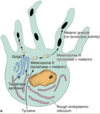
Starting with tyrosine, how is melanin made?

A

Tyrosine – (tyrosinase) –> DOPA —-> melanin

40
Q

What enzyme converts tyrosine to DOPA? This then becomes what?

A

Tyrosinase;

melanin

41
Q

The tyrosine-to-DOPA reaction occurs in what organelle?

A

The promelanosome

42
Q

What are melanosomes?

A

Precursors to melanin granules

43
Q

How do keratinocytes get melanin from melanocytes?

A

They envelop melanocyte projections

(cytocrine secretion by melanocytes)

44
Q

What is the purpose of melanin?

A

To protect nucleic acid from UV rays

45
Q

How do melanocytes appear on histology?

A

As very translucent cells with dark centers

46
Q

What are the APCs of the skin?

A

Langerhans cells

47
Q

The dermis comes from what embryonic derivative?

A

The dermatomyotome

(a portion of the somite, which is derived from para-axial mesoderm)

48
Q

What is the dermis like in regards to innervation, vascularization, and lymphatic supply?

A

Highly supplied in all 3

49
Q

Where can postganglionic sympathetic fibers be easily found clustering in the skin?

A

Surrounding hair follicles

(Note: I would assume, some can be found around arteries and sweat glands as well)

50
Q

Describe the papillary layer of the dermis.

A

Loose connective tissue:

collagen types I and III, some elastic fibers

51
Q

Hemidesmosomes anchor what cytoskeletal component to the basal lamina?

A

Intermediate filaments

52
Q

What two fiber types are most prevalent in the reticular layer of the dermis?

A

Type I collagen;

elastic fibers

53
Q

What are the purpose of the collagen fibers in the reticular layer of the dermis?

What are the purpose of the elastic fibers in the reticular layer of the dermis?

What are the purpose of the reticular fibers in the reticular layer of the dermis?

A

Strength;

flexibility and recoil;

there are very few (reticular layer = misnomer)

54
Q

Where are apocrine sweat glands found in the body?

A

The axillary and perineal regions

(also, the eyelids)

55
Q

Is the stratum lucidum often very thick when compared to the stratum corneum?

A

No;

it is a thin, grayish-white layer superficial to the stratum granulosum

56
Q

What type of secretion do both eccrine and apocrine sweat glands use?

A

Merocrine secretion

(confusingly, apocrine sweat glands do NOT use apocrine secretion)

57
Q

What type of sweat glands use merocrine secretion?

What type of sweat glands use apocrine secretion?

What type of sweat glands use holocrine secretion?

A

Eccrine, apocrine;

none;

none

(Note: apocrine sweat glands due not use apocrine secretion)

58
Q

What type of cell is found in all epidermal layers?

A

Keratinocytes

59
Q

What three cell types are predominantly found in the stratum basale?

A

Melanocytes, germinating keratinocytes, merkel cells

60
Q

Which cell type is predominantly found in the stratum spinosum (amongst keratinocytes)?

A

Langerhan’s cells

61
Q

Identify each layer of epidermis in this image.

A
62
Q

What is another name for the stratum basale of the epidermis?

A

The stratum germinativum

63
Q

What structure of stratum lucidum creates its lighter appearance?

A

The protein eleidin

64
Q

Which cell type(s) of the non-keratinized epidermis has hemidesmosomes and desmosomes?

Which cell type(s) of the non-keratinized epidermis has hemidesmosomes but no desmosomes?

Which cell type(s) of the non-keratinized epidermis has desmosomes but no hemidesmosomes?

A

Stratum basale keratinocytes and merkel cells;

melanocytes;

stratum spinosum keratinocytes

65
Q

Promelanosomes are derived from what organelle?

What reaction takes place in promelanosomes?

A

The Golgi apparatus;

tyrosine to DOPA via tyrosinase

66
Q

Where do melanocytes send melanin?

A

Into their cytoplasmic projections which keratinocytes engulf

(cytocrine secretion)

67
Q

What two structures are almost always associated with hair follicles?

A

A sebaceous gland, an arrector pili muscle

68
Q

Melanin provides pigment to what bodily tissues in particular?

A

The skin, hair, and irises

69
Q

Describe the life of a keratinocyte.

A

It beings as a basal cell in the stratum basale;

it moves into the stratum spinosum and begins making keratin;

it enters the stratum granulosum and filaggrin is released from keratohyalin granules as lipid-rich organelles are extruded to form a water-proofing barrier;

the cells move into the stratum corneum and become keratinized, they lose their desmosomes and flake off the surface

70
Q

What are lamellar bodies and from where are they secreted?

A

Secreted lipid-rich organelles;

type II pneumocytes, keratinocytes of the stratum granulosum

71
Q

What do keratinocytes of the stratum granulosum secrete to water-proof the skin?

A

Lamellar bodies (lipid-rich organelles)

72
Q

Is the epidermis:dermis ratio near-constant throughout the body surface?

A

No

73
Q

What are the two plexi supplying blood to the integument?

A

Larger vessels between the dermis and subcutaneous layers;

smaller vessels infiltrating the papillary layer of the dermis

74
Q

What is the importance of the shunts between the deep and superficial blood vessels of the skin?

A

This is the site of autonomic regulation of blood flow to the skin

(and associated temperature regulation)

75
Q

A pacinian corpusle has a capsule made up of what type of cell?

How does this corpuscle appear on histology?

A

Schwann cells;

cross-section of an onion

76
Q

What type of sensation do pacinian corpuscles detect?

A

Vibration, deep pressure

77
Q

How does a pacinian corpuscle detect any coarse pressure or vibration?

A

Distension of its capsule

78
Q

A Meissner corpusle has a capsule made up of what type of cell?

How does it appear on histology?

A

Schwann cells;

a stack of ethereal coins

80
Q

What type of sensation do Meissner corpuscles detect?

A

Light touch

81
Q

How does a Meissner corpuscle detect light touch?

A

Distension of its capsule

82
Q

Where are Meissner corpuscles found?

A

The papillary dermis

(perpendicular to the epidermis)

83
Q

What non-encapsulated sensory fibers are found extensively in the dermis?

A

Free nerve endings

84
Q

What are the only two sensory receptors easily seen on an H&E stain of the dermis?

A

Pacinian corpuscles;

Meissner’s corpuscles

85
Q

What type of sensory receptor senses torque and stretching of the dermis?

(e.g. during an ‘Indian burn’ on the arm)

A

Ruffini corpuscles

86
Q

How do Ruffini corpuscles sense twisting and torsion of the dermis?

A

Collagen fibers extend into the Ruffini corpuscle, connecting it to other structures of the dermis

87
Q

What do Ruffini corpuscles sense?

A

Twisting/torsion of the dermis

88
Q

What do Krause end bulbs sense?

A

Low frequency vibration

89
Q

What type of sensory receptor transmits dermal information as to extreme temperatures and/or damage?

What type of sensory receptor transmits dermal information of light touch?

What type of sensory receptor transmits dermal information of twisting and/or torsion?

What type of sensory receptor transmits dermal information of vibration and deep pressure?

What type of sensory receptor transmits dermal information of low frequency vibration?

What type of sensory receptor transmits dermal information of fine-detail touch and two-point discrimination?

A

Free nerve endings;

Meissner’s corpuscles;

Ruffini corpuscles;

Pacinian corpuscles;

Krause end bulbs;

Merkel cells

90
Q

What type of sensory receptor transmits dermal information as to extreme temperatures and/or damage?

A

Free nerve endings

91
Q

What type of sensory receptor transmits information of dermal light touch?

A

Meissner’s corpuscles

92
Q

What type of sensory receptor transmits dermal information of stretching and/or torsion?

A

Ruffini corpuscles

93
Q

What type of sensory receptor transmits dermal information of vibration and deep pressure?

A

Pacinian corpuscles

94
Q

What type of sensory receptor transmits dermal information of low frequency vibration?

A

Krause end bulbs

95
Q

What type of sensory receptor transmits dermal information of fine-detail touch and two-point discrimination?

A

Merkel cells

96
Q

For what type of dermal sensory information are Merkel cells responsible?

A

Finely detailed touch, two-point discrimination

97
Q

For what type of dermal sensory information are Pacinian corpuscles responsible?

A

Deep pressure, vibration

98
Q

For what type of dermal sensory information are Meissner corpuscles responsible?

A

Light touch

99
Q

For what type of dermal sensory information are free nerve endings responsible?

A

Noxious stimuli

100
Q

For what type of dermal sensory information are Ruffini corpuscles responsible?

A

Torsion and stretching

101
Q

For what type of dermal sensory information are Krause end bulbs responsible?

A

Low frequency vibrations

102
Q

What type of sensory cell lines the basal laminar surface to provide finely detailed tactile sensation and two-point discrimination to the skin?

A

Merkel cells

103
Q

When stimulated, calcium enters Merkel cells and what happens?

A

Neurosecretory granules are released

104
Q

What is the basic structure of a Ruffini corpuscle?

A

A fluid filled space through which collagen fibers pass

105
Q

Where are Ruffini corpuscles located?

A

The junction of the dermis and subcutaneous tissue

106
Q

Krause end bulbs are primarily found in skin covering what regions of the body?

A

Skin of the clitoris and penis

107
Q

Where are Merkel cells found?

A

Superficial to the basal lamina

108
Q

Where are Meissner’s corpuscles found?

A

The papillary dermis

109
Q

Where are Pacinian corpuscles found?

A

The dermis/subcutaneous tissues

110
Q

Where are Ruffini corpuscles found?

A

Between the dermis and subcutaneous tissues

111
Q

Where are free nerve endings found?

A

The dermis/subcutaneous tissues

112
Q

Where are Krause end bulbs found (area of the body)?

A

Skin of the clitoris and penis

113
Q

Where are Merkel cells found?

Where are Meissner’s corpuscles found?

Where are Pacinian corpuscles found?

Where are Ruffini corpuscles found?

Where are free nerve endings found?

Where are Krause end bulbs found (area of the body)?

A

Superficial to the basal lamina;

the papillary dermis;

the dermis/subcutaneous tissues;

between the dermis and subcutaneous tissues;

the dermis/subcutaneous tissues;

skin of the clitoris and penis

114
Q

Where is(are) the hair follicle(s) in this slide?

A
115
Q

Where is(are) the sebaceous gland(s) in this slide?

A
116
Q

Where is(are) the arrector pili muscle(s) in this slide?

A
117
Q

What is the purpose of the arrector pili?

A

To straighten the hair (and slow down air movement –> resulting in warming);

to help empty sebaceous glands

118
Q

What connective tissue structure rises up into and supplies each hair follicle?

A

A dermal papilla

119
Q

What is the inner part of a hair follicle called?

What is the outer part of a hair follicle called?

A

Medulla (M);

cortex (CO)

  • (Note: obliquely cut hair follicle on left;*
  • cross-section of a hair follicle on right)*
120
Q

What thin layer of keratin follows up the outside of the hair follicle cortex?

A

A cuticle (CU)

  • (Note: obliquely cut hair follicle on left;*
  • cross-section of a hair follicle on right)*
121
Q

Which part of the hair follicle contains the hardest keratin?

Which part of the hair follicle contains the softest keratin?

(Of the medulla, cortex, and cuticle)

A

Cuticle (CU);

Medulla (M)

  • (Note: obliquely cut hair follicle on left;*
  • cross-section of a hair follicle on right)*
122
Q

The epidermal matrix at the root of each hair follicle is producing keratin and extending upwards cellularly to create what layer around the hair follicle that contains keratinocytes and melanocytes?

This portion of the root sheath ends mid-way up the root shaft.

A

The internal root sheath (IRS)

  • (Note: obliquely cut hair follicle on left;*
  • cross-section of a hair follicle on right)*
123
Q

What layer of the hair follicle root sheath extends upwards and is continous with the other cells of the epidermis at the integument’s surface?

A

The external root sheath (ERS)

  • (Note: obliquely cut hair follicle on left;*
  • cross-section of a hair follicle on right)*
124
Q

What separates the epidermally derived root of the hair follicle matrix from the surrounding dermis?

A

The glassy membrane (G)

  • (Note: obliquely cut hair follicle on left;*
  • cross-section of a hair follicle on right)*
125
Q

Name the portions of the hair follicle root from innermost to outermost.

A

Medulla (M);

cortex (CO);

cuticle (CU);

internal root sheath (IRS);

external root sheath (ERS);

glassy membrane (G)

  • (Note: obliquely cut hair follicle on left;*
  • cross-section of a hair follicle on right)*
126
Q

What protein is the main structural component of hair and nails?

A

Keratin

127
Q

Sebaceous glands are outgrowths of what portion of the hair follicle?

A

The external root sheath (ERS)

128
Q

Where do arrector pili originate?

A

The subcutaneous layer

129
Q

What pattern does the hair cuticle have?

A

A shingle-like pattern

130
Q

What are the portions of nail that we can see when we look down at our hands?

A

The nail plate (main portion);

the cuticle (eponychium)

131
Q

The nail cuticle (eponychium) is an extension of what?

A

The nail matrix (DNM)

132
Q

The portion of the nail plate that is still inside the finger (arising from the nail matrix) is called what?

A

The nail root (NR)

133
Q

The nail root sits deep to the cuticle (eponychium) and superficial to what surface?

A

The nail bed

134
Q

What type of secretion do sebaceous glands use?

What type of secretion do eccrine sweat glands use?

What type of secretion do apocrine sweat glands use?

A

Holocrine;

merocrine;

merocrine

135
Q

What type of secretion do sebaceous glands use?

A

Holocrine

136
Q

What do sebaceous glands secrete?

A

Sebum

137
Q

What three types of cells are found in an sweat glands?

A

Clear cells (sweat), dark cells (proteinous), myoepithelial cells (contractile)

138
Q

Describe the epithelial arrangement of sweat glands.

A

Stratified cuboidal

139
Q

Eccrine sweat glands are innervated by ________ nerve endings.

Apocrine sweat glands are innervated by ________ nerve endings.

A

Cholinergic;

adrenergic

140
Q

Describe the size of apocrine sweat gland lumens.

A
141
Q

What are the first and second stages of integument healing of an open wound?

A

Clot formation;

leukocyte cleaning of debris

142
Q

What are the third and fourth stages of integument healing of an open wound?

A

Angiogenesis and granulation tissue formation;

epithelium regenerates, connective tissue fibrosis occurs

143
Q

Melanin is mainly derived from what amino acid?

A

Tyrosine

144
Q

Describe, in detail, the spatial protein arrangement that allows basal keratinocytes and basement membrane layers to adhere to one another.

Use the following terms:

  • Type VII collagen*
  • BPAG1, BPAG2*
  • keratin 5, keratin 14*
  • β4-integrin*
  • Type IV collagen*
  • anchoring filaments*
A