MSK - Histology - Cartilage; Bone Flashcards
What are the three main types of cartilage?
Hyaline;
elastic;
fibrous
What type of fibers are most prevalent in hyaline cartilage?
What type of fibers are most prevalent in elastic cartilage?
What type of fibers are most prevalent in fibrocartilage?
Type II collagen;
elastic fibers and type II collagen;
types I and II collagen
What type of fibers are most prevalent in hyaline cartilage?
Type II collagen
What type of fibers are most prevalent in elastic cartilage?
Elastic fibers; type II collagen
What type(s) of fibers are most prevalent in fibrocartilage?
Types I and II collagen
What are some examples of hyaline cartilage in the body?
Costal cartilage;
articular cartilage;
tracheal rings
What are some examples of elastic cartilage in the body?
Ears,
epiglottis,
part of the larynx,
auditory tubes
What are some examples of fibrocartilage in the body?
Intervertebral discs;
pubic symphysis;
menisci of the knee joint
What type of cell is most prevalent in hyaline cartilage?
What type of cell is most prevalent in elastic cartilage?
What type of cell is most prevalent in fibrocartilage?
Chondrocytes;
chondrocytes;
fibroblasts
What percentage of cartilage is typically made up by ground substance?
90 - 95%
Chondrocytes receive their nutrients via:
Diffusion
(cartilage is an avascular tissue)
The functional component of cartilage is:
the ECM
What type of cartilage is in the ear?
What type of cartilage are the menisci of the knee?
What type of cartilage is the epiglottis?
What type of cartilage are the intervertebral discs?
Elastic;
fibrocartilage;
elastic;
fibrocartilage
What two types of cartilage are found in the knee?
Articular (lining the joint);
fibrocartilage (the menisci)
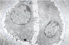
Chondrocytes sit in hollow spaces within the ground substance called:
Lacunae
Is ground substance eosinophilic or basophilic?
Basophilic (blue or dark purple)
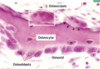
What are the three layers of ECM in the cartilage surrounding chondroblasts?
(How strongly does each stain?)
What type of fiber predominates here?
The pericellular matrix (visible with special staining);
the territorial matrix (lightly stained);
the interterritorial (interstitial) matrix (heavily stained);
type II collagen
What two stains are used for elastic fibers?
Verhoeff’s;
orcein
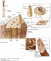
What are the two types of cartilaginous growth?
Appositional (from the perichondrium);
interstitial (within the tissue by existing chondrocytes)
How many chondrocytes are typically found in each lacunae?
1 - 2
If there are two or more chondrocytes in one lacunae, these chondrocytes are termed:
Isogenous groups
What are the three types of cartilage?
Hyaline,
elastic,
fibrocartilage
Which types of cartilage have a surrounding perichondial layer?
Hyaline (excepting articular cartilage);
elastic
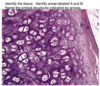
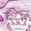
Describe the layers of hyaline cartilage from superficial to deep.
Fibrous layer (perichondrium)
Chondrogenic layer (perichondrium)
Inner chondrogenic layer (chondroblast differentiation)
Mature cartilage

The outer (fibrous) layer of cartilaginous perichondrium found in hyaline cartilage is what type of tissue?
Dense irregular connective tissue (type I collagen)

What is the major proteoglycan of hyaline cartilage?
What is the major glycoprotein of hyaline cartilage?
Aggrecan;
chondronectin
What substance forms the fetal skeleton?
How does it form bone?
Hyaline cartilage;
endochondrial ossification
Which type of cartilage has no perichondrial layer?
Fibrocartilage
What type of cartilage is shown here?

Elastic cartilage
What is shown in this transmission electron micrograph?

Two chondrocytes within in a lacunae
(blue line = lacunae;
RER very abundant)
What is the predominant type of collagen is found in fibrocartilage?
What is the predominant type of collagen is found in hyaline cartilage?
What is the predominant type of collagen is found in elastic cartilage?
Type I (some type II present);
type II;
type II (elastic fibers also present)
What type of cartilage does NOT calcify with age?
Elastic cartilage
What type of tissue is this?

Elastic cartilage
What type of tissue is this?

Fibrocartilage
(note the lack of distinct layers and presence of interspersed type I collagen with ground substance)
What is unique about the orientation of the chondrocytes in fibrocartilage?
They usually occur in rows

(see attached image)
Fibrocartilage is resistant to what two types of force?
Compressive;
shear
What type of tissue is this?

Fibrocartilage
Damaged perichondrium will likely have what response?
Fibroblast proliferation / scar formation
What type of tissue is this?

Fibrocartilage
What type of cartilage has no perichondrium?
Fibrocartilage


Fibrocartilage

Hyaline cartilage;
isogenous groups

A - fibrous perichondrium
B - chondrogenic perichondrium
Arrows - isogenous groups
Name the three types of cartilage and the predominant connective tissue fibers found in each.
Hyaline cartilage - collagen type II
Elastic cartilage - collagen type II and elastic fibers
Fibrocartilage - collagen type I and II
Describe the general makeup of hyaline cartilage ground substance.
Glycosaminoglycans (GAGs) attached to proteoglycan core proteins that are linked together by long chains of hyaluronic acid

Is cartilage basophilic or eosinophilic?
Why?
High prevalence of sulfate and carboxyl groups in glycosaminoglycans (GAGs)
What is the precursor cell for chondroblasts?
Mesenchymal cells
Which ECM is closest to chondrocytes and enriched in glycosaminoglycans (GAGs)?
Territorial matrix
What part of cartilage is basophilic?
What part of cartilage is eosinophilic?
Glycosaminoglycans (sulfate and carboxy groups, in particular);
collagen fibers
Which is more basophilic, the interterritorial (interstitial) matrix or the territorial (pericellular) matrix?
Why?
Territorial
(higher ratio of glycosaminoglycans to collagen fibers)
What type of strength does hyaline cartilage have due to its meshwork of type II collagen?
Tensile strength
Why is it important that ground substance is so good at storing water?
It enables diffusion of nutrients through hydrated matrix to chondrocytes
What type of cartilaginous growth involves new chondroblasts differentiating from the inner chondrogenic perichondrial layer?
Appositional growth
(as opposed to interstitial)
What type of cartilaginous growth involves mature chondrocyte replication within lacunae?
Interstitial growth
(as opposed to appositional)
What is a damaging effect of cartilage calcification?
Decreased nutrient diffusion
(and resulting chondrocyte apoptosis)
What natural process can limit diffusion of nutrients through cartilage and result in chondrocyte apoptosis?
Calcification
What type of cartilage is the cartilage of the nose?
Describe the pieces of cartilage present in the nose.
Hyaline

What type of tissue is the precursor of bone at the endochondral plates?
Hyaline cartilage
Does articular cartilage have a perichondrium?
No;
although other hyaline cartilage does
Articular cartilage replicates via what type(s) of growth?
Interstitial only
(no perichondrium = no appositional growth)
Why does articular cartilage not rapidly regenerate in cases of osteoarthritis?
Articular cartilage has no perichondrium and relies on the very limited interstitial growth to regenerate
Verhoeff stain is especially useful in visualising what type of fiber?
Elastic
Orcein stain is especially useful in visualising what type of fiber?
Elastic
What is the main feature of elastic cartilage as compared to the other types?
It’s flexibility and ability to return to its original shape
What type of cartilage is the most eosinophilic?
Why?
Fibrocartilage;
higher ratio of collagen fibers to ground substance
What is the strongest of the three types of cartilage?
Fibrocartilage
What type of cartilage is often found on portions of tendons or ligaments that insert into cartilage?
Fibrocartilage
What are the two macroscopic types of mature bone?
Compact (dense);
cancellous (spongy; trabecular)
What lines the outside of bones?
What type of tissue is it?
Periosteum;
dense irregular connective tissue
What are the three main regions seen in a long bone?
Epiphysis (ends)
Metaphysis
Diaphysis (shaft)
Metaphysis
Epiphysis (ends)

What are the two microscopic types of mature bone?
Mature (Lamellar; compact);
primary (woven)
What are the two macroscopic types of mature bone?
What are the two microscopic types of bone?
Do the two macroscopic types correspond to the two microscopic types?
Compact (dense), cancellous (spongy, trabecular);
mature (compact, lamellar), primary (woven);
Sort of.
Both type of macroscopic structure refer to mature bone (both compact or cancellous would be considered lamellar, although compact in the microscopic sense tends to correspond with compact in the macroscopic sense)
What is woven bone?
A microscopic type of immature bone consisting of irregularly aligned collagen fibers
True/False.
Spongy bone can also be lamellar as a cross-section of one of the trabecula will show lamellar arrangement.
True.

What is the functional unit of compact bone?
The osteon (Haversian system)

What are the microscopic layers of a long bone from superficial to deep?
Periosteum (DICT);
external circumferential lamellae;
osteons (Haversian systems) + interstitial lamellae);
internal circumferential lamellae;
trabeculae

What are the three types of lamellae encasing osteons in long bone?
External circumferential lamellae;
interstitial lamellae;
internal circumferential lamellae;

What lumen runs down Haversian systems (osteons)?
What tube connects these lumens?
Haversian (central) canals;
Volkmann’s (perforating) canals

What sits inside each bony lacunae?
What sits inside the cannaliculi extending out from these lacunae?
An osteocyte;
cytoplasmic osteocyte projections

Volkmann’s (peripheral) canals connect what bony structures?
Haversian (central) canals
What runs through Haversian and Volkmann canals?
Vessels (bony VANs)
What are the inner and outer layer of periosteum, respectively?
Fibrous (Sharpey’s layer);
osteoprogenitor (osteogenic)
True/False.
The adjacent concentric circles (lamellae) of each osteon are oriented so that their collagen fibers are parallel to one another for strength?
False.
they are perpendicular to one another
(see upper right portion of image)

What thin layer lines the internal surface of trabecular (spongy) bone?
It is similar to what other bony structure?
Endosteum;
the progenitor layer of the periosteum
True/False.
Bony cannaliculi open from trabecular bone into the marrow space.
True.

In what key ways is bone different from cartilage?
Mineralized ground substance:
mainly eosinophilic (due to calcification) instead of basophilic;
it is vascular;
osteocytes instead of chondrocytes
In what ways are bone and cartilage very similar?
They consist of cells sitting in lacunae surrounded by ECM;
the ground substance / matrix is the functional unit
Osteoprogenitor cells are derived from what type of cell?
Mesenchymal cells
What type of cell is found in the endosteum?
Osteoprogenitor cells (osteoblast precursors)
Osteoprogenitor cells are specialized:
They become:
Fibroblasts;
osteoblasts
What type of cell is found in the inner layer of the periosteum?
Osteoprogenitor cells (osteoblast precursors)
What are the four main types of cell found in bone?
Osteoprogenitor cells;
osteoblasts;
osteocytes;
osteoclasts
Describe the differentiation of a mature bone cell.
Mesenchymal cell –>
osteoprogenitor cell (specialized fibroblast) –>
osteoblast –>
osteocyte
What do osteoblasts secrete (4)?
Collagen (type I),
matrix proteins,
matrix vesicles,
osteoid
What is found in wide abundance in the matrix vesicles secreted by osteoblasts?
Alkaline phosphatase (for mineralization purposes)
True/False.
Osteocytes are highly branched.
True.
Where are osteocyte processes found?
Within bony cannaliculi

Osteoclasts are derived from what type of tissue?
Hematopoeitic stem cells
Osteoclasts are basically modified:
Macrophages
What do osteoclasts do?
Where are they found?
Resorb bone.
Howship’s lacunae (holes in the endosteum)

How do osteoclasts appear on light microscopy?
Large, eosinophilic, multinucleated

What is the thin grayish layer seen covering the osteoblastic surface?
Osteoid

How does a slide of a bony structure have to be prepared in order to appear as it does in this micrograph?

It is decalcified
Bony cannaliculi allow for communication between adjacent:
Osteocytes
How does a slide of a bony structure have to be prepared in order to appear as it does in this micrograph?

It must be ground
What type of cells are found in the fibrous (outer; Sharpey’s) layer of periosteum?
What type of cells are found in the cellular (inner; osteogenic) layer of periosteum?
Fibroblasts;
osteoprogenitor (periosteal) cells
What percentage of bone matrix is inorganic?
Of what does it consist?
65%;
mostly calcium phosphate deposits
+
hydroxyapatite - Ca5(PO4)<span>3</span>OH
What percentage of bone matrix is organic?
Of what does it consist?
35%;
type I collagen (90%),
proteoglycans (chondroitin sulfate, keratan sulfate, hyaluronic acid),
other proteins
65% of bone matrix is:
35% of bone matrix is:
Inorganic (hydroxyapatite);
organic (collagen + proteoglycans)
Once calcium and phosphate levels have risen to a sufficient level, what protein do osteoblasts secrete to further increase calcium levels?
Osteocalcin

What is the result of osteocalcin secretion by osteoblasts?
Calcium levels increase,
auto-inducing matrix vesicle release

After osteoblast osteocalcin release, calcium levels rise and matrix vesicles are secreted by the osteoblasts.
What is the result?
This is due to increased levels/activity of what enzyme?
Phosphate levels rise;
alkaline phosphatase

What three substances released by osteoblasts are major promoters of bone mineralization?
True/False.
The increasing calcium and phosphate further increase osteoblastic secretions in a positive feedback loop.
Osteocalcin (increases Ca2+),
collagen (type I),
alkaline phosphate within matrix vesicles (increases PO43-);
True.

How do the following two osteoblastic secretions increase bone mineralization?
Osteocalcin
Alkaline phosphate within matrix vesicles
What else is secreted by osteoblasts during this process?
Osteocalcin - increases Ca2+,
alkaline phosphate within matrix vesicles - increases PO43-;
type I collagen

What osteoblastic secretion promotes bone mineralization by increasing phosphate levels?
What osteoblastic secretion promotes bone mineralization by increasing calcium levels?
Alkaline phosphatase (within matrix vesicles);
osteocalcin (protein)
What are the two types of embryonic osteogenesis?
Intramembranous ossification (flat bones of the skull);
endochondral ossification (long and short bones)
Which type of embryonic osteogenesis requires a perichondrium to initiate?
Endochondral ossification
Intramembranous ossification does not require what structure to be present that endochondral ossification does require?
A perichondrium
Without the progenitor cells present in the perichondrium, what cell type gives rise to bone in intramembranous ossification?
Mesenchymal cells
Which type of embryonic osteogenesis does not require osteoprogenitor cells?
What happens instead?
Intramembranous ossification;
mesenchymal cells differentiate directly into osteoblasts (skipping the osteoprogenitor step)

What type of embryonic osteogenesis is depicted here?
For what type of bone?

Intramembranous ossification;
flat bones of the skull
(note: the spicules of bone and absence of perichondrium)
Mesenchyme is found on which side of the immature bone in intramembranous ossification?

Both sides
(giving rise to osteoblasts)
Which part of the micrograph shows the developing bone?
Is it mature yet?

The red in the middle;
no, it is being remodeled by osteoclasts
In endochondral ossification, only a _________ ___________ center exists in the diaphysis until after birth when __________ ___________ centers form in the _________.
Primary ossification (center);
secondary ossification (center);
epiphyses

When do secondary ossification centers form? Where?
This is part of what type of osteogenesis?
After birth, the epiphyses;
endochondral

What substance is used as a template for endochondral ossification?
Hyaline cartilage

Describe the general process of endochondral ossification.
Hyaline cartilage is used as a template;
perichondrial progenitor cells (which originated from mesenchyme) hypertrophy and calcify in the diaphysis;
between the sides of the developing periosteal bone collar (also from mesenchyme), a primary ossification center forms;
this bony structure now extends outwards towards the epiphyses, where secondary ossification sites form and complete the process

When do blood vessels first pierce the bone during development?
After the primary ossification center forms
(before birth)

What portion of long and short bones is not covered by periosteum?
That covered by articular cartilage
What types of growth does bone exhibit?
Only appositional
(interstitial osteocyte division not possible due to hardness of the lacunae)
What layer of tissue allows for bony appositional growth?
The periosteum
Endochondral ossification involves what types of cartilaginous growth?
Interstitial,
appositional
The bone collar is the developing ___________ found in _____________ ossification.
Periosteum;
endochondral
True/False.
Chondrocytes leave behind calcified tissue as they move from deep to superficial during endochondral ossification.
False.
Chondrocytes calcify and die as they move closer to the deepest part of the developing bone.
How does calcified cartilage get turned into bony tissue during endochondral ossification?
Osteoclasts break down the calcified material,
osteoblasts use these materials to lay down osteoid,
osteoclasts remodel the osteoid and osteoblasts

From closest to the epiphyseal plate to closest to the developing interior bony layer, how many stages of hyaline cartilage are found in endochondral ossification?
What are they?
4;
reserve,
proliferation,
hypertrophy,
calcification
(and then osteoclastic resorption and ossification after that)

What form of hyaline cartilage is found at the epiphyseal plate during endochondral ossification?
Reserve cartilage

Reserve cartilage (at the epiphyseal plate) does what three things as a bone lengthens due to endochondral ossification?
Proliferates, hypertrophies, calcifies

As the bone in this image grows longer (towards the epiphyseal plate at the top of the attached image), there are a few stages of chondrocyte change that occur.
Name them in accordance with the red, yellow, green, and white circles.

Red - reserve chondrocytes
Yellow - proliferating chondrocytes
green - hypertrophying chondrocytes
white - calcified, dead chondrocytes
What types of cell are busiest at the calcified cartilage layer in the attached image?

Osteoclasts (breaking down the calcified material);
osteoblasts (creating osteoid);
osteoclasts (remodeling the immature bone)
What is the zone beneath the zone of calcified cartilage in endochondral bone formation?
The zone of resoprtion
Influence of what puberty-associated substance causes rapid bone growth?
It is causing increased proliferation of what types of cell?
Growth hormone;
chondrocytes, osteoblasts
(although osteoclasts are performing at their original speed, so growth increases; this is your ‘growth spurt’)
Growth hormone causes what effect on endochondral ossification during puberty?
Increased chondrocyte proliferation and osteoblast function
What hormone increases chondrocyte proliferation and osteoblast function during puberty?
What hormone closes the epiphyseal plate and ends this process?
Growth hormone;
estrogen
(converted from testosterone via aromatase)
Does cartilage directly transform into bone in endochondral ossification?
No, woven bone is placed on cartilage spicules
Increases in long bone length are caused by ___________ growth of __________.
Increases in bone width are caused by ___________ growth of __________.
interstitial, chondrocytes;
appositional, periosteal (osteoprogenitor) cells
What is the epiphyseal line?
The closed epiphyseal plate
What is the main hormone regulating blood calcium levels?
What other hormones are involved?
PTH;
calcitonin, vitamin D
What is parathyroid hormone’s method of action?
Stimulates osteoblasts to stimulate osteoclasts
(indirect osteoclast activation)
What is calcitonin’s method of action?
Stimulates osteoblasts
Which of the following stimulates osteoblasts, PTH or calcitonin?
BOTH;
calcitonin stimulates osteoblastic activity,
PTH stimulates osteoblasts to activate osteoclasts
Which of the following directly stimulates osteoclasts,
PTH or calcitonin?
NEITHER
PTH stimulates osteoblasts to activate osteoclasts
(indirect action)
What receptor is found on osteoclastic membranes that binds osteoblastic ligands during osteoclastic activation?
RANK

Osteoblasts increase osteoclast differentiation due to what hormone?
Osteoblasts increase osteoclast differentiation due to what environmental condition?
PTH;
increased pH
Vitamin D causes an increase of osteoblastic secretion of what substance?
Osteocalcin
(increasing calcium, phosphate, and bone growth)
What vitamin is able to increase osteocalcin release?
What cell releases osteocalcin?
Vitamin D;
osteoblasts
Which of the following differentiations of an osteoclast require signaling from an osteoblast?
Monocyte –> macrophage –> osteoclast precursor –> osteoclast
What signaling?
Macrophage (1) –> Osteoclast precursor (2) –> osteoclast;
M-CSF signaling (1), RANK signaling (2)

What ligand do (__________) secrete to cause macrophages to differentiate into osteoclast precursors?
What ligand do (__________) express to cause osteoclast precursors to differentiate into osteoclasts?
(Osteoblasts)
M-CSF;
(Osteoblasts)
RANK;

What are the stages of fracture repair?
Hematoma –>
callus formation (periosteal reconnection; hyaline cartilage formation) –>
spongy bone formation –>
healed fracture
Fracture repair is similar to what type of osteogenesis?
Endochondral osteogenesis
What is indicated by the small arrows and large arrowheads, respectively?

Small - osteoclasts
Large - osteoblasts
The black arrowheads are indicating:
The white arrowheads are indicating:

Osteoblasts;
osteocytes (within lacunae)
Identify A - E.

A - Osteogenic periosteum
B - Fibrogenic periosteum
C - Immature bone
D - Calcified cartilage
E - Mesenchyme
What is indicated in this image of developing bone?
This is what type of bone development?

A primary ossification center;
endochondral ossification
Identify A - C and the structure indicated by the arrows in this image of bone development.

A - Fibrous periosteum
B - Bone collar
C - Primary center of ossification.
Arrows - Osteogenic periosteum with osteoblasts.

Black arrow - Osteoblasts
Blue arrow - Osteocytes in lacunae
Blue arrowhead - Osteoclast
A - Mesenchyme
B - Immature bone
Identify the tissue and indicated sections.

Compact, dense or cortical bone.
Green - Outer circumferential lamellae
Yellow - Inner circumferential lamellae
Blue bar - contains osteons and interstitial lamellae.
Identify the tissue and labeled structures.

Compact bone
A - Haversian canal
B - Interstitial lamellae
Arrows - Osteon or Haversian system
What is indicated by the arrows and ‘A’s?

Arrows - Mature spongy bone
A - Adipose tissue in yellow bone marrow
Identify the labeled structures.

A - Red bone marrow
B - Inner circumferential lamellae
Black arrows - Haversian canals
Blue arrows - Osteon
Blue arrowheads - Volkmann’s canals

Black arrows - Osteoblasts
Black arrowheads - Osteoclasts
A - Mesenchyme
* - Osteocytes

Black arrows - Osteoblasts
* - Osteoid
Blue arrows - Osteocytes within lacunae
What developmental process is occurring?
Identify the cells indicated by small black arrows.
Identify the cells indicated by small black arrowheads.
Identify the tissue labeled ‘A’.

Intramembraneous bone development
Black arrows - Osteoblasts
Black arrowheads - Osteoclasts
A - Mesenchyme

A - Mature bone
B - Immature bone

A - Fibrous periosteum
B - Osteogenic periosteum
C - Bone collar
D - Primary marrow cavity

Arrows - Periosteal bud
A - Fibrous periosteum
B - Osteogenic periosteum
C - Bone collar

A - Haversian canal
* - Lacunae
Arrows - Canalliculi

* - Calcified cartilage
Arrows - Immatue bone
Bar - Zone of hypertrophy and maturation
What is the general name for organic bone matrix?
Osteoid
(largely type I collagen)
What are Sharpey’s fibers?
Collagen fibers that extend from the periosteum into the bone ECM that provide a strong anchoring for the periosteum, ligaments, and tendons to bone
The epiphyseal plate is at the top of the attached image.
Name the layers of hyaline cartilage from top to bottom (each layer is a section between two orange lines).

Reserve (quiescent)
Proliferative
Hypertrophy
Calcification
(and resoprtion beneath that)
What is the term for a bone fracture caused by disease that led to weakness of the bony structure?
A pathologic fracture
What is often the first sign or symptom of osteoporosis?
Pathologic fracture
What is the cellular cause of osteoporosis?
Osteoclastic activity > osteoblastic activity
What is the most common disorder of the joints?
Osteoarthritis
(degenerative joint disease)
What term refers to the rubbing off of articular cartilage to expose subchondral bone?
Eburnation
What are joint mice?
Small fractures dislodge tiny pieces of bone or cartilage into the joint
What is the most common type of bone tumor?
Metastases
(much more common than primary bone tumors)
What is the most common location for osteoma formation?
Head and neck
What is the most common primary malignant bone tumor?
Where is it most common?
In what age group?
Primary osteosarcoma;
the metaphyseal plates of the knee joint, hip joint, or humerus;
< 20 years of age (75%)
A pathologic fracture leads to the finding of the attached X-ray of a lytic lesion and a raised periosteum.
What is the blue arrow indicating?
What disease is this?

Codman’s triangle (shadow under raised periosteum)
primary osteosarcoma

Describe Codman’s triangle.
What causes this appearance?
A raised triangular shadow indicative of primary osteosarcoma;
expanding tissue eats through the cortex and pushes the periosteum away from the bone

Primary osteosarcomas typically spread how?
How good is long-term survival with aggressive treatment?(chemo and limb salvage)
Hematogenously (as most sarcomas);
60 - 70%
What do primary osteosarcomas secrete that causes them to expand and push the periosteum away from the bone?
(resulting in Codman’s triangle)
Osteoid (which is organic and not as calcified - doesn’t show up well on x-ray) or inorganic bone matrix
(these are osteogenic tumors)
The tumor shown in this radiograph has a cartilage cap connected to the bone by a bony connection.
What is it?

Osteochondroma
Osteochondromas are benign or malignant?
They only occur in what types of bone?
Do they ever proceed to chondrosarcomas?
Benign;
those of endochondral origin (long and short);
very, very rarely
The RANK-ligand is found in ___________, and the RANK-receptor is found in __________.
The mCSF-ligand is secreted from ___________, and the mCSF-receptor is found in __________.
Osteoblasts, osteoblast precursors;
osteoblasts, macrophages

What disease, in particular, is characterized by very low levels of vitamin D in adults?
Osteomalacia