Cardio - Physiology - Excitation & Contraction; EKG; Cardiac Cycle Flashcards

1
Q

In a normal heart, what percentage of cardiac output comes from atrial contraction?

A

10%

2
Q

The AV node has less of what type of channel and what type of cell junction than surrounding ventricular myocytes?

A

Sodium channels (those that are present are mostly inactive);

gap junctions

3
Q

True/False.

Gap junctions are opened and closed based on intracellular conditions?

A

True

(they are regulated by intracellular pH and Ca2+ levels)

4
Q

What intracellular conditions cause closure of cardiac connexons (gap junctions)?

A

High calcium levels;

low pH

5
Q

What intracellular conditions cause opening of cardiac connexons (gap junctions)?

A

Low calcium;

normal pH

6
Q

Describe the structure of a gap junction.

A

6 connexins coming together to make one connexon

7
Q

In what state will cardiac gap junctions (connexons) be if the cell is at normal physiological conditions (e.g. low calcium levels, pH of 7.2)?

A

Open

8
Q

In what state will cardiac gap junctions (connexons) be if the cell is undergoing ischemic conditions (e.g. high calcium levels, pH of ~6.4)?

A

Closed

9
Q

What structure is responsible for the electrocardiogram P-R interval?

A

The AV node

10
Q

The AV node is responsible for what part of a normal EKG reading?

A

The P-R interval

11
Q

What type of cholinergic receptor do the vagal nerves activate on the heart?

What effect does it have on ion channels?

A

M2 (G-protein);

activating K+ leak channels (promoting K+ efflux),

inhibiting adenylyl cyclase (and thus inhibiting Ca2+ influx)

12
Q

What type of adrenergic receptor do sympathetic nerves activate on the heart?

What effect does it have on ion channels?

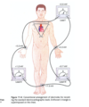
A

β1 (G-protein);

activating adenylyl cyclase (promoting Ca2+ influx)

13
Q

Vagal nerves activate M2 receptors on the heart. These receptors activate what type of G-protein subunits?

A

Gi

14
Q

Sympathetic nerves activate β1 receptors on the heart. These receptors activate what type of G-protein subunits?

A

Gs

15
Q

The channels in the AV node are mostly:

A

L-type calcium channels

16
Q

What is a normal time range for the PR interval?

What is the normal time value for the QRS complex?

A

0.12 - 0.20 sec;

< 0.12 sec

17
Q

What prevents atrial depolarization from bypassing the AV node and directly leading to depolarization of the ventricles?

A

A fibrous ‘skeletal’ ring separating the atria from the ventricles

18
Q

Why is it important that gap junctions close during ischemic conditions?

A

To try to isolate the ischemic tissues from the healthy

19
Q

Where does repolarization occur first, the epicardium or endocardium?

A

Epicardium

20
Q

Describe the differences in action potential between the following:

SA node pacemaker cells

Atrial myocytes

AV node pacemaker cells

Purkinje fibers

Ventricular myocytes (epicardium and endocardium)

A
21
Q

What type of cell are SA node and AV node pacemaker cells?

A

Modified cardiomyocytes

22
Q

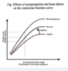
Arrange the following types of cell from longest to shortest action potential:

Atrial

Purkinje

Ventricular

A

Purkinje > Ventricular >>> Atrial

23
Q

Which has more of a phase 1 ‘notch,’ the epicardium or endocardium?

A

Epicardium

24
Q

Arrange the following types of cell from most to least unstable disatolic potential:

SA node pacemakers

AV node pacemakers

Purkinje fibers

A

SA node pacemakers >

AV node pacemakers >

Purkinje fibers

25
Q

Describe when each of the following types of ionic channel in the cardiac myocytes is active during an action potential.

A
26
Q

Name the ionic channel responsible for each of the following segments of the cardiac myocyte action potential.

A
27
Q

What is the specific name of the ionic channel responsible for phase 0 of the cardiac myocyte action potential shown below?

A

Na channel

28
Q

What is the specific name of the ionic channel responsible for phase 1 of the cardiac myocyte action potential shown below?

A

Transient outward channel

(K+)

29
Q

What is the specific name of the ionic channel responsible for phase 2 of the cardiac myocyte action potential shown below?

A

L-type Ca2+ channel

30
Q

What are the specific names of the ionic channel responsible for phases 3, early 4, and late 4 of the cardiac myocyte action potential shown below?

A

Delayed inward rectifier (K+);

Inward rectifier (K+)

31
Q

Conduction velocity in the cardiac myocytes is proportional to the flow of which ion?

This will affect the steepness of which phase?

A

Na+;

phase 0

32
Q

What is the effective refractory period (ERP) in terms of cardiac myocyte action potentials?

A

The inexcitable period until ~50% of Na channels are able to reopen

33
Q

If a cardiac myocyte is stimulated during the effective refractory period (ERP), what occurs?

If a cardiac myocyte is stimulated during the relative refractory period (RRP), what occurs?

If a cardiac myocyte is stimulated after the relative refractory period (RRP), what occurs?

A

Nothing;

a blunt, shortened action potential;

a normal action potential

34
Q

What parts of the heart are influenced by vagal stimulation?

A

SA node (right vagus n.)

AV node + Purkinje fibers (left vagus n.)

(Note: ventricular myocytes are not directly innervated by the parasympathetic system)

35
Q

The right vagus nerve innervates which particular portion(s) of the heart?

A

The SA node

36
Q

The left vagus nerve innervates which particular portion(s) of the heart?

A

The AV node and Purkinje fibers

37
Q

Which vagus nerve (right or left), or neither or both, innervates the ventricular myocytes?

A

Neither

38
Q

What parts of the heart are influenced by sympathetic stimulation?

A

Virtually all portions

(SA node, AV node, Purkinje fibers, ventricular myocytes)

39
Q

What cardiac GPCR is activated by norepinephrine?

What cardiac GPCR is activated by acetylcholine?

A

β1

M2

40
Q

SA and AV node pacemaker cells have action potentials that are missing which phases normally found in other contractile myocytes?

A

Phases 1 and 2

41
Q

AV nodal conduction is centered around flow of what ion?

A

Ca2+

42
Q

What effect will sympathetic stimulation have on the PR interval?

A

Shorter PR

(increased ICa)

43
Q

What effect will vagal stimulation have on the PR interval?

A

Longer PR

(increased IK; decreased ICa)

44
Q

What effect does adenylyl cyclase activation have on cardiac calcium channels?

A

They are phosphorylated and activated

(allowing calcium influx)

45
Q

Are cardiac calcium channels similar to sodium channels?

How are they different in regards to time?

How are they different in regards to refractory periods?

A

Yes;

they are open longer;

the refractory period lasts longer than repolarization

46
Q

Increased potassium conductance (IK) will have what effect on effective refractory periods in cardiac tissue?

What might cause this increase in IK?

A

The ERP increases;

vagal simulation

47
Q

Why is it so important that the AV node be regulated by calcium conductance?

A

It slows down the current –> this allows time for ventricular filling

48
Q

Which parts of the heart have automaticity?

At how many BPM each?

A

SA node (60 - 90 min-1)

AV node (40 - 60 min-1)

Purkinje fibers (15 - 40 min-1)

49
Q

What is the term for cardiac cells that have the potential for pacemaking but are not normally displaying automaticity?

What is the term for cardiac locations that are abnormal sites of pacemaking?

A

Latent pacemakers;

ectopic pacemakers

50
Q

What is the importance of the If (Ih) channel in cardiac tissue in terms of what types of ion it allows through?

A

It allows for either Na+ influx or K+ efflux

(either monovalent cation, depending on which is needed)

51
Q

At an Em close to EK, will the If channel allow sodium or potassium through?

A

Sodium (slowly depolarizing the cell)

52
Q

What differentiates the SA node from the Purkinje fiber pacemaker cells in terms of types of channel?

A

There are very few IK1 channels.

If thus becomes much more important

53
Q

If the SA node (60 - 90 min-1) pacemakers fail, what pacemaker cells take over?

If the SA node pacemakers and the backup above fail, what pacemakers take over?

A

The AV node (40 - 60 min-1);

Purkinje fibers (15 - 40 min-1)

54
Q

Acetylcholine has what effect on cardiac If channels and IKAch channels?

A

Decreased functional If channels (dephosphoylated);

increased functional IKAch channels

55
Q

An increase in PK (via IKAch) has what effect on cardiac automaticity?

An increase in PNa (via If) (or PCa) has what effect on cardiac automaticity?

A

A decrease in automaticity (typically acetylcholine-induced);

an increase in automaticity (typically norepinephrine-induced)

56
Q

Changes in which phase of the cardiac action potential will change heart rate?

A

Phase 4

(from repolarization leading up to threshold)

57
Q

What effect does hyperkalemia have on SA node automaticity?

What effect does hyperkalemia have on Purkinje fiber automaticity?

A

Little to no effect (fail-safe mechanisms);

decrease in automaticity

58
Q

What effect does hyperkalemia have on cardiac automaticity?

What effect does hypokalemia have on cardiac automaticity?

A

Decrease;

increase

59
Q

What change in extracellular potassium causes a decrease in cardiac automaticity?

What change in extracellular potassium causes an increase in cardiac automaticity?

A

Hyperkalemia;

hypokalemia

60
Q

Incoming calcium causes what to happen in cardiac muscle?

A

Sarcoplasmic reticulum release of calcium

(calcium-induced calcium release)

61
Q

Hyperkalemia stimulates which transmembrane protein(s) on the heart (in particular, the one that digoxin inhibits)?

What effect does this have on the Na/Ca exchanger?

What effect does this have on contractility?

A

The Na/K pump;

increases activity (calcium is ejected);

less intracellular calcium = myocyte contractility decreases;

62
Q

What drug inhibits the cardiac Na/K pump (thus increasing intracellular calcium via blockage of the Na/Ca exchanger)?

What effect does this have on contractility?

A

Digoxin;

increased

63
Q

In considering hyper- or hypokalemia, what two ion transporters are most important to consider?

A

The Na/K pump;

the Na/Ca exchanger

64
Q

Whether calcium entering a cardiac myocyte is due to any of the following: (1) sympathetic activation of calcium channels, (2) influx from neighboring myocytes through gap junctions (3) decreased activity of the Na/Ca exchanger, what will the effect be?

A

Calcium-induced calcium release from the SR

(increased contractility)

65
Q

Identify which of the following will result in increased cardiac myocyte contractility:

Increase in intracellular calcium

Increase in extracellular calcium

Either

A

Either

66
Q

How does inhibition of the Na/K pump lead to increased cardiac myocyte contractility?

What drug has this effect?

What endogenous substance has this effect?

A

Decreased Na/Ca exchange

(intracellular calcium increases);

digoxin,

ouabain

67
Q

What is the most basic setup for an electrocardiogram (EKG)?

A

A lead

(a pair of bipolar electrodes - one positive, one negative)

+

a voltimeter

68
Q

Einthoven’s triangle is made of leads in which locations?

A

The right wrist,

the left wrist,

the left leg

69
Q

In Einthoven’s triangle, identify if the left wrist has positive electrodes, negative electrodes, or one of each.

A

One of each

70
Q

In Einthoven’s triangle, identify if the right wrist has positive electrodes, negative electrodes, or one of each.

A

Both negative

71
Q

In Einthoven’s triangle, identify if the left foot has positive electrodes, negative electrodes, or one of each.

A

Both positive

72
Q

In Einthoven’s triangle, identify if the following locations each has positive electrodes, negative electrodes, or one of each.

Left foot

Right wrist

Left wrist

A

Left foot - both positive

Right wrist - both negative

Left wrist - one of each

73
Q

In Einthoven’s triangle, lead _ connects the wrists.

In Einthoven’s triangle, lead _ connects the left wrist and left foot.

In Einthoven’s triangle, lead _ connects the right wrist and left foot.

A

I (left arm to right arm)

III (left arm to left leg)

II (right arm to left leg)

74
Q

Describe how a single PQRST complex on an EKG relates to which part of the heart is being stimulated at any given time.

A
75
Q

Depolarization towards the positive electrode results in what on an EKG?

A

Upwards deflection

76
Q

Repolarization towards the positive electrode results in what on an EKG?

A

Downwards deflection

77
Q

Depolarization away from the positive electrode results in what on an EKG?

A

Downwards deflection

78
Q

Repolarization away from the positive electrode results in what on an EKG?

A

Upwards deflection

79
Q

What changes in depolarization and repolarization (in relation to a positive electrode) will cause upward deflection on EKG?

A

Depolarization towards;

repolarization away from

80
Q

What changes in depolarization and repolarization (in relation to a positive electrode) will cause downward deflection on EKG?

A

Depolarization away from;

repolarization towards

81
Q

What three factors affect the amplitude of EKG deflection?

A
  1. Amount of muscle tissue involved
  2. Synchrony of the tissue
  3. Angle of the polarization events relative to the leads
82
Q

Depolarization of cardiac tissue begins in which layer, the endocardium or epicardium?

Repolarization of cardiac tissue begins in which layer, the endocardium or epicardium?

A

Endocardium;

epicardium

83
Q

True/False.

The QRS complex is analogous to a ventricular myocyte’s action potential.

A

False.

The QRS complex is only indicating the depolarizing phase

(the T wave shows the repolarizing phase)

84
Q

Is depolarization of cardiac tissue from:

  1. endocardium to epicardium (in to out)

OR

  1. epicardium to endocardium (out to in)?
A
  1. endocardium to epicardium (in to out)
85
Q

Is repolarization of cardiac tissue from:

  1. endocardium to epicardium (in to out)

OR

  1. epicardium to endocardium (out to in)?
A
  1. epicardium to endocardium (out to in)
86
Q

What does the PR interval tell us?

What does the QT interval tell us?

A

Time between the atrial and ventricular action potentials

(AV node conduction);

duration of the ventricular muscle action potential

87
Q

Why does the S wave exist (downward deflection after R)?

A

A few small ventricular areas are activated at a late stage

88
Q

Which portion of the cardiac ventricular septum is activated first?

A

The left half (via the left bundle branch)

89
Q

Describe the differences in timing between the endocardium and the epicardium for depolarization and repolarization.

A
90
Q

Which EKG leads are in the frontal plane?

(I.e. in a coronal plane)

A

Leads I - III

91
Q

What is the mean electrical axis (MEA)?

A

The average of the electrical vectors of the heart

92
Q

Even the slightest discrepencies in cardiac output between the right and left sides of the heart can have what effects?

A

Very quick overload of either the pulmonary circuit or systemic circuit

(an uninhibited 1% difference would empty out one of the circuits in 100 minutes)

93
Q

If both propranolol and atropine are administered to an individual (blocking both parasympathetic and sympathetic influences on cardiac tissue), what will occur?

A

The heart rate will increase to the intrinsic rate of SA node automaticity (~100 BPM)

94
Q

Which has a stronger effect on heart activity during normal physiological conditions, vagal or sympathetic outflow?

A

Vagal

(note in the image how blocking parasympathetic outflow has larger effects than blocking sympathetic outflow)

95
Q

In ‘forward’ heart failure, the heart does not produce enough cardiac output to what?

In ‘backward’ heart failure, the heart does not produce enough cardiac output to what?

A

Meet bodily demands (e.g. reduced pump activity in an AMI);

sufficiently empty the ventricles (congestive failure)

96
Q

True/False.

Forward and backward heart failures are mutually exclusive of one another.

A

False.

97
Q

True/False.

The forward heart failure (insufficient output to meet bodily demands) can lead to backward failure in chronic situations.

A

True.

98
Q

What is the difference between systolic and diastolic congestive heart failures in terms of cardiac structural changes?

(Note: the terms are not mutually exclusive.)

A
99
Q

What are the two most common causes of heart failure?

A
  1. AMI
  2. Chronic hypertension
100
Q

If the heart can’t contract forcefully enough, this is termed _________ dysfunction.

If the heart can’t fill enough (due to hypertrophy or other reasons), this is termed _________ dysfunction.

A

Systolic;

diastolic

101
Q

How is stroke volume calculated?

A

EDV- ESV

102
Q

How is cardiac output calculated?

A

SV x HR

103
Q

How is the cardiac ejection fraction calculated?

A

SV / EDV

(SV = EDV - ESV)

104
Q

What three factors affect cardiac pump activity?

A

Contractility

Afterload

Preload

105
Q

How is stroke volume affected by preload?

A

Frank-Starling mechanism

(contractility increases as fiber length increases as preload increases)

106
Q

If aortic or pulmonic pressures are elevated, this will affect what factor determining stroke volume (contractility, preload, afterload)?

A

Afterload

107
Q

Preload = end-________ volume.

Afterload = the _________ against which the ventricles contract

A

Diastolic;

pressure

108
Q

What is the most important factor for determining stroke volume?

(Factors affecting SV: contractility, preload, afterload)

A

Preload

109
Q

Describe the Frank-Starling curve.

A

(more of a hockey stick than a curve)

110
Q

Why do very high preloads not result in increased stroke volumes?

A

The actin-myosin fibers are stretched out past one another to a poor relationship of actin:myosin

111
Q

Describe the normal operating range of the heart in terms of preload and stroke volume.

A

P: 100 - 150 mL

SV: 60 - 100 mL

112
Q

The stroke volume is directly proportional to:

A

Cardiac myofiber length (determined by end-diastolic volume).

113
Q

What is the major mechanism by which left ventricular output and right ventricular output are kept equal with one another?

A

The Frank-Starling law

114
Q

True/False.

Contractility as a factor of stroke volume is independent of preload and afterload and refers more to the ionic state of the myocytes.

A

True.

115
Q

What is the normal value for cardiac ejection fraction?

A

60 - 65%

116
Q

An increase in afterload has what effect on stroke volume?

An increase in preload has what effect on stroke volume?

An increase in contractility has what effect on stroke volume?

A

Increased afterload = decreased SV

Increased preload = increased SV

Increased contractility = increased SV

117
Q

What are a few causes of rightward deviation in mean electrical axis?

A

Right ventricular hypertrophy

Acute right strain

Left posterior fascicular block

118
Q

What are a few causes of leftward deviation in mean electrical axis?

A

Left ventricular hypertrophy (sometimes)

Inferior wall MI

Left anterior fascicular block

119
Q

What do absent P waves indicate?

A

Lack of SA pacemaking –> some other portion of the heart takes on pacemaking activity

120
Q

What does a prolonged PR interval indicate?

What does a shortened PR interval indicate?

A

AV node blockage (or just increased parasympathetic activity);

abnormal route bypassing the AV node

(e.g. Wolf-Parkinson-White syndrome)

121
Q

What does a prolonged QRS complex indicate?

A

Abnormal conduction and/or delay through the ventricles

122
Q

What do a pronounced Q wave and ST elevation indicate?

A

Transmural infarct

(STEMI)

123
Q

What does ST segment depression indicate?

A

Ischemia / angina

124
Q

Describe how a normal Frank-Starling curve will change if an individual has heart failure.

Describe how a normal Frank-Starling curve will change if norepinephrine levels increase in a healthy individual.

A
125
Q

In terms of contractility and stroke volume, what effect can digitalis have on an individual with heart failure?

A
126
Q

What change in afterload causes an increase in stroke volume?

What change in preload causes an increase in stroke volume?

What change in contractility causes an increase in stroke volume?

A

Decreased afterload = increased SV

Increased preload = increased SV

Increased contractility = increased SV

127
Q

What is the definition of afterload?

A

“It is systolic load on the ventricle after contraction has begun. It is the resistance that must be overcome in order for the ventricle to eject its contents during contraction.”

128
Q

Of the three factors affecting stroke volume [preload, afterload, contractility], which is the most influential?

A

Preload

(Frank-Starling law)

129
Q

Why might an individual with left-sided heart failure be short of breath?

A

Fluid accumulation in the lungs

130
Q

Why might an individual with heart failure be tachycardic?

A

Compensatory increase in HR to make up for the decrease in stroke volume and maintain cardiac output

(baroreceptor reflex)

(CO = SV x HR)

131
Q

What is one cardiac cycle?

A

The time from the beginning of systole to the beginning of the next systole

132
Q

List the six steps of the cardiac cycle.

(assume the mitral and tricuspid valves have just closed)

A

Isovolumetric contraction (systole)

Rapid ejection (systole)

Reduced ejection (systole)

Isovolumetric relaxation (diastole)

Rapid filling (diastole)

Reduced filling (diastole)

– followed by atrial systole –

133
Q

What three steps of the cardiac cycle make up systole?

A

Isovolumetric contraction

Rapid ejection

Reduced ejection

134
Q

What three steps of the cardiac cycle make up diastole?

A

Isovolumetric relaxation

Rapid filling

Reduced filling

135
Q

When in the cardiac cycle does S1 occur?

When in the cardiac cycle does S2 occur?

When in the cardiac cycle can an S3 (sometimes pathologic) sometimes be heard?

When in the cardiac cycle can an S4 (pathologic) sometimes be heard?

A

Closure of the AV valves –> isovolumetric contraction

Closure of the semilunar valves –> isovolumetric relaxation

Increased ventricular filling –> rapid filling

Forced flow into stiff ventricle –> atrial systole

136
Q

When in the cardiac cycle can an S3 (sometimes pathologic) sometimes be heard?

When in the cardiac cycle can an S4 (pathologic) sometimes be heard?

A

Increased ventricular filling –> rapid filling

Forced flow into stiff ventricle –> atrial systole

137
Q

In the attached Wigger’s diagram, identify the point (a, b, c, or d) on the pressure lines where, respectively, the mitral valve opens, the mitral valve closes, the aortic valve opens, and the aortic valve closes.

A

Mitral valve

opens - d

closes - a

Aortic valve

opens - b

closes - c

138
Q

In the attached Wigger’s diagram, describe what valvular events occur at points a, b, c, and d.

A

a - mitral valve closes

b - aortic valve opens

c - aortic valve closes

d - mitral valve opens

139
Q

Identify what occurs at each corner in this pressure-volume diagram for the left ventricle.

A
140
Q

Which line on a left ventricular pressure-volume diagram indicates preload?

Which line on a left ventricular pressure-volume diagram indicates the end-systolic volume?

What on the diagram represents stroke volume?

A

EDV - red

ESV - blue

the space between the two

141
Q

Describe how an increase in preload would affect a pressure-volume diagram for the left ventricle.

A

(dashed line = change)

142
Q

Describe how an increase in afterload would affect a pressure-volume diagram for the left ventricle.

A

(dashed line = change)

143
Q

Describe how an increase in contractility would affect a pressure-volume diagram for the left ventricle.

A

(dashed line = change)

144
Q

What does a notched P wave indicate?

A

Left atrial dilation/enlargement

(typically due to mitral stenosis)

145
Q

What does the QT interval best represent about the myocardium?

A

Ventricular repolarization