Meninges and Ventricles Flashcards

1
Q

What do the meninges refer to?

A

the membranous coverings of the brain and spinal cord

2
Q

How many layers of meninges are there? What are they called?

A

3 layers:

  1. Dura mater (outermost)
  2. Arachnoid mater (middle)
  3. Pia mater (innermost)
3
Q

What are the 2 major functions of the meninges?

A
  1. Provide a supportive framework for the cerebral and cranial vasculature.
  2. Acting with cerebrospinal fluid to protect the CNS from mechanical damage.
4
Q

How are the meninges often involved in cerebral pathology?

A

a common site of infection (meningitis), and intracranial bleeds.

5
Q

Diagram of the meninges around the brain

A
6
Q

Which layer of the meninges lies directly underneath the bones of the skull and vertebral column?

A

Dura mater

7
Q

Describe the dura mater

A

It is thick, tough and inextensible (protective)

8
Q

Describe arachnoid mater

A

Thin, delicate layer, spider’s web-like, vascular

9
Q

Describe the pia mater

A

Microscopically thin, follows gyri and sulci (tightly adhered to surface of brain)

10
Q

Where does the dura mater receive its blood supply from?

A

primarily from the middle meningeal artery and vein.

11
Q

Within the cranial cavity, the dura contains two connective tissue sheets. What are these called?

A
  1. Periosteal layer
  2. Meningeal layer
12
Q

What does the periosteal dura mater layer line?

A

The inner surface of the bones of the cranium (tightly adhered to)

13
Q

What does the meningeal dura mater layer line?

A

Deep to the periosteal layer inside the cranial cavity - tightly adhered to the arachnoid mater. (It is the only layer present in the vertebral column)

14
Q

Where is the only space in the layers of the meninges?

A

Below the arachnoid mater –> subarachnoid space

15
Q

What is found in the subarachnoid space?

A

Cerebrospinal fluid

16
Q

Function of cerebrospinal fluid?

A

acts to cushion the brain

17
Q

Only at specific areas, the inner dural layer separates from the outer layer and protrude into the cranial cavity. What does this form?

A

Forming double-layered dural folds (also called partitions or septae)

18
Q

Dural infoldings diagram

A

These dural infoldings partition the brain, and divide the cranial cavity into several compartments.

19
Q

How many dural infoldings are there? What are they called?

A

3 folds:

  1. Falx cerebri
  2. Tentorium cerebelli
  3. Falx cerebelli
20
Q

Where is the falx cerebri located?

A

Between the two cerebral hemispheres (forms a sickle shape from front to back)

21
Q

Where is the tentorium cerebelli located?

A

Separates the cerebral hemispheres from the cerebellum below (tent over the cerebellum) –> divides the cranial cavity into supratentorial and infratentorial compartments

22
Q

Where is the falx cerebelli located?

A

It is a small infolding of the dura in the sagittal plane over the floor of the posterior cranial fossa. Separates the two cerebellar hemispheres

23
Q

What are the attachments of the falx cerebri?

A

Anterior: crista galli of ethmoid bone

Posterior: inner surface of occipital bone (internal occipital protuberance)

24
Q

Superior view of the floor of the cranial cavity

A
25
Q

What are the names of these 3 structures?

A
  • Top - falx cerebri
  • Left - tentorium cerebelli
  • Right - tentorium cerebelli
26
Q

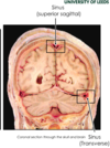
Venous sinuses are also found between the two layers of the dura mater. How are these formed?

A

Spaces created at specific locations by the separation of the dural layers
27
Q

What are the dural venous sinuses responsible for?

A
  • Venous blood from brain is collected here and then drained into the internal jugular veins
  • Cerebrospinal fluid is reabsorbed
28
Q

Spinal cord enclosed within dural sac

A
29
Q

What happens regarding the meninges when the spinal nerve roots leave the spinal cord?

A

They take layers of meninges with them - spinal nerve roots ensheathed within dura

30
Q

How do the meninges of the spinal cord differ from the brain? 2 points

A
  • Dura mater surrounding spinal cord is only composed of one layer –> the meningeal layer only (i.e. no periosteal layer)
  • Space between spinal cord and dura (epidural space) but dura mater of brain is tightly adhered to skull
31
Q

For the spinal cord, what is found between the spinal cord and the dura?

A

Epidural space occupied by fat

32
Q

What is found below the arachnoid layer of meninges of the spinal cord?

A

Subarachnoid space (CSF)

33
Q

What are denticulate ligaments? Function?

A
  • Projections of the pia mater (pia ligament) that anchor the spinal cord to the arachnoid matter and to the dura mater each side
  • Only found in spinal cord
34
Q

Where are the denticulate ligaments found?

A

Interspersed between the spinal nerves

35
Q

What is a potential space?

A

A potential space is a space between two adjacent structures that are normally pressed together (directly apposed) –> only becomes a space in pathological conditions

E.g. pleural space is a potential space

36
Q

Is the epidural space in the skull (between the skull and the dura) real or potential?

A

Potential (normally dura should be tightly adhered)

37
Q

Is the subdural space in the skull (between the dura and the arachnoid) real or potential?

A

Potential

38
Q

Is the subarachnoid space in the skull (between the arachnoid and pia mater) real or potential?

A

Real - cerebrospinal fluid contained here

39
Q

Diagram of cranial real vs potential

A
40
Q

Is the spinal epidural space (between the spinal cord and dura) real or potential?

A

Real - filled with fat

41
Q

Is the spinal subdural space (between the dura and arachnoid) real or potential?

A

Potential

42
Q

Is the spinal subarachnoid space (between the arachnoid and pia mater) real or potential?

A

Real

43
Q

Diagram of real vs potential spinal spaces

A
44
Q

Function of CSF?

A
  • Protection: acts as a cushion for the brain, limiting neural damage in cranial injuries.
  • Buoyancy: by being immersed in CSF, the net weight of the brain is reduced to approximately 25 grams. This prevents excessive pressure on the base of the brain.
  • Chemical stability: the CSF creates an environment to allow for proper functioning of the brain, e.g. maintaining low extracellular K+ for synaptic transmission.
45
Q

Where is CSF derived from?

A

Blood derived (but doesn’t contain blood) - is a clear, colourless fluid

46
Q

What does CSF contain?

A

Contains glucose and inorganic salts, little protein, few cells.

47
Q

What space does CSF fill?

A

Subarachnoid space and ventricular system of brain

48
Q

Where is CSF produced? What is it produced by?

A

Produced in ventricles by specialised areas of ventricular lining: choroid plexus.

49
Q

Summary - strength of dura vs arachnoid vs pia mater

A

Dura - Tough, fibrous membrane

Arachnoid - Soft translucent membrane

Pia - Microscopically thin, delicate membrane

50
Q

The cranial periosteal and meningeal layers of dura are are tightly fused together except at which 2 locations?

A
  1. venous sinuses
  2. various infoldings
51
Q

Name of space between dura and arachnoid?

A

Subdural space

52
Q

Name of space between Pia and Arachnoid?

A

Subarachnoid space

53
Q

What are the ventricles of the brain?

A

Fluid filled communicating cavities inside the adult brain

54
Q

Function of the ventricular system?

A

These structures are responsible for the production, transport and removal of cerebrospinal fluid

55
Q

What are the 5 secondary brain vesicles (during development of the human brain)?

A
  1. Telencephalon
  2. Diencephalon
  3. Mesencephalon
  4. Metencephalon
  5. Myelencephalon
56
Q

The embryonic tube has a lumen (fluid filled space).

i.e. telecephalon has a fluid filled space.

What do these spaces give rise to?

A

The ventricles of the brain

57
Q

Which ventricle does the fluid filled cavity in the telencephalon give rise to?

A

Lateral ventricle

58
Q

Which ventricle does the fluid filled cavity in the diencephalon give rise to?

A

Third ventricle

59
Q

Which cavity does the fluid filled cavity in the mesencephalon give rise to?

A

Cerebral aqueduct

60
Q

What is the cerebral aqueduct?

A

a narrow conduit that allows for CSF to flow between the third ventricle and the fourth ventricle

61
Q

Which ventricle does the fluid filled cavity in the metencephalon give rise to?

A

Upper part of 4th ventricle

62
Q

Which cavity does the fluid filled cavity in the myelencephalon give rise to?

A

Lower part of 4th ventricle / central canal of spinal cord

63
Q

How does the telencephalon grow in comparison to the rest of the embryonic tube?

A

Grows much faster

64
Q

What does this fast growth rate of the telencephalon result in?

A

Telencephalon folds over the developing diencephalon –> causing characteristic C shape of cerebral hemispheres (as well as many internal structures)

65
Q

Sagittal MRI view of ventricle

A
66
Q

Horizontal MRI of ventricles (green)

A
67
Q

Coronal MRI view of ventricles (green)

A
68
Q

Superior (bird’s eye) view of ventricular system

A
69
Q

What do the ventricles produce?

A

Within the choroid plexus that CSF is produced - and transport it around the cranial cavity

70
Q

What cells are the ventricles of the brain lined by? What structure do they form?

A

Ependymal cells –> form a structure called the choriod plexus

71
Q

Embryologically, where is the ventricular system is derived from?

A

The lumen of the neural tube

72
Q

In total, how many ventricles of the brain are there? What are they called?

A

4

  • 2 lateral ventricles (left and right)
  • Third ventricle
  • Fourth ventricle
73
Q

Where are the left and right lateral ventricles located?

A

Within their respective hemispheres of the cerebrum.

74
Q

Describe the shape of each lateral ventricle

A
  • Follow a C-shaped course through the lobes of a cerebral hemisphere
  • Have ‘horns’ which project into the frontal, occipital and temporal bones
75
Q

What are the horns of the lateral ventricle? What bone does each project into?

A
  • Anterior horn –> projects into frontal lobe
  • Posterior horn –> projects into occipital lobe
  • Inferior horn –> projects into temporal lobe
76
Q

Detailed diagram of lateral ventricle

A
77
Q

Where is the 3rd ventricle located?

A

Between the right and left thalamus

78
Q

What is the thalamus?

A

a mostly gray matter structure (egg shaped) of the diencephalon

79
Q

View of 3rd ventricle (coloured in blue is where it would be if we had the other half of the brain)

A
80
Q

Where is the 4th ventricle?

A

Space between the brainstem anteriorly and cerebellum posteriorly

81
Q

Diagram of 4th ventricle (in red)

A
82
Q

Diagram of all the ventricles

A
83
Q

how does the 4th ventricle receive CSF from the 3rd ventricle?

A

Via the cerebral aqueduct

84
Q

What are the features of the lateral ventricle?

A
  • Anterior, posterior inferior horn
  • Body
  • Antrum
85
Q

How do all 3 horns of the lateral ventricle communicate?

A

At the antrum of the lateral ventricle

86
Q

How are the lateral ventricles connected to the 3rd ventricle?

A

At the interventricular foramina / Foramen of Monro (one for each lateral ventricle)

87
Q

What connects the 3rd and 4th ventricle?

A

Cerebral aqueduct (found at the level of the midbrain)

88
Q

How does fluid leave the 4th ventricle and enter the subarachnoid space (between the arachnoid mater and pia mater)?

A
  • At 2 lateral apertures (foramina of Luschka)
  • At 1 median aperture (foramen of Magendie)
89
Q

Ventricular system summary

A
90
Q

Each ventricle contains a choroid plexus. What is this?

A

A collection of blood vessels and ependymal cells

91
Q

Function of the ependymal cells? How do they produce CSF?

A

Filter blood from capillaries, remove some proteins –> left with CSF

92
Q

Where does CSF produced by the choroid plexus of the lateral ventricle flow?

A

Flow through the foramina of monroe / interventricular foramina to the third ventricle

93
Q

Once in the 3rd ventricle, together with the CSF produced by the choroid plexus in the 3rd ventricle, where does the CSF then flow?

A

Through the cerebral aquaduct to enter the 4th ventricle

94
Q

Once in the 4th ventricle, together with the CSF produced by the choroid plexus in the 4th ventricle, where does the CSF then flow?

A

Flows through the apertures (lateral and median apertures) to escape into the subarachnoid space

This then flows all around the subarachoid space (covering the brain AND spinal cord)

95
Q

Regarding the apertures that CSF leaves the 4th ventricle via, what are the lateral apertures called? The median?

A

Lateral - Foramina of Luschka

Median - Foramen of Magendie

96
Q

How is CSF in the subarachnoid space reabsorbed into the circulation?

A

Small projections of arachnoid mater (arachnoid granulations/villi) protrude into the dura mater sinuses. They allow the fluid to drain into the dural venous sinuses (mainly the superior sagittal sinus)

CSF allows CSF to pass from the subarachnoid space into the venous system

97
Q

In the subarachnoid space, there are some areas that are a little wider. What are these called?

A

Subarachnoid cisterns (located in brain and spinal cord)

98
Q

What is the lumbar cistern?

A

The lumbar cistern refers to an enlargement of subarachnoid space in the lower lumbar spinal canal

99
Q

Why is the lumbar cistern clinically relevant?

A

The lumbar cistern is the favoured site for sampling CSF

100
Q

Why is the lumbar cistern the favoured site for sampling CSF?

A

Because a needle can be inserted here with relatively little risk of damaging the CNS (puncture below end of spinal cord but can still access subarachnoid space)

101
Q

What is hydrocephalus? What causes it?

A

A condition in which an accumulation of cerebrospinal fluid (CSF) occurs within the brain (ventricles become enlarged) –> caused if CSF circulation is blocked and CSF accumulates upstream of the blockage

102
Q

What can cause an accumulation of CSF circulation?

A
  • Blockage in ventricles
  • Overproduction of CSF
  • Lack of absorption of CSF
103
Q

Why can hydrocephalus lead to an enlarged head in new borns?

A

Skull isn’t properly fused

104
Q

In adults, as the skull cannot expand during hydrocephalus, what happens?

A

Compression into brain tissue itself

105
Q

E.g. if there is blockage in cerebral aquaduct, where does CSF accumulate?

A

Lateral ventricle and the 3rd ventricle (upstream)

106
Q

E.g. if there is a blockage in one of the interventricular foramina, where does CSF accumulate?

A

In the corresponding lateral ventricle (upstream)