Cranial Nerves Flashcards

1
Q

Where do 10/12 cranial nerves emerge from? What system does this make them part of?

What are the 2 exceptions to this?

A
  • Emerge from brainstem - part of peripheral nervous system
  • CN I (olfactory) and CN II (optic) emerge from the cerebrum - part of the CNS
2
Q

Where did CN II (optic nerve) actually develop from?

A

The diencephalon

3
Q

Where did CN I (olfactory) actually develop from?

A

From the telencephalon

4
Q

The oflactory bulb and the olfactory tract are components of the olfactory nerve. What are these components?

A
  • As the olfactory nerve leaves the nasal epithelium to enter the cranial cavity, they synapse with the olfactory bulb and trac
  • The bulb and tract then bring the information into the cerebral hemispheres
5
Q

Where are the oflactory bulb and tract extensions from?

A

The telencephalon

6
Q

Cross-section of midbrain

A
  • Upside down mickey mouse shape
    • Ears corresponding to cerebral peduncles
  • Grey matter appears white
  • White matter appears dark
7
Q

For each fibre type within a cranial nerve, there is a corresponding nucleus in the brainstem. I.e. these fibres arise from the nuclei

Example:

  • How many fibre types does CN III (occulomotor) carry?
  • What are these fibre types
  • How and where are the corresponding nuclei?
  • What are the corresponding nuclei called?
A
  • CN III carries 2 fibre types
    • Somatic motor fibres to extraocular muscles
    • Visceral motor (autonomic) fibres (parasympathetic) to sphincter pupillae and ciliary muscles
  • 2 fibres there 2 corresponding nuclei in the midbrain (where CN III emerges)
    • Oculomotor nucelus corresponds to somatic motor fibre
    • Edinger-Westphal nucleus corresponds to visceral motor fibre
8
Q

What are the 2 corresponding nuclei of CN III in the midbrain called?

A
  1. Oculomotor nucleus
  2. Edinger-Westphal nucleus
9
Q

What is the Edinger-Westphal nucleus? Location? What does it innervate?

A
  • A small parasympathetic motor nucleus in the midbrain
    • Parasympathetic fibres emerge from this nucleus
  • One of the two nuclei for the oculomotor nerve
  • Innervates the iris sphincter muscle and the ciliary muscle
10
Q

What is the oculomotor nucleus? Location? What does it innervate?

A
  • A small somatic motor nucleus in the midbrain
    • Somatic motor fibres emerge from here
  • One of the two nuclei for the oculomotor nerve
  • The fibers of the oculomotor nerve arise from a nucleus in the midbrain
11
Q

How do fibres from both the Edinger-Westphal and Oculomotor nucelus form the Oculomotor nerve?

A

Fibres arise from both of these nuclei in the midbrain (at the level of the interpeduncular fossa between the 2 cerebral peduncles) and combine to form the Oculomotor nerve (CN III)

12
Q

Cranial nerve nuclei are scattered throughout the brainstem.

Spread out in the midbrain (III, IV), the pons (V, VI, VII, VIII), and the medulla (IX, X, XI, XII).

  • Where are the sensory nuclei located?
  • Where are the motor nuclei located?
A
  • Sensory nuclei are located lateral
  • Motor nuclei are located medial

Notice how cranial nerve nuclei are located anterior to the ventricular system, but not tightly against the anterior border. Ie. located in tegmentum.

13
Q

General organisation of CN nuclei in brainstem.

This is a cross-section through open medulla oblongata (can see 4th ventricle at the back).

A
  • Sensory nuclei more lateral
    • Visceral sensory
    • Special sensory
    • Somatic sensory
  • Motor nuclei more medial
    • Somatic motor
    • Parasympathetic motor
    • Head and neck motor
14
Q

Each cranial nerve can be described as being sensory, motor or both. They can more specifically transmit seven types of information; three are unique to cranial nerves (SSS, SVS and SVM).

  • What are the sensory modalities?
  • What are the motor modalities?
A
  • Sensory:
    • Special –> special senses (vision, taste, olfactory, hearing, balance)
    • Somatic –> somatic sensation
    • Visceral –> monitors states of internal organs and glands
  • Motor:
    • Somatic –> for skeletal muscles
    • Parasympathetic –> e.g for secretions from glands
    • Head and neck –> some muscles of head and neck
15
Q

Cranial Nerve Summary:

A
  • Part of the PNS (except I & II), which are extensions of the forebrain
  • Nerves of head and neck carry either sensory (afferent) and/or motor (efferent) fibres:
    • eg. Sensory from skin of face
    • eg. Taste sense from tongue
    • eg. Motor to eye muscles
    • eg. Motor to neck muscles
    • eg. Visceral motor to salivary glands
    • eg. Visceral sensory from organs
  • Each cranial nerve may carry several different fibre types
    • Can be purely motor, purely sensory or mixed
  • For each fibre type within a cranial nerve, there is a corresponding nucleus in the brainstem
16
Q

How many cranial nerves are purely motor (efferent)? Purely sensory (afferent)? Mixed?

A

5 cranial nerves are purely motor (efferent); 3 are purely sensory (afferent); the rest are mixed

17
Q

What is a nucelus?

A

A nucleus is a functional group of neurons within the CNS

18
Q

Cranial Nerve Summary 2:

A
  • Cranial nerve nuclei are located within the tegmentum of the brainstem
  • Found in all brainstem parts (midbrain, pons and medulla)
  • Generally, two types of cranial nerve nuclei:
    • Sensory: Receive information from fibres entering brainstem in cranial nerves which synapse here, and nuclei then send fibres to appropriate higher centre
    • Motor: Receive motor information from higher centre which synapse in nuclei which then give rise to motor fibres that leave the brainstem in cranial nerves
19
Q

How are cranial nerves I and II different?

A

Derived from forebrain not brainstem - part of CNS

20
Q

What is CN I?

A

Olfactory nerve

21
Q

Olfactory nerve (CN I):

  • Where is the true location of CN I
  • Function?
  • Foramina?
  • Modality?
  • Test?
A
  • Location: from nasal mucosa, merge with olfactory bulbs and olfactory tracts (extensions of the telencephalon)
  • Function : smell
  • *Foramina*:cribriform plate** of ethmoid bone
  • Modality: special sensory
  • Test:
    • Offer a familiar smelling item (e.g. orange)
    • Any changes in sense of smell?
22
Q

What is anosmia?

A

Loss of smell

23
Q

Optic nerve (CN II):

  • Location of CN II?
  • Function?
  • Foramina?
  • Modality?
  • Test?
A
  • Location: One leaves each eyeball, emerging from the retina to the optic chiasma. Optic tract then carry visual info to the thalamus
    • Optic chiasma is where info from the two optic nerves merge
  • Function: Vision
  • Foramina: Optic canals
  • Modality: Special sensory
  • Test: Visual tests e.g. fundoscopy
24
Q

What is the only sensory modality to not pass through the thalamus?

A

Sense of smell

25
Q

what is the optic chiasm?

A

Optic chiasma is where info from the two optic nerves merge

26
Q

Describe CN I pathway

A
  • Olfactory receptors expressed in olfactory epithelium at back of nasal cavity
  • Receptors pierce through cribriform plate of ethmoid bone which then synapses with the olfactory bulb.
  • Then sends fibres using optic tract to reach the areas of cortex responsible for processing smell.
27
Q

Describe pathway of CN II (optic)

A
  • Emerges from back of retina
  • Make way through optic canal to enter cranial cavity
  • Two optic nerves merge at optic chiasma
  • Optic tracts bring info to thalamus
28
Q

What is the combined function of CN III, CN IV and CN VI?

A

Innervate the extraocular muscles (that move eyeballs)

29
Q

Through which foramina do CN III, CN IV and VI pass through to reach the orbit?

A

The superior orbital fissure

30
Q

Oculomotor (CN III):

  • Location?
  • Function?
  • Foramina?
  • Modality?
  • Test?
A
  • Location: Emerges at pontomesencephalic junction/interpeduncular fossa (between midbrain and pons)
  • Modality:
    • Somatic motor
    • Visceral (parasympathetic) motor
  • Function:
    • Somatic motor: to four extraocular muscles
    • Visceral (parasympathetic) motor: to ciliary muscle and sphincter pupillae
  • Foramina: Superior orbital fissure
  • Test: Follow finger test
31
Q

What test is used to check the function of CN III, IV and VI?

A

Follow finger test

32
Q

Trochlear (IV):

  • Location?
  • Function?
  • Foramina?
  • Modality?
  • Test?
A
  • Location: Dorsal midbrain
  • Modality: Somatic motor
  • Function: Somatic motor to superior oblique (SO4)
  • Foramina: Superior orbital fissure
  • Test: Follow finger (H) test
33
Q

Abducens nerve (VI):

  • Location?
  • Function?
  • Foramina?
  • Modality?
  • Test?
A
  • Location: Pontomedullary junction
  • Modality: Somatic motor
  • Function: Somatic motor to lateral rectus (LR6)
  • Foramina: Superior orbital fissure
  • Test: Follow finger (H) test
34
Q

Describe the innervation of the extraocular nerves

A

There are 6 extraocular muscles; 4 recti muscles and 2 oblique muscles.

  • Superior rectus, medial rectus, inferior rectus and inferior oblique are all innervated by oculomotor nerve (CN III)
  • Lateral rectus is innervated by abducens nerve (CN VI) –> LR6
  • Superior oblique is innervated by trochlear nerve (CN IV) –> SO4

LR6SO4 to remember this innervation

35
Q

CN III (oculomotor) passes through superior orbital fissure to enter orbit. What does it innervate?

This is the somatic motor portion of the nerve

A
  • Along this path it innervates levator palpebrae superioris which elevates the eyelid
  • The superior rectus
  • Medial rectus
  • Inferior rectus
  • Inferior oblique
36
Q

What does the visceral motor portion of the oculomotor nerve innervate?

A

Autonomic (parasympathetic) fibres that innervate the ciliary muscle and the sphincter pupillae (intraocular muscles)

37
Q

What are the modalities of the oculomotor nerve (CN III)?

A
  • Somatic motor –> innervates 4 extraocular muscles and levator palpebrae superioris
  • Visceral motor (autonomic parasympathetic fibres) –> innervates ciliary muscles & sphincter pupillae
38
Q

Movement of the eyes and the corresponding muscles

A
39
Q

Which muscle closes the eyelid? What is this innervated by?

A
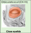
  • Orbicularis oculi muscle
    • Sphincter around eyelid
    • Superficial muscle of facial expression
  • Innervated by CN VII (facial)
40
Q

Which muscles perform dilation (opening) of eyelids? Innervations?

A

2 muscles:

  • Superior tarsal muscle
    • This muscle is just underneath levator palpebrae superioris
    • Keeps the eyelid open
    • Innervation from the sympathetic nervous system
  • Levator palpebrae superioris
    • Opens superior eyelid
    • Innervated by CN III motor fibres (oculomotor)
41
Q

What controls the pupil and lens of the eye?

A

Intraocular muscles

42
Q

Which muscles dilate the pupil? Innervation?

A
  • Dilators of iris
    • Innervated by sympathetic nervous system
43
Q

Which muscles constrict the pupil? Innervation?

A
  • Sphincter pupillae
    • Innervated by CN III parasympathetic fibres (oculomotor)
44
Q

Which muscle changes the lens? Innervation?

A
  • Ciliary muscle (by pulling on ligamnet
    • Change shape of lens by pulling on ligaments attached to lens
      • Make lens more flat or round
    • Innervated by CN III parasympathetic fibres (oculomotor)
45
Q

What is the biggest cranial nerve? Why?

A

CN V trigeminal nerve - has 3 divisions

46
Q

What are th 3 divisions of CN V?

A

V1 opthalmic

V2 maxillary

V3 mandibular

47
Q

V1 Opthalmic division:

  • Location?
  • Function?
  • Foramina?
  • Modality?
  • Test?
A
  • Location: Emerges from pons
  • Foramen: superior orbital fissure to enter orbit
  • Modality: Somatic sensory
  • Function: Somatic sensation from upper face (above eye, including upper eyelid)
  • Test:
    • Cotton wool bud on forehead and ask patient if they can feel it
    • Corneal reflex test (as cornea innervated by this nerve)
      • Lightly touch a wisp of cotton on the patient’s cornea. This foreign body sensation should cause the patient to reflexively blink.
48
Q

V2 Maxillary division:

  • Location?
  • Function?
  • Foramina?
  • Modality?
  • Test?
A
  • Location: Pons
  • Foramen: Exits cranial cavity via foramen rotundum
  • Modality: Somatic sensory
  • Function: Somatic sensation from middle face (between eye and mouth including lower eyelid and upper lip)
  • Test: Cotton wool bud on cheek to test sensation
49
Q

V3 Mandibular division:

  • Location?
  • Function?
  • Foramina?
  • Modality?
  • Test?
A
  • Location: Pons
  • Foramen: exits cranial cavity via foramen ovale
  • Modality:
    • Somatic sensory
    • Special visceral motor
  • Function:
    • Somatic sensation from lower face mandible and anterior 2/3 tongue
    • Motor fibres to muscles of mastication, anterior belly of digastric, tensor tympani (ear) etc
  • Test:
    • Cotton wool bud on jaw
    • Clench teeth, feel muscle mass
    • Move jaw side to side against resistance (pterygoids)
    • Protrude jaw
    • Jaw-jerk reflex
50
Q

Distributio of Cranial Nerve V

A
51
Q

Foramen for CN V

A
52
Q

Areas of sensory innervation of CN V

A
53
Q

Where do the three major branches of the trigeminal nerve converge?

A

On the trigeminal ganglion

54
Q

Associated with the trigeminal nerve are the four autonomic ganglia of the head and neck. What are these?

A
  1. Ciliary
  2. Pterygopalatine
  3. Submandibular
  4. Otic
55
Q

CN VII Facial Nerve:

  • Location?
  • Function?
  • Foramina?
  • Modality?
  • Test?
A
  • Location: Cerebellopontine angle
  • Foramina:
    • internal acoutsic meatus to enter the cranial cavity
    • stylomastoid foramen to exit
  • Modality:
    • Somatic sensory
    • Visceral sensory
    • Special sensory
    • Visceral motor
  • Function:
    • Special visceral motor –> to muscles of facial expression, stapedius muscle, posterior belly of digastric
    • Special sensory –> as taste to anterior 2/3rds tongue (chorda tympani)
    • Somatic sensory –> to skin of ear
    • Visceral motor (parasympathetic) –> to all glands except parotid
  • Test:
    • Facial movements, taste, salivation
56
Q

CN VIII Vestibulocochlear Nerve:

  • Location?
  • Function?
  • Foramina?
  • Modality?
  • Test?
A
  • Location: Cerebellopontine angle (just lateral and inferior to CN VII)
  • Foramen: Internal acoustic meatus
  • Modality: Special sensory
  • Function –> hearing and balance
  • Test:
    • Bedside hearing tests; Rinne’s and Weber’s test
57
Q

How do CN VII and CN VIII both enter the cranial cavity? Describe pathway after entering

A

Internal auditory meatus

  • CN VII –> travels to petrous temporal bone where it gives off branches
  • CN VIII –> goes to vestibular apparatus and cochlear apparatus in middle ear
58
Q

What branches does CN VII give off in the petrous temporal bone? What do these branches innervate?

A

The nerve arises in the pons and begins as two roots; a large motor root, and a small sensory root. The two roots travel through the internal acoustic meatus and fuse. The nerve gives rise to:

  • Greater petrosal nerve
    • Parasympathetic fibres to mucous glands and lacrimal gland.
  • Nerve to stapedius muscle
    • motor fibres to stapedius muscle of the middle ear.
  • Chorda tympani
    • special sensory fibres to the anterior 2/3 tongue and parasympathetic fibres to the submandibular and sublingual glands.
59
Q

How does the facial nerve exist the cranial cavity?

A

Via the stylomastoid foramen. This is an exit located just posterior to the styloid process of the temporal bone.

60
Q

The vestibular and cochlear portions of the vestibulocochlear nerve are functionally discrete, and so originate from different nuclei in the brain.

What is the vestibular and cochlear component responsible for?

A
  • Vestibular component –> responsible for the special senses of balance
  • Cochlear component –> responsible for the special senses of hearing
61
Q

CN IX Glossopharyngeal Nerve:

  • Location?
  • Function?
  • Foramina?
  • Modality?
  • Test?
A
  • Location: Posterolateral sulcus of medulla, lateral to olives
  • Foramen: Jugular foramen
  • Modality:
    • Special motor
    • Visceral motor (parasympathetic)
    • Special sensory
    • Somatic sensory
    • Visceral sensory
  • Function:
    • Special motor –> branchial motor for swallowing (pharynx)
    • Visceral motor (parasympathetic) to parotid gland
    • Special sensory –> for taste to posterior 1/3rd tongue
    • Somatic sensory –> to middle ear, pharynx, posterior 1/3rd tongue
    • Visceral sensation –> from carotid body and carotid sinus monitoring O2 and BP
  • Test: Gag reflex
62
Q

CN X Vagus

  • Location?
  • Function?
  • Foramina?
  • Modality?
  • Test?
A
  • Location: Posterolateral sulcus of medulla, lateral to olives
  • Foramen: jugular foramen
  • Modality:
    • Motor
    • Visceral motor (parasympathetic)
    • Sensory
    • Special sensory
  • Function:
    • Branchial motor to muscles of pharynx and larynx, muscles of soft palate (uvula)
    • Sensory:
      • Innervates the skin of the external acoustic meatus and the internal surfaces of the laryngopharynx and larynx.
      • Provides visceral sensation to the heart and abdominal viscera.
    • Special sensory –> Provides taste sensation to the epiglottis and root of the tongue.
    • Visceral motor (parasympathetic) –> Innervates the smooth muscle of the trachea, bronchi and gastro-intestinal tract and regulates heart rhythm.
  • Test:
    • Speaking, movement of uvula
63
Q

How does CN IX and CN X pass from the medulla into the cranial cavity?

A

Via the jugular foramen

64
Q

What do the carotid body and sinus monitor? Which nerve innervates these?

A
  • BP and O2 levels
  • CN IX innervates these
65
Q

CN XI Accessory:

  • Location?
  • Function?
  • Foramina?
  • Modality?
  • Test?
A
  • Location: Caudal medulla and rostral spinal cord
  • Foramen: jugular foramen
  • Modality: Somatic motor
  • Function: Somatic motor to sternocleidomastoid and trapezius
  • Test: Shrug shoulder and rotate head against resistance
66
Q

CN XII Hypoglossal:

  • Location?
  • Function?
  • Foramina?
  • Modality?
  • Test?
A
  • Location: Anterolateral sulcus (between pyramid and olives)
  • Foramen: Hypoglossal canal
  • Modality: Somatic motor
  • Function: Somatic motor to muscles of tongue
  • Test: Protrude tongue (does it deviate to one side)
67
Q

Jugular foramen and hypoglossal canal

A
68
Q

What can the accessory nerve be divided into? Where odes each part arise from?

A

Spinal and cranial parts:

  • Cranial part arises from lateral aspect of the medulla oblongata.
  • The spinal portion arises from neurones of the upper spinal cord, specifically C1-C5/C6 spinal nerve roots.

Both pass through the jugular foramen

69
Q

Emerging of CN XII

A
70
Q

What innervates the extrinsic muscles of the tongue. What do these muscles do?

A
  • Hypoglossal nerve CN XII
  • Changes shape and direction of tongue
71
Q

If the hypoglossal nerve is paralysed, how will this affect the movements of the tongue?

A
  • No paralysis –> tongue can protrude straight forward
  • Injury to CN XII –> deviation of tongue towards the paralysed side
72
Q

Innervation of tongue: taste and somatic

A

Taste:

  • Anterior 2/3: facial nerve (chorda tympani)
  • Posterior 1/3: glossopharyngeal nerve
  • Epiglottis: vagus nerve

Somatic:

  • Anterior 2/3: mandibular nerve (lingual nerve)
  • Posterior 1/3: glossopharyngeal
  • Epiglottis: vagus nerve
73
Q

Motor innervation of tongue?

A

Hypoglossal nerve

74
Q

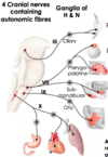
There are some autonomic fibres running through cranial nerves. Which nerves are these?

A
  • III, VII, IX and X
  • These are associated with ganglia in the head and neck:
    • Ciliary
    • Pterygopalatine
    • Submandibular
    • Otic
  • All these fibres pass from their ganglia to the respective organ by “piggy-backing” on branches of the trigeminal nerve. (Head and neck only)
75
Q

Function of the ciliary ganglia?

A

Pupil constriction (Sphincter pupillae)

76
Q

Function of the pterygopalatine ganglia?

A

Lacrimation (lacrimal gland)

77
Q

Function of the submandibular ganglia?

A

Salivation (salivary glands):

  • Submandibular
  • Sublingual
78
Q

Function of the otic ganglia?

A

Salivation (Salivary glands):

  • Parotid
79
Q

Complete the table

A
80
Q

Which cranial nerves contain autonomic fibres? Which ganglia does each contribute to?

A

CN III

  • Gives rise to presynaptic fibres that pass towards the ciliary ganglion
  • Here, these fibres synapse with postsynaptic fibres
  • Pass to the sphincter pupillae of the eye via a branch of the 1st divison (opthalmic) of the trigeminal nerve (CN V1)

CN VII

  • Gives rise to presynaptic fibres that pass towards and synapse in the pterygopalatine ganglion
    • ​Post synaptic fibres then pass towards the lacrimal gland via the 2nd division (maxillary) of the trigeminal nerve (CN V2)
  • Also gives rise to presynaptic fibres that pass towards and synapse in the submandibular ganglion
    • Post synaptic fibres then pass towards the submandibular and sublinguinal gland via the 3rd division (mandibular) of the trigeminal nerve (CN V3)

CN IX:

  • Gives rise to presynaptic fibres that pass towards and synapse in the otic ganglion
  • Postsynaptic fibres then pass to the parotid gland via the 3rd division (mandibular) of the trigeminal nerve (CN V3)

CN X

N.B. these are all parasympathetic

81
Q

Where does the vagus nerve send the majority of its autonomic fibres?

A

To thoracic and abdominal viscera

82
Q

What contributes the sympathetic input to the autonomic ganglia of the head and neck?

A
  • Comes from the superior cervical ganglion (upper limit of sympathetic chain)
  • Postsynaptic fibres leave and pass towards the common carotid artery
    • As the common carotid artery bifurcates, its going to receive these branches from the superior cervical ganglion
    • These are then going to run up through the arteries and innervate the various target organs of the head
83
Q

Describe the innervation of the tongue regarding:

a) taste (special sensory)
b) general sensation (somatic sensory)

A

a) ant 2/3rd tongue –> chorda tympani (branch of CN VII), post 1/3rd tongue –> CN IX
b) ant 2/3rd tongue –> lingual nerve (branch of CN V3), post 1/3rd tongue –> CN IX