3.3.2 Thyroid Gland Pathology Flashcards
What are some of the sources of hypothyroidism?
Autoimmune disease
Endemic iodine deficiency
Exogeneous goitrogenic agents
Diffuse non-toxic goiter
Ablation of thyroid gland
What is this an image of?

Follicular Adenoma of Thyroid
Microscopically, the constituent cells often form uniform-appearing follicles that contain colloid (Fig. 24-15). The follicular growth pattern within the adenoma is usually quite distinct from the adjacent non-neoplastic thyroid. This is another feature distinguishing adenomas from multinodular goiters, in which nodular and uninvolved thyroid parenchyma may have similar growth patterns.
What is this an image of?

Riedel struma, gross
Why is medullary carcinoma a WMBK?
It has familial tendencies and can be prevented by prophylactic surgery
What is granulomatous thyroiditis?
granulomatous thyroiditis or De Quervain thyroiditis, occurs much less frequently than does Hashimoto disease
Sub-acute thyroiditis is believed to be caused by a viral infection or a post-viral inflammatory process (molecular mimicry? – see I2 notes on theories of autoimmunity). The majority of patients have a history of an upper respiratory infection just before the onset of thyroiditis.
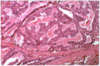
What is this an image of?

Riedel struma, micro slide
What type of HF can be a result of graves?
High output HF
Ventricles beating too fast leading to inability of atria to refill
What is this an image of?

Diffuse toxic goiter – Graves Disease.
What is Hashimoto’s thyroidosis?
Most common goitrous hypothyroidism in areas with sufficienct iodine
The vast majority of cases of autoimmune hypothyroidism are due to Hashimoto thyroiditis. Circulating autoantibodies, including anti-TSH receptor autoantibodies, are commonly found in Hashimoto thyroiditis.
What is this an image of?

Follicular carcinoma
It is important to note the absence of these details because some papillary carcinomas may appear histologically to be almost entirely follicular
What is this an image of?

The typical follicular thyroid adenoma is a solitary, spherical, encapsulated lesion that is well demarcated from the surrounding thyroid parenchyma (Fig. 24-14). Follicular adenomas average about 3 cm in diameter, but some are smaller and others are much larger (up to 10 cm in diameter). In freshly resected specimens, the adenoma bulges from the cut surface and compresses the adjacent thyroid. The color ranges from gray-white to red-brown, depending on the cellularity of the adenoma and its colloid content. The neoplastic cells are demarcated from the adjacent parenchyma by a well-defined, intact capsule. These features are important in making the distinction from multinodular goiters, which contain multiple nodules on their cut surface (even though the patient may present clinically with a solitary dominant nodule), produce less compression of the adjacent thyroid parenchyma, and lack a well-formed capsule.
What is this an image of?

Medullary carcinoma of the thyroid
Medullary carcinoma, high power. Typical C cells, which would stain for calcitonin with an immunostain.
What is this an image of?

Hashimotos Thyroiditis
Anti-microsomal antibody
What is this an image of?

Papillary carcinoma
Morphology. Papillary carcinomas are solitary or multifocal lesions. Some tumors may be well-circumscribed and even encapsulated; others may infiltrate the adjacent parenchyma with ill-defined margins. The lesions may contain areas of fibrosis and calcification and are often cystic. On the cut surface, they may appear granular and may sometimes contain grossly discernible papillary foci. The definitive diagnosis of papillary carcinoma can be made only after microscopic examination. The characteristic hallmarks of papillary neoplasms include the following (Fig. 24-17):
Papillary carcinomas can contain branching papillae having a fibrovascular stalk covered by a single to multiple layers of cuboidal epithelial cells.
“Orphan Annie” or “optically clear” nuclei
Concentrically calcified structures termed psammoma bodies are often present within the lesion, usually within the cores of papillae.
What is this an image of?

Hashimoto thyroiditis is an autoimmune disease in which the immune system reacts against a variety of thyroid antigens. The overriding feature of Hashimoto thyroiditis is progressive depletion of thyroid epithelial cells (thyrocytes), which are gradually replaced by mononuclear cell infiltration and fibrosis. Multiple immunologic mechanisms may contribute to the death of thyrocytes (Fig. 24-9). Sensitization of autoreactive CD4+ T-helper cells to thyroid antigens appears to be the initiating event. The effector mechanisms for thyrocyte death include the following: CD8+ cytotoxic T cell-mediated cell death: CD8+ cytotoxic T cells may cause thyrocyte destruction by one of two pathways: exocytosis of perforin/granzyme granules or engagement of death receptors, specifically CD95 (also known as Fas) on the target cell
What is the most common invasion location of anaplastic carcinoma?
Trachea invasion is most common
Endocrine hyperfunction tends to be due to?
Increased trophic hormone
Neoplasms of the pituitary or target gland
Exogeneous artificial hormone
What is endemic cretenism?
Hypothyroidism in newborns and infants
Pathogenesis - Iodine deficiency during intraurerine life and infancy
What is this an image of?

Anaplastic carcinomas of the thyroid are undifferentiated tumors of the thyroid follicular epithelium. In striking contrast to the differentiated thyroid carcinomas, anaplastic carcinomas are aggressive tumors, with a mortality rate approaching 100%.
Typically occurs with a p53 mutation
What is Riedel’s thyroiditis?
Fibrous proliferation of connective tissue of thyroid extending into surrounding tissues
What is this an image of?

Follicular Carcinoma
Follicular carcinomas are single nodules that may be well circumscribed or widely infiltrative (Fig. 24-18). Sharply demarcated lesions may be exceedingly difficult to distinguish from follicular adenomas by gross examination.
What is this an image of?

Microscopically, medullary carcinomas (Linked to MEN2) are composed of polygonal to spindle-shaped cells, which may form nests, trabeculae, and even follicles. Small, more anaplastic cells are present in some tumors and may be the predominant cell type. Acellular amyloid deposits, derived from altered calcitonin molecules, are present in the adjacent stroma in many cases
What is this an image of?

Papillary Carcinoma of the Thyroid
What is this an image of?
Graves disease, micro. Note hyperplastic follicular cells, with resorption of colloid.


