Week 3: Celiac Disease Flashcards
What is celiac disease?

How is the autoimmune response triggered in celiac disease

HLA types common in Celiac disease
HLA DQ2
HLA DQ8
Different serological types of celiac disease

Question

D. Submucosa
Question

B. Lamina Propria
What is the main difference between small intestinal mucosa and colonic mucosa?
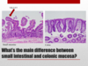
Differences in function of small intestine and colon

Describe histologic features of small intestine

Describe histologic features of the large intestine

Question

D. Goblet cell
Villi compared to crypts

Celiac Disease Diagnostic criteria

Who diagnoses Celiac disease

E.
Pathologic histologic features of celiac disease

Small intestinal CD3 stain

Question

A. the one on the left
Histologic features of celiac disease

What is the preferred biopsy location for celiac disease

Describe histological features of 1st and 2nd portions of duodenum
- 1st is more sensitive for celiac has a lot of Brunners glands
- 2nd part is more specific for celiac

Mucosal damage and symptomology or serology

MARSH Classifications

Describe these features


Compare celiac histology to normal small intestine

Compare celiac histology to colon

Question


Anti-tTG levels can be elevated in?

Other serological tests for celiac disease

HLA testing benefits for celiac disease

Assume a different scenario

No, lack of blah blah

Are IELs and villous blunting pathognomonic of celiac

Differentials of celiac disease

How common are increased IELs in duodenal biopsies?

What is the best initial test for celiac disease?
Anti-tTG test
Summary of celiac disease

Algorithm of celiac disease testing

Question

Looks normal
B. Villi are the right height
Question

too many lymphocytes in the villus tip
CD3 stain
way too many lymphocytes at the villus tips

Question
Is this celiac disease?

C Maybe poster child for gluten diet restriction

Describe

stomach
antral mucosa 2x
lymphoplasmacytic infiltrate along superficial aspect of antral mucosa

Diagnosis

H. pylori
Celiac disease complications

What is refractory celiac disease?

