STEEPLECHASE - MUSCULOSKELETAL Flashcards

1
Q
A

Brachygnathia

2
Q
A

Complete palatoschisis

3
Q
A

Prognathia

4
Q
A

Pectus excavatum

5
Q
A

Kyphosis

6
Q
A

Scoliosis

7
Q
A

Block vertebrae

8
Q
A

Butterfly/hemivertebrae

9
Q
A

Lordosis

10
Q
A

Manatee - confined in small aquarium, developed severe scoliosis (lateral deviation)

11
Q
A

Spina bifida in dog

12
Q
A

Mircomelia

13
Q
A

Peromelia

14
Q
A

Notomelia

15
Q
A

Hemimelia

16
Q
A

Amelia

17
Q
A

Polydactyly

18
Q
A

Syndactyly

19
Q
A

Ectrodactyly

20
Q
A

Dactylomegaly

21
Q
A

Adactyly

22
Q
A
23
Q
A

Osteogenesis impefecta - lateral deviation in joints, collagen I mutation

24
Q
A

Osteogenesis imperfecta

25
Q
A
26
Q
A

Osteopetrosis

27
Q
A
28
Q
A

Congenital cortical hyperostosis

29
Q
A

Congenital hyperostosis

30
Q
A

‘Lion jaw’ craniomandibular osteopathy

31
Q
A

Craniomandibular osteopathy ‘Lion jaw’

32
Q
A

Chondrodysplasia

33
Q
A
34
Q
A

Disproportionate dwarfism - Grey alpine calf w/ Ellis van Creveld syndrome (chondrodysplasia)

35
Q
A

Manganese deficiency, not congenital, acquired/nutritional deficiency (chondrodysplasia)

36
Q
A

Texel lamb - disproportionate dwarfism, shortened limbs and wide base (chondrodysplasia)

37
Q
A

(Chondrodysplasia)

38
Q
A

Osteochondrosis

39
Q
A

Osteochondrosis latens - well-demarcated area of necrosis of cartilage

40
Q
A

Osteochondrosis manifesta - arrows = associated with areas of foci of necrosis - areas of retained cartilage = hypertrophic chondrocytes

41
Q
A

Osteochondrosis dissecans - arrow pointing to ‘joint mice’

42
Q
A

Vertebral myelopathy

43
Q
A

Dynamic vertebral myelopathy - flexion

44
Q
A
45
Q
A

Osteoporosis

46
Q
A

Rickets and osteomalacia

47
Q
A
48
Q
A

Head brachygnathia superior

49
Q
A

Dental dysplasia, with small pink-grey barely erupted teeth in a two-week old Holstein calf with osteogenesis imperfecta (right), left = normal calf

50
Q
A
51
Q
A

Osteopetrosis

52
Q
A

Cervical vertebral myelopathy “Wobbler syndrome”

53
Q
A

Cervical vertebral malformation-malarticulation in a horse. Flexion of the cervical vertebrae results in stenosis of the spinal canal (asterisk). Flaring of caudal epiphysis (arrowhead), can also contribute to spinal compression

54
Q
A

Cervical vertebral malformation-matriculation in a horse, Osteochondrosis of the articular facet joints can contribute to the intervertebral joint instability
55
Q
A

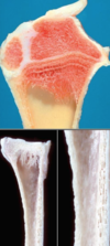
Osteoporosis

56
Q
A

Osteoporosis

57
Q
A

Osteoporosis

58
Q
A

Osteoporosis

59
Q
A

Costochondral junction, rickets

60
Q
A

Pig, rib, dysplasia

61
Q
A

Fibrous osteodystrophy, “Rubber jaw”

62
Q
A

Fibrous osteodystrophy

63
Q
A

Fibrous osteodystrophy

64
Q
A

Fibrous osteodystrophy

65
Q
A
66
Q
A
67
Q
A
68
Q
A
69
Q
A

Congenital porphyria

70
Q
A

Porphyria

71
Q
A

Erythropoietic porphyria

72
Q
A

Exostosis

73
Q
A

Enostosis

74
Q
A

Osteophyte

75
Q
A

Enthesophyte

76
Q
A

Hyperostosis

77
Q
A

Hypertrophic osteopathy

78
Q
A

Bone, hypertrophic pulmonary osteoarthropathy

79
Q
A
80
Q
A

Osteochondromatosis feline leucopenia

81
Q
A
82
Q
A

Subchondral cyst

83
Q
A

Compression force

84
Q
A

Avulsion/distraction forces

85
Q
A

Shear forces @ mid diaphysis

86
Q
A

Bending forces - two forces opposed to mid-diaphysis

87
Q
A

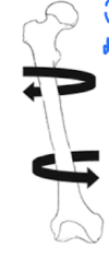
Torsion force - twisting on longitudinal axis

88
Q
A

Compound FX

89
Q
A

Transverse FX - perpendicular to long axis of bone

90
Q
A

Linear FX - parallel to long axis

91
Q
A

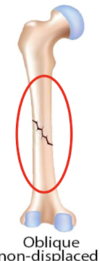
Oblique non-displaced - tangential (along), remain relatively aligned

92
Q
A

Oblique displaced FX

93
Q
A

Spiral 3D FX

94
Q
A

Greenstick FX - cortical bone, minimal involvement of underlying trabeculae bone

95
Q
A

Comminuted FX - severe forces (shatter FX fragments)

96
Q
A
97
Q
A
98
Q
A

Direct or contact healing

99
Q
A

Small gap healing

100
Q
A
101
Q
A

Thorax gunshot perforations

102
Q
A

FX fixation devices - wires, screws, plates + intramedullary nails

103
Q
A

Epiphysiolysis - dog

104
Q
A

Valgus deformity - deviation of limb laterally, damage on lateral aspect

105
Q
A

Varus deformity - deviation of limb medially, damage occurred on medial aspect of physis

106
Q
A

Sesamoid FX

107
Q
A

Sesamoid FX

108
Q
A
109
Q
A

Chronic pyogranulomatous osteomyelitis in cow - Actinomyces bovis = lumpy jaw

110
Q
A

Sequestrum in a foal with Salmonella osteomyelitis

111
Q
A

Suppurative vertebral osteomyelitis in pig, usually Streptococcus suis

112
Q
A

Highly suggestive histologic feature = Splendore-Hoeppli phenomenon (intense eosinophilic material)

113
Q
A
114
Q
A
115
Q
A
116
Q
A
117
Q
A
118
Q
A

Bone osteomyelitis physis

119
Q
A

Bone abscess

120
Q
A

Large dog breeds

121
Q
A

Panosteitis in a German Shepherd dog. Patchy foci of increased bone density within the medullary cavity (arrows)

122
Q
A

Panosteitis

123
Q
A
124
Q
A

Aseptic necrosis

125
Q
A

Cachexia (atrophy)

126
Q
A

Muscle atrophy

127
Q
A

Hyperadrenocorticism (Cushing’s) - ‘pot belly’ due to abdominal muscle atrophy

128
Q
A

Left superficial cricoarytenoideus dorsalis muscle in horse - denervation atrophy

129
Q
A

Compensatory response to injury

130
Q
A

Vacuolar change

131
Q
A

Internal nuclei

132
Q
A

Whorled and ring fibres

133
Q
A
134
Q
A

Distinct groups of atrophic and hypertrophic fibres in child w/ spinal muscular atrophy, hypertrophic fibres up to 100 micrometres

135
Q
A

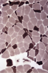
Atrophy restricted to darkly stained type 2 fibres induced by steroid therapy

136
Q
A

ATPase staining following preincubation at pH 4.3 showing atrophy selectively affecting darkly stained type 1 fibres in a case of myotubular myopathy + hypertrophy of pale type 2 fibres

137
Q
A

Internal nuclei (arrow) within fibres of varying size, Duchenne muscular dystrophy

138
Q
A

Chain of internal nuclei in longitudinally sectioned fibre 48 micrometres in diameter

139
Q
A

An area of multiple splitting = small cluster of fibres

140
Q
A

Hypertrophic fibres w/ multiple internal splits - Duchenne muscular dystrophy, small arrow = nuclei along splits, variation in fibre size due to branching of fibres (large arrow)

141
Q
A

Whorled fibre w/ twisted myofibrils

142
Q
A
143
Q
A
144
Q
A
145
Q
A

Segmental necrosis

146
Q
A

Segmental necrosis

147
Q
A

Segmental necrosis

148
Q
A

Muscle necrosis and regeneration

149
Q
A

Muscle regeneration - myotubule grows + differentiates + develops sarcomeres (striation) and nuclei move to the periphery (requires intact basal lamina)

150
Q
A
151
Q
A
152
Q
A

Arthrogryposis - failure of innervation of muscles

153
Q
A

Hypermuscular Belgian blue bull

154
Q
A

Splay leg in pigs, myofibrillar hypoplasia

155
Q
A
156
Q
A

Muscle dystrophy

157
Q
A

Myotonia (channelopathies) - inability of skeletal muscle to relax

158
Q
A
159
Q
A

Splay leg syndrome

160
Q
A

Muscle - lumbar porcine stress syndrome, malignant hyperthermia

161
Q
A
162
Q
A
163
Q
A

Degenerative myopathy - disturbance of circulation -> saddle thrombus

164
Q
A

Degenerative myopathy - disturbance of circulation

165
Q
A
166
Q
A

Ischaemic muscle necrosis - cow

167
Q
A

Rear leg muscle - coagulative necrosis of cow

168
Q
A
169
Q
A

White muscle disease, selenium and vit E deficiency
170
Q
A

White muscle selenium deficiency

171
Q
A

Toxic myopathy - monophasic/polyphasic necrosis

172
Q
A
173
Q
A

Myodegeneration - cassia toxicity

174
Q
A

Exertional myopathy

175
Q
A

Exertional myopathy or ‘capture myopathy’ in wild animals

176
Q
A

Traumatic diaphragmatic hernia

177
Q
A

Myositis ossificans

178
Q
A

Suppurative myositis

179
Q
A

Inflammatory diseases

180
Q
A

Clostridium chauvoei, clostridial myositis

181
Q
A

Pyogenic bacteria, localised suppurative + necrotising myositis - injection site in a cow

182
Q
A

Inflammatory disease

183
Q
A
184
Q
A

FMDV - pale multifocal, irregular, linear stripping (piglet)

185
Q
A
186
Q
A