Neurology - Anatomy and Physiology (2) Flashcards
Cerebral arteries—cortical distribution (459)
- Anterior cerebral artery
- Middle cerebral artery
- Posterior cerebral artery
- Anterior cerebral artery
- Supplies anteromedial surface
- Middle cerebral artery
- Supplies lateral surface
- Posterior cerebral artery
- Supplies posterior and inferior surfaces

Watershed zones
- Between anterior cerebral/middle cerebral, posterior cerebral/middle cerebral arteries.
- Damage in severe hypotension –> upper leg/upper arm weakness, defects in higher-order visual processing.
Regulation of cerebral perfusion (459)
- Brain perfusion relies on tight autoregulation.
- Cerebral perfusion is primarily driven by Pco2
- Po2 also modulates perfusion in severe hypoxia
- Therapeutic hyperventilation (decreased Pco2) helps decrease intracranial pressure in cases of acute cerebral edema (stroke, trauma) via decreased cerebral perfusion by vasoconstriction.

Effects of strokes:
Middle Cerebral Artery (MCA)
- Type of artery / circulation
- Area of lesion
- Symptoms
- Type of artery / circulation
- Anterior circulation
- Area of lesion
- (1) Motor cortex—upper limb and face.
- (2) Sensory cortex—upper limb and face.
- (3) Temporal lobe (Wernicke area); frontal lobe (Broca area).
- Symptoms
- (1) Contralateral paralysis
- Upper limb and face.
- (2) Contralateral loss of sensation
- Upper and lower limbs, and face.
- (3) Aphasia if in dominant (usuallyleft) hemisphere.
- Hemineglect if lesion affects nondominant (usually right) side.
- (1) Contralateral paralysis
Effects of strokes:
Anterior Cerebral Artery (ACA)
- Type of artery / circulation
- Area of lesion
- Symptoms
- Type of artery / circulation
- Anterior circulation
- Area of lesion
- (1) Motor cortex—lower limb.
- (2) Sensory cortex—lower limb.
- Symptoms
- (1) Contralateral paralysis—lower limb.
- (2) Contralateral loss of sensation—lower limb.
Effects of strokes:
Lenticulo-striate artery
- Type of artery / circulation
- Area of lesion
- Symptoms
- Notes
- Type of artery / circulation
- Anterior circulation
- Area of lesion
- Striatum, internal capsule.
- Symptoms
- Contralateral hemiparesis / hemiplegia.
- Notes
- Common location of lacunar infarcts, 2° to unmanaged hypertension.
Effects of strokes:
Anterior Spinal Artery (ASA)
- Type of artery / circulation
- Area of lesion
- Symptoms
- Notes
- Type of artery / circulation
- Posterior circulation
- Area of lesion
- (1) Lateral corticospinal tract.
- (2) Medial lemniscus.
- (3) Caudal medulla—hypoglossal nerve.
- Symptoms
- (1) Contralateral hemiparesis—upper and lower limbs.
- (2) Decreased contralateral proprioception.
- (3) Ipsilateral hypoglossal dysfunction (tongue deviates ipsilaterally).
- Notes
- Stroke commonly bilateral.
-
Medial medullary syndrome
- Caused by infarct of paramedian branches of ASA and vertebral arteries.
Effects of strokes:
Posterior Inferior Cerebellar Artery (PICA)
- Type of artery / circulation
- Area of lesion
- Symptoms
- Notes
- Type of artery / circulation
- Posterior circulation
- Area of lesion
- Lateral medulla—vestibular nuclei, lateral spinothalamic tract, spinal trigeminal nucleus, nucleus ambiguus, sympathetic fibers, inferior cerebellar peduncle.
- Symptoms
- Vomiting, vertigo, nystagmus
- Decreased pain and temperature sensation from ipsilateral face and contralateral body
- Dysphagia, hoarseness, decreased gag reflex
- Ipsilateral Horner syndrome
- Ataxia, dysmetria.
- Notes
- Lateral medullary (Wallenberg) syndrome.
- Nucleus ambiguus effects are specific to PICA lesions.
- “Don’t pick a (PICA) horse (hoarseness) that can’t eat (dysphagia).”
Effects of strokes:
Anterior Inferior Cerebellar Artery (AICA)
- Type of artery / circulation
- Area of lesion
- Symptoms
- Notes
- Type of artery / circulation
- Posterior circulation
- Area of lesion
- (1) Lateral pons—cranial nerve nuclei, vestibular nuclei, facial nucleus, spinal trigeminal nucleus, cochlear nuclei, sympathetic fibers.
- (2) Middle and inferior cerebellar peduncles.
- Symptoms
- (1) Vomiting, vertigo, nystagmus.
- Paralysis of face, decreased lacrimation, salivation, decreased taste from anterior 2⁄3 of tongue, decreased corneal reflex.
- Face—decreased pain and temperature sensation.
- Ipsilateral decreased hearing.
- Ipsilateral Horner syndrome.
- (2) Ataxia, dysmetria.
- (1) Vomiting, vertigo, nystagmus.
- Notes
-
Lateral pontine syndrome.
- Facial nucleus effects are specific to AICA lesions.
- “Facial droop means AICA’s pooped.”
-
Lateral pontine syndrome.
Effects of strokes:
Posterior Cerebral Artery (PCA)
- Type of artery / circulation
- Area of lesion
- Symptoms
- Type of artery / circulation
- Posterior circulation
- Area of lesion
- Occipital cortex, visual cortex.
- Symptoms
- Contralateral hemianopia with macular sparing.
Effects of strokes:
Basilar artery (BA)
- Type of artery / circulation
- Area of lesion
- Symptoms
- Notes
- Type of artery / circulation
- Posterior circulation
- Area of lesion
- Pons, medulla, lower midbrain, corticospinal and corticobulbar tracts, ocular cranial nerve nuclei, paramedian pontine reticular formation.
- Symptoms
- Preserved consciousness and blinking, quadriplegia, loss of voluntary facial, mouth, and tongue movements.
- Notes
- “Locked-in syndrome.”
Effects of strokes:
Anterior Communicationg Artery (ACom)
- Type of artery / circulation
- Area of lesion
- Symptoms
- Notes
- Type of artery / circulation
- Communicating artery
- Area of lesion
- Most common lesion is aneurysm.
- Can lead to stroke.
- Saccular (berry) aneurysm can impinge cranial nerves.
- Symptoms
- Visual field defects.
- Notes
- Lesions are typically aneurysms, not strokes.
Effects of strokes:
Posterior Communicating Artery (PCom)
- Type of artery / circulation
- Area of lesion
- Symptoms
- Notes
- Type of artery / circulation
- Communicating artery
- Area of lesion
- Common site of saccular aneurysm.
- Symptoms
- CN III palsy—eye is “down and out” with ptosis and pupil dilation.
- Notes
- Lesions are typically aneurysms, not strokes.
Aneurysms
- Definition
- Berry aneurysm
- Location
- Findings
- Associated with…
- Other risk factors
- Charcot-Bouchard microaneurysm
- Definition
- In general, an abnormal dilation of artery due to weakening of vessel wall.
-
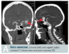
Berry aneurysm
- Location
- Occurs at the bifurcations in the circle of Willis [A].
- Most common site is junction of the anterior communicating artery and anterior cerebral artery.
- Findings
- Rupture (most common complication) leads to subarachnoid hemorrhage (“worst headache of life”) or hemorrhagic stroke.
- Can also cause bitemporal hemianopia via compression of optic chiasm.
- Associated with…
- ADPKD, Ehlers-Danlos syndrome, and Marfan syndrome.
- Other risk factors
- Advanced age, hypertension, smoking, race (increased risk in blacks).
- Location
-
Charcot-Bouchard microaneurysm
- Associated with chronic hypertension
- Affects small vessels (e.g., in basal ganglia, thalamus).
Central post-stroke pain syndrome
- Neuropathic pain due to thalamic lesions.
- Initial sensation of numbness and tingling followed in weeks to months by allodynia (ordinarily painless stimuli cause pain) and dysaesthesia.
- Occurs in 10% of stroke patients.
Epidural hematoma
- Type of hemorrhage
- Definition
- CT
- Type of hemorrhage
- Intracranial hemorrhage
- Definition
- Rupture of middle meningeal artery (branch of maxillary artery), often 2° to fracture of temporal bone.
- Lucid interval.
- Rapid expansion under systemic arterial pressure –> transtentorial herniation, CN III palsy.
- CT
- CT shows biconvex (lentiform), hyperdense blood collection [A] not crossing suture lines.
- Can cross falx, tentorium.

Subdural hematoma
- Type of hemorrhage
- Definition
- CT
- Type of hemorrhage
- Intracranial hemorrhage
- Definition
- Rupture of bridging veins.
- Slow venous bleeding (less pressure = hematoma develops over time).
- Seen in elderly individuals, alcoholics, blunt trauma, shaken baby
- Predisposing factors: brain atrophy, shaking, whiplash
- CT
- Crescent-shaped hemorrhage that crosses suture lines [B].
- Midline shift.
- Cannot cross falx, tentorium.

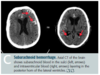
Subarachnoid hemorrhage
- Type of hemorrhage
- Definition
- Type of hemorrhage
- Intracranial hemorrhage
- Definition
- Rupture of an aneurysm (such as a berry [saccular] aneurysm, as seen in Marfan, Ehlers-Danlos, ADPKD) or an AVM.
- Rapid time course.
- Patients complain of “worst headache of my life (WHOML).”
- Bloody or yellow (xanthochromic) spinal tap.
- 2–3 days afterward, risk of vasospasm due to blood breakdown (not visible on CT, treat with nimodipine) and rebleed (visible on CT) [C].
Intraparenchymal (hypertensive) hemorrhage
- Type of hemorrhage
- Definition
- Type of hemorrhage
- Intracranial hemorrhage
- Definition
- Most commonly caused by systemic hypertension [D].
- Also seen with amyloid angiopathy, vasculitis, and neoplasm.
- Typically occurs in basal ganglia and internal capsule (Charcot-Bouchard aneurysm of lenticulostriate vessels), but can be lobar.

Ischemic brain disease/stroke
- Definition
- Stroke imaging
- Histologic features
- 12-48 hours
- 24-72 hours
- 3-5 days
- 1-2 weeks
- >2 weeks
- Definition
- Irreversible damage begins after 5 minutes of hypoxia.
- Most vulnerable—hippocampus, neocortex, cerebellum, watershed areas.
- Ischemic hypoxia—“hypocampus” is most vulnerable.
- Irreversible neuronal injury.
- Stroke imaging
- Bright on diffusion-weighted MRI in 3–30 minutes (highest sensitivity for early ischemia), dark abnormality on noncontrast CT in ~ 12–24 hours.
- Absence of bright areas on noncontrast CT highly accurate to exclude hemorrhage (contraindication for tPA).
- Histologic features
- 12-48 hours: Red neurons
- 24-72 hours: Necrosis + neutrophils
- 3-5 days: Macrophages
- 1-2 weeks: Reactive gliosis + vascular proliferation
- >2 weeks: Glial scar
Hemorrhagic stroke
- Intracerebral bleeding
- Often due to hypertension, anticoagulation, and cancer (abnormal vessels can bleed).
- May be 2° to ischemic stroke followed by reperfusion (increased vessel fragility).
- Basal ganglia are most common site of intracerebral hemorrhage.
Ischemic stroke
- Definition
- 3 types
- Thrombotic
- Embolic
- Hypoxic
- Treatment
- Definition
- Acute blockage of vessels –> disruption of blood flow and subsequent ischemia.
- Results in liquefactive necrosis.
- 3 types
- Thrombotic
- Due to a clot forming directly at the site of infarction (commonly the MCA [A]), usually over an atherosclerotic plaque.
- Embolic
- An embolus from another part of the body obstructs a vessel.
- Can affect multiple vascular territories.
- Often cardioembolic.
- Hypoxic
- Due to hypoperfusion or hypoxemia.
- Common during cardiovascular surgeries, tends to affect watershed areas.
- Thrombotic
- Treatment
- tPA (if within 3–4.5 hr of onset and no hemorrhage/risk of hemorrhage).
- Reduce risk with medical therapy (e.g., aspirin, clopidogrel)
- Optimum control of blood pressure, blood sugars, and lipids
- Treat conditions that increase risk (e.g., atrial fibrillation).

Transient ischemic attack
- Brief, reversible episode of focal neurologic dysfunction
- Last < 24 hours without acute infarction ((-) MRI), with the majority resolving in < 15 minutes
- Deficits due to focal ischemia.
Dural venous sinuses
- Large venous channels that run through the dura.
- Drain blood from cerebral veins and receive CSF from arachnoid granulations.
- Empty into internal jugular vein.

Ventricular system
- Lateral ventricle –>
- 3rd ventricle –>
- 4th ventricle –>
- CSF
- Lateral ventricle –> 3rd ventricle via right and left interventricular foramina of Monro.
- 3rd ventricle –> 4th ventricle via cerebral aqueduct (of Sylvius).
- 4th ventricle –> subarachnoid space via:
- Foramina of Luschka = Lateral.
- Foramen of Magendie = Medial.
- CSF
- Made by ependymal cells of choroid plexus
- Reabsorbed by arachnoid granulations
- Drains into dural venous sinuses.

Hydrocephalus
- Communicating hydrocephalus
- Normal pressure hydrocephalus
- Communicating hydrocephalus
- Communicating (nonobstructive)
- Decreased CSF absorption by arachnoid granulations, which can lead to increased intracranial pressure, papilledema, and herniation (e.g., arachnoid scarring post-meningitis).
- Normal pressure hydrocephalus
- Communicating (nonobstructive)
- Does not result in increased subarachnoid space volume.
- Expansion of ventricles [A] distorts the fibers of the corona radiata and leads to clinical triad of urinary incontinence, ataxia, and cognitive dysfunction (sometimes reversible).
- “Wet, wobbly, and wacky.”

Hydrocephalus
- Hydrocephalus ex vacuo
- Noncommunicating hydrocephalus
- Hydrocephalus ex vacuo
- Communicating (nonobstructive)
- Appearance of increased CSF in atrophy (e.g., Alzheimer disease, advanced HIV, Pick disease).
- Intracranial pressure is normal
- Triad (urinary incontinenc, ataxia, and cognitive dysfunction) is not seen.
- Apparent increase in CSF observed on imaging is actually result of decreased neural tissue due to neuronal atrophy.
- Noncommunicating hydrocephalus
- Noncommunicating (obstructive)
- Caused by a structural blockage of CSF circulation within the ventricular system (e.g., stenosis of the aqueduct of Sylvius).
Spinal nerves
- Number
- Where they exit
- Vertebral disc herniation
- There are 31 spinal nerves in total
- 8 cervical, 12 thoracic, 5 lumbar, 5 sacral, 1 coccygeal.
- 31, just like 31 flavors of Baskin-Robbins ice cream
- Where they exit
- Nerves C1–C7 exit above the corresponding vertebra.
- All other nerves exit below (e.g., C3 exits above the 3rd cervical vertebra; L2 exits below the 2nd lumbar vertebra).
-
Vertebral disc herniation
- Nucleus pulposus (soft central disc) herniates through annulus fibrosus (outer ring)
- Usually occurs posterolaterally at L4–L5 or L5–S1.
Spinal cord—lower extent
- Lower borders
- Spinal cord
- Subarachnoid space
- Lumbar puncture
- Lower borders
- In adults, spinal cord extends to lower border of L1–L2 vertebrae.
- Subarachnoid space (which contains the CSF) extends to lower border of S2 vertebra.
- Lumbar puncture
- Lumbar puncture is usually performed between L3–L4 or L4–L5 (level of cauda equina).
- Goal of lumbar puncture is to obtain sample of CSF without damaging spinal cord.
- To keep the cord alive, keep the spinal needle between L3 and L5.
Spinal cord and associated tracts (465)
- Corticospinal and spinothalamic tracts
- Dorsal column
- Corticospinal and spinothalamic tracts
- Legs (Lumbosacral) are Lateral in Lateral corticospinal, spinothalamic tracts.
- Dorsal column
- Organized as you are, with hands at sides.
- Arms outside, legs inside.

