Biology 10: Homeostasis Flashcards
renal hilum
a deep slit in the center of the kidney’s medial surface
where the renal artery, renal vein, and ureter pass

portal system
system which consists of two capillary beds in series through which blood must travel before returning to the heart
renal portal system pathway
renal artery –> afferent arterioles –> glomerulus –> efferent arteriole –> vasa recta
detrusor muscle
muscular lining around the bladder
innervated by parasympathetic system to contract
what are the two sphincters that urine must pass through to leave the body?
internal urethral sphincter - smooth muscle - normally contracted - involuntary
external urethral sphincter - skeletal muscle - voluntary
micturition reflex
when the bladder is full…
- stretch receptors send message to nervous system
- parasympathetic signaling causes detrusor muscle to contract
- internal urethral sphincter relaxes
individual chooses to relax the external urethral sphincter
filtrate
fluid collected from the passage of blood through the glomerulus into Bowman’s space
does not contain cells or proteins due to size of glomerular pores

what happens to blood that is not filtered at the Bowman’s capsule?
blood remaining in the glomerulus travels into afferent arterioles, which empty into vasa recta

filtration
movement of solutes from blood to filtrate at Bowman’s capsule

secretion
movement of solutes from blood to filtrate anywhere besides Bowman’s capsule
allow kidneys to eliminate ions or other substances when present in excess amounts in the blood
allows kidneys to excrete wastes that are too large to pass through glomerular pores
reabsorption
movement of solutes from filtrate to blood
compounds filtered/secreted can be taken back up for use
glucose, amino acids, vitamins are always reabsorbed
water reabsorbed depending on ADH or aldosterone
proximal convoluted tubule
filtrate’s first stop after the Bowman’s capsule
amino acids, glucose, water-soluble vitamins, salts, water are reabsorbed
solutes enter the interstitium and are picked up by the vasa recta to be returned to the bloodstream
secretion of H+, K+, NH3+, urea

interstitium
the connective tissue surrounding the nephron

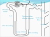
descending loop of Henle
receives filtrate from the proximal convoluted tubule
dives deep into the medulla with an increasing osmolarity
permeable only to water
as it gets deeper, the interstitial concentration favors the outflow of water which is reabsorbed in the vasa recta

countercurrent multiplier system
created by the vasa recta and nephron
the flow of filtrate through the loop of Henle is in the opposite direction from the flow of blood through the vasa recta
filtrate is constantly exposed to hypertonic blood, allows for maximal reabsorption of water

ascending limb of Henle
portion of the loop of Henle that is only permeable to salts, impermeable to water
at deeper parts of the medulla, salt concentrations are high but decrease as the ascending limb rises
increasing amounts of salts are removed from the filtrate as it travels up the loop of Henle

diluting segment
portion of the loop of Henle at the transition of the loop of Henle from the inner to outer medulla
thicker because the cells lining the tube are larger
cells have lots of mitochondria, reabsorption of Na and Cl by active transport
only portion of the nephron that can produce urine that is more dilute than the blood
how does filtrate change as it moves through the loop of henle?
at the beginning of the loop of henle, filtrate is isotonic to interstitium
at the end, there is a slight degree of dilution
volume of filtrate is significantly reduced - lots of water reabsorbed
distal convoluted tubule
portion of the nephron after the ascending limb of the loop of henle
responds to aldosterone, which promotes sodium reabsorption - water follows
the urine is concentrated and its volume is decreased
also the site of waste product secretion
collecting duct
final section of the nephron
responsive to both aldosterone and ADH to change permeability of water
as permeability of the collecting duct increases, water is reabsorbed, further concentrating the urine
if well hydrated, collecting duct is fairly impermeable to salt and water
how does aldosterone work to decrease blood pressure?
- decreased blood pressure stimulates release of renin from juxtaglomerular cells in kidney
- renin cleaves angiotensin (liver protein) –> angiotensin I
- angiotensin-converting enzyme (ACE) in lungs cleaves A1 –> angiotensin II
angiotensin II promotes the release of aldosterone from the adrenal cortex
aldosterone causes distal convoluted tubule and collecting duct to reabsorb calcium, causing water reabsorption, increasing blood volume and pressure
how does ADH work to decrease blood pressure?
released by posterior pituitary in response to high blood osmolarity
cause more water to be reabsorbed by making the cell junctions of the collecting duct leaky
high conc of interstitium causes water to follow
osmotic pressure
the “sucking” pressure that draws water into the vasculature caused by all dissolved particles
oncotic pressure
osmotic pressure attributable to dissolved proteins specifically
what are the three layers of the skin from the deepest to outermost layer?
hypodermis (subcutaneous layer)
dermis
epidermis
from deepest layer outward, what are the strata of the epidermis?
stratum basale
stratum spinosum
stratum granulosum
stratum lucidum
stratum corneum
Come Lets Get Sun Burned

stratum basale
deepest layer of the epidermis
contains stem cells
responsible for the proliferation of keratinocytes (produce keratin)

stratum spinosum
second-most deepest epidermal layer
cells are connected to each other
site of Langerhans cells

stratum granulosa
third-most deepest layer in epidermis
keratinocytes die and lose their nuclei

stratum lucidum
fourth-deepest layer of the epidermis
only present in thick, hairless skin (sole of feet, palm of hands)
nearly transparent

stratum corneum
outermost layer of epidermis
several layers of flattened keratinocytes
forming a barrier that prevents invasion by pathogens
helps to prevent loss of fluids and salt

calluses
regions of the epidermis that form from excessive keratin deposition in areas of repeated strain due to friction
melanocytes
cell type derived from neural crest cells, found in stratum basale
produce melanin that is passed onto keratinocytes
langerhans cells
special macrophages that reside within the stratum spinosum
capable of presenting antigens to T-cells in order to activate the immune system
what are the layers of the dermis?
papillary layer (upper) of loose connective tissue
denser reticular layer (lower)
epidermis
top layer of skin
main cells are keratinocytes
divided into five strata
dermis
second layer of the skin
sweat glands, blood vessels, hair follicles originate here
merkel cells (discs)
sensory receptors present at epidermal-dermal junction
responsible for deep pressure and texture sensation within the skin
free nerve endings
sensory receptors in the skin that respond to pain
meissner’s corpuscles
sensory receptors in the skin that respond to light touch
ruffini endings
sensory receptors in the skin that respond to stretch
Pacinian corpuscles
sensory receptors that respond to deep pressure and vibration
hypodermis
innermost layer of skin
layer of connective tissue that connects the skin to the rest of the body
contains fat and fibrous tissue
sweating
cooling mechanism that is contrlled by the autonomic nervous system
body temp rises above set point, hypthalamus starts thermoregulation
postganglionic sympathetic neurons that use acetylcholine innervate sweat glands
evaporation of water from skin absorbs body heat
how does the body react to cold conditions?
-
arrector pili muscles contract - causing piloerection (hairs of skin to stand up)
- traps layer of heated air near skin
- arterioles that feed capillaries of skin constrict, keeping blood from reaching skin
- skeletal muscles contract rapidly - shivering
white fat
a layer of fat just below the skin that helps to insulate the body
brown fat
fat present in infants
less efficient ETC - more heat energy released as fuel burned
